UNIT I CELL OF INFLAMMATION INFLAMMATION THE INFLAMMATORY


 Initial")

 • Basophils comprise about 1% of circulating leucocytes and are")


 • From 2 sources: with common morphology, function and")

 Products of complement d)Some coagulation factors (factor V and thromboplastin) which convert fibrinogen")

 Foreign body giant cells. contain numerous nuclei (up")

 Anaplastic cancer giant cells. • larger, have numerous")
, JAYPEE Publication")
- Slides: 18
UNIT I CELL OF INFLAMMATION
INFLAMMATION
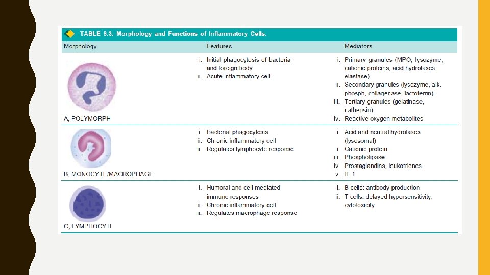
THE INFLAMMATORY CELLS • The cells participating in acute and chronic inflammation are circulating leucocytes, plasma cells and tissue macrophages. • Role of these cells in inflammation. 1. Polymorphonuclear Neutrophils (PMNs) • Commonly called as neutrophils or polymorphs, these cells along with basophils and eosinophils are known as granulocytes due to the presence of granules in the cytoplasm. • These granules contain many substances like proteases, myeloperoxidase, lysozyme, esterase, aryl sulfatase, acid and alkaline phosphatase, and cationic proteins. • These cells comprise 40 -75% of circulating leucocytes and their number is increased in blood (neutrophilia) and tissues in acute bacterial infections. • These cells arise in the bone marrow from stem cells.
• The functions of neutrophils in inflammation are as follows: • i) Initial phagocytosis of microorganisms as they form the first line of body defence in bacterial infection. • The steps involved are adhesion of neutrophils to vascular endothelium, emigration through the vessel wall, chemotaxis, engulfment, degranulation, killing and degradation of the foreign material. • ii) Engulfment of antigen-antibody complexes and material. on microbial • iii) Harmful effect of neutrophils in causing basement membrane destruction of the glomeruli and small blood vessels
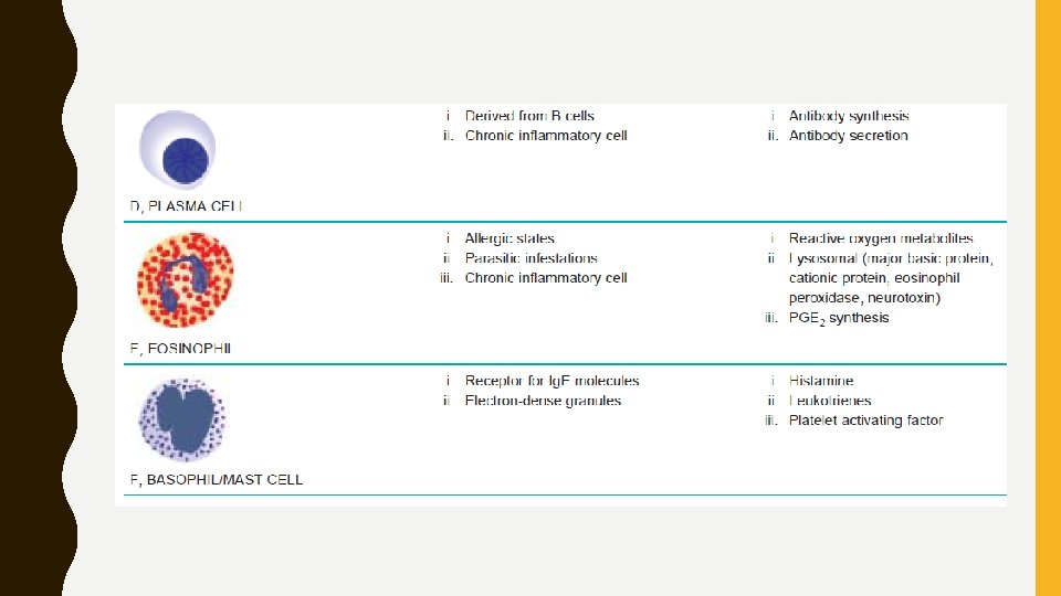
2. Eosinophils • Larger than neutrophils, fewer in no. , 1 -6% of total blood leucocytes • Structure and function similar with neutrophils like production in bone marrow, locomotion, phagocytosis, lobed nucleus and presence of granules in cytoplasm (having variety of enzymes) • Proteins: bactericidal and toxic against helminthic parasites • Granules of eosinophils- richer in myeloperoxidase than neutrophils & lack lysozyme. • High steroid hormones leads to fall in number of eosinophils and even disappearance from blood • Increase in following conditions : i. Allergic conditions ii. Parasitic infestations iii. Skin diseases
3. Basophils (Mast Cells) • Basophils comprise about 1% of circulating leucocytes and are morphologically and pharmacologically similar to mast cells of tissue. • Contains coarse basophilic granules in cytoplasm and a polymorphonuclear nucleus • Granules: Contain heparin and histamine • Basophils and mast cells have receptors for Ig. E • Degranulate on cross-linking with antigen • The role of these cells in inflammation are: i. In immediate and delayed type of hypersensitivity reactions ii. Release of histamine by Ig. E-sensitised basophils
4. Lymphocytes • Next to neutrophils, these cells are the most numerous of the circulating leucocytes (20 -45%). • Present in blood (Highest numbers), spleen, thymus, lymph nodes and mucosa-associated lymphoid tissue (MALT). • Scanty cytoplasm and consist almost entirely of nucleus • B lymphocytes: Antibody formation • T lymphocytes: Cell-mediated immunity • Participate in the following types of inflammatory responses: i. In tissues, chronic inflammation and late stage acute inflammation. ii. In blood, increased tuberculosis (lymphocytosis) in chronic infections like
5. Plasma Cells • larger than lymphocytes- with more abundant cytoplasm and an eccentric nucleus • Normally not seen in peripheral blood • Develop from B lymphocytes • Rich γ-globulin in their cytoplasm • Thus, interrelationship between plasmacytosis and hyperglobulinaemia • Most active in antibody synthesis • Increased in the following conditions: i. prolonged infection with immunological responses e. g. in syphilis, rheumatoid arthritis, tuberculosis ii. hypersensitivity states iii. multiple myeloma
6. Mononuclear-Phagocyte System (Reticuloendothelial System) • From 2 sources: with common morphology, function and origin • These are as under: • Blood monocytes. comprise 4 -8% of circulating leucocytes. • Tissue macrophages. following cells in different tissues: i. Macrophages in inflammation ii. Histiocytes - macrophages present in connective tissues iii. Kupffer cells- liver cells. iv. Alveolar macrophages (type II pneumocytes) in lungs v. Macrophages/histiocytes of the bone marrow vi. Tingible cellsofoflymph nodes germinalbody centres vii. Littoral cells of splenic sinusoids viii. Osteoclasts in the bones. ix. Microglial cells of the brain x. Langerhans’ cells/dendritic histiocytes of the skin xi. Hoffbauer cells of the placenta xii. Mesangial cells of glomerulus
• Mononuclear phagocytes-participate in immune system • Role in inflammation are as under: i. Phagocytosis (cell eating) and pinocytosis (cell drinking) ii. Macrophages on activation by lymphokines released by T lymphocytes or by non-immunologic stimuli release a variety of biologically active substances as under: a) Proteases like collagenase and elastase- degrade collagen and elastic tissue b) Plasminogen activator- activates the fibrinolytic system
c) Products of complement d)Some coagulation factors (factor V and thromboplastin) which convert fibrinogen to fibrin e) Chemotactic agents for other leucocytes f) Metabolites of arachidonic acid
7. Giant Cells • multinucleate giant cells exist in normal tissues (e. g. osteoclasts in the bones, trophoblasts in placenta, megakaryocytes in the bone marrow). • In chronic inflammation when the macrophages fail to deal with particles to be removed, they fuse together and form multinucleated giant cells. • Morphologically distinct giant cells appear in some tumours also. • Some of the common types of giant cells are described below:
A. Giant cells in inflammation: i) Foreign body giant cells. contain numerous nuclei (up to 100) size and shape uniform in resemble nuclei of macrophages • nuclei scattered throughout the cytoplasm • seen in chronic infective granulomas, leprosy and tuberculosis ii) Langhans’ giant cells. seen in tuberculosis and sarcoidosis • nuclei resembles nuclei of macrophages and epithelioid cells. • nuclei re-arranged- either around periphery in the form of horseshoe or ring, or clustered at the two poles of giant cell iii) Touton giant cells. multinucleated cells have vacuolated to lipid content e. g. in xanthoma (Fatty growth under skin) cytoplasm due iv) Aschoff giant cells. multinucleate giant cells are derived from histiocytes- seen in rheumatic nodule cardiac
Giant cells of various types. A, Foreign body giant cell with uniform nuclei dispersed throughout cytoplasm B, Langhans’ giant cells with uniform nuclei arranged peripherally or clustered at the two poles C, Touton giant cell with circular pattern of nuclei and vacuolated cytoplasm D, Anaplastic tumour giant cell with nuclei of variable size and shape. E, Reed-Sternberg cell. F, Osteoclastic tumour giant cell
B. Giant cells in tumours: i) Anaplastic cancer giant cells. • larger, have numerous nuclei • Giant cells are not derived from macrophages-formed from dividing nuclei of neoplastic cells e. g. carcinoma of liver, various soft tissue sarcomas etc. ii) Reed-Sternberg cells. are also malignant tumour giant cellsgenerally binucleate seen in various Hodgkin’s lymphomas. iii) Giant cell tumour of bone. tumour of the bones has uniform distribution of osteoclastic giant cells spread in stroma
• Reference: Textbook of Pathology, by Harsh Mohan (Author), JAYPEE Publication