UNIT 10 THE DIGESTIVE SYSTEM Functions of the


, vitamins, and water across the digestive")


 from")


 Functions when you eat: Lubricating the mouth Moistening and lubricating")

 Breaks down connective tissues in meat and plant fibers in")


 Intestinal phase Trigger")


- Slides: 84
UNIT 10 – THE DIGESTIVE SYSTEM
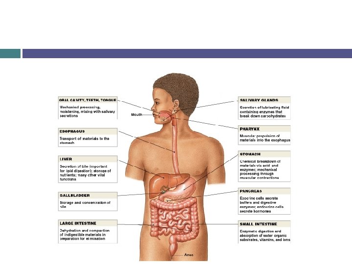
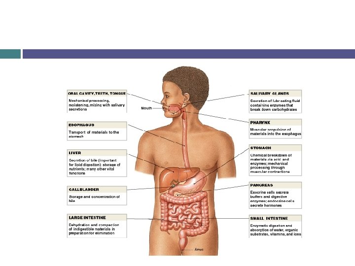
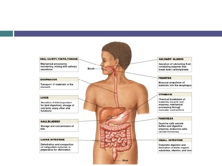
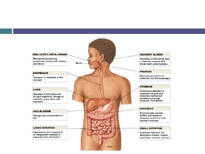
Functions of the Digestive System
Ingestion Materials enter the digestive tract via the mouth
Mechanical Processing Crushing and shearing that make materials easier to propel along the digestive tract. Increases surface area Makes material more susceptible to enzymes Examples are tearing and mashing with teeth and squashing and compaction by tongue. Swirling, mixing and churning of stomach provides mechanical processing after ingestion.
Digestion The chemical breakdown of food into small fragments suitable for absorption. Polysaccharides into monosaccharides, large proteins into small proteins and amino acids, triglycerides into small lipid molecules.
Secretion Release of water, acids, enzymes, buffers, and salts by digestive epithelium and glandular organs.
Absorption Movement of organic substrates, electrolytes (inorganic ions), vitamins, and water across the digestive epithelium into the body.
Excretion Removal of waste products from the body. The process is called defecation, and the product is called feces.
Additional Functions Lining of the digestive tract also protects body tissues from: Corrosive digestive acids Mechanical stresses Bacteria swallowed in food or those that reside in the digestive tract
The Peritoneum
Peritoneal Cavity Contains the organs of the abdominopelvic cavity.
Mesentaries Sheets of serous membrane that suspend portions of the digestive tract. Stabilize the positions of organs and prevent the intestines from becoming entangled during digestive movements or sudden changes in body position.
Histological Organization of Digestive Tract
The Lining of the Digestive Tract Digestive Epithelium Oral cavity, pharynx, and esophagus are lined with stratified squamous epithelium Stomach, small intestine, and most of the large intestine are lined with simple columnar epithelium Has folds, called plicae Increases surface area for absorption
The Lining of the Digestive Tract
The Outer Layer Dominated by smooth muscle Play a key role in mechanical processing and movement of material along the tract.
The Movement of Digestive Materials
Peristalsis Contractions of smooth muscle that propels small oval masses of material (bolus) from one portion of the digestive tract to another. Circular muscles behind the bolus contract, while circular muscles ahead of the bolus relax. Longitudinal muscles ahead of the bolus contract, shortening adjacent segments. A wave of contraction in the circular muscles then forces the bolus forward.
Peristalsis
Segmentation Cycles of contraction in the small intestine that churn and fragment the bolus. Does not follow a set pattern. Does not push the bolus in any one direction.
The Structures of the Digestive System
Oral Cavity that mouth opens into. Functions: Sensory analysis of material before swallowing Mechanical processing through actions of teeth, tongue, and palatal surfaces Lubrication by mixing with mucous and salivary gland secretions Limited digestion of carbohydrates and lipids
Oral Cavity
Oral Cavity Lined by stratified squamous epithelium Nutrients are not absorbed in oral cavity, but lining inferior to tongue is thin enough and vascular enough to allow rapid absorption lipidsoluble drugs Nitroglycerine Uvula – dangling process that helps prevent food from entering the pharynx prematurely
Tongue Manipulates material inside mouth and occasionally used to bring food into the oral cavity (ice cream) Functions: Mechanical processing by compression, abrasion, and distorting Manipulation to assist in chewing and to prepare materials for swallowing Sensory analysis by touch, temp, and taste receptors Secretion of mucins and the enzyme lingual lipase Lipid breakdown
Tongue
Salivary Glands Three pairs secrete into oral cavity: Parotid salivary glands – located in the superior portion of your cheeks Secrete salivary amylase – breaks down starches Sublingual salivary gland – located in floor of mouth, under tongue Produce lubricant mucous secretion that acts as a buffer and Submandibular salivary glands – located in floor of mouth along inner surfaces of mandible Secrete buffers, mucins, and salivary amylase
Salivary Glands Saliva 1 -1. 5 liters are produced each day 99. 4% water 0. 6% is and assortment of electrolytes (Na+, Cl-, and HCO 3 -), buffers, mucins, antibodies, enzymes and waste products. Mucins are responsible for lubrication Buffers help keep p. H of mouth around 7. 0 Helps control bacteria growth
Salivary Glands Saliva (con’t) Functions when you eat: Lubricating the mouth Moistening and lubricating the materials in your mouth Dissolving chemicals that can stimulate taste buds and provide sensory info about the material Initiating the digestion of complex carbohydrates with salivary amylase and lipids with lingual lipase.
Salivary Glands
Teeth Perform mastication (chewing) Breaks down connective tissues in meat and plant fibers in vegetable matter Matrix is similar to that of bones except that there are no cells in teeth. The enamel that covers the teeth is made up of crystallized calcium phosphate Hardest biologically manufactured substance
Pharynx Common passageway for solid food, liquid, and air
The Esophagus Hollow muscular tube that begins posterior to the cricoid cartilage, runs posterior to the trachea, and ends at the stomach Upper esophageal shpincter prevents air from entering the esophagus Lower esophagus sphincter (cardiac sphincter) prevents backflow from stomach into esophagus
The Esophagus
The Esophagus Bolus makes the trip through the esophagus in about 9 seconds Drier bolus takes longer and may require a second peristaltic wave Liquids make the journey in a few seconds
The Esophagus
The Stomach Functions: Storage of ingested food Mechanical processing of ingested food Breakdown of chemical bonds in food through the actions of acids and enzymes Produces intrinsic factor – required for absorption of vitamin B 12 Chyme – a viscous, highly acidic soupy mixture of partially digested food Formed from ingested food mixing with the secretions of the stomach
The Stomach
The Stomach Anatomy: Lesser curvature forms medial surface Greater curvature forms lateral surface Stomach contains rugae, or folds, that allows the stomach to expand The lining of the stomach is simple columnar epithelium Has a mucous layer that protects the cells from stomach acid
The Stomach Anatomy: Regions Cardia – superior, medial part Junction between esophagus and stomach Fundus – superior and lateral to the cardia Body – area between fundus and pylorus Mixing tank for ingested food Contains gastric glands Pylorus – most inferior part (form the “J” shape) Secrets gastrin
The Stomach
The Stomach
The Stomach Gastric glands Produce 2 about 1500 ml of gastric juice/day types Parietal Secrete intrinsic factor – needed for vitamin B 12 absorption Secrete HCl – keeps stomach at a p. H of 1. 5 -2 Chief cells Secrete pepsinogen – turns to pepsin in presence of HCl Breaks down proteins Secretes rennin in infants – breaks down milk proteins Secretes gastric lipase – breaks down milk fats
The Stomach Pyloric glands Produce mucous Produce gastrin – stimulates the parietal and chief cells, as well as contractions in the gastric wall
The Stomach Regulation of Gastric Activity Controlled by the CNS, regulated by short reflexes, and regulated by hormones of the digestive tract. Divided into 3 phases Cephalic Trigger – see, smell, taste, think of food Result – gastric juice production (500 ml/h) Gastric Trigger – arrival of food in stomach Result – contraction of stomach muscles to mix food, stimulates gastrin release, stimulates acid release
The Stomach Regulation of Gastric Activity Divided into 3 phases (con’t) Intestinal phase Trigger – chyme enters the small intestine Result – release of secretin; allows small amounts of chyme enter the small intestine This allows for proper digestive, secretory, and absorptive actions of the small intestine
The Stomach
The Stomach Digestion in the stomach Preliminary digestion of proteins by pepsin, carbohydrates by salivary amylase, and lipids by lingual lipase NO NUTRIENT ABSORPTION TAKES PLACE IN THE STOMACH!!!! Although ethyl alcohol and aspirin can be absorbed in the stomach Fats slow the absorption of alcohol in the stomach
The Small Intestine and Associated Organs The small intestine is the key player in digestion and nutrient absorption 90% of nutrients are absorbed in the small intestine Averages 20 ft in length
The Small Intestine and Associated Organs The small intestine has three segments Duodenum – closest to stomach (10 in. long) Acts as a mixing bowl Jejunum – most of the chemical digestion and absorption take place here 8 ft long Ileum – longest segment (12 ft) Has ileocecal valve – controls contents entering the large intestine
The Small Intestine and Associated Organs
The Small Intestine and Associated Organs The small intestine contains folds, called plicae Increases surface area for absorption Also contain villi Finger-like projections that also increase surface area Lined with simple columnar epithleium. Secrete the brush border enzymes Small intestine has 2200 ft 2 of surface area Lined with smooth muscle, which when contracted in the proper sequence, propels the material through the tract
The Small Intestine and Associated Organs
The Small Intestine and Associated Organs Intestinal Movements Initially in the duodenum, weak peristalsis moves the chyme The gastroenteric reflex is responsible for the movement of material through the small intestine. Also The responsible for secretions in the small intestine. gastroileal reflex relaxes the ileocecal valve This allows material to enter the large intestine
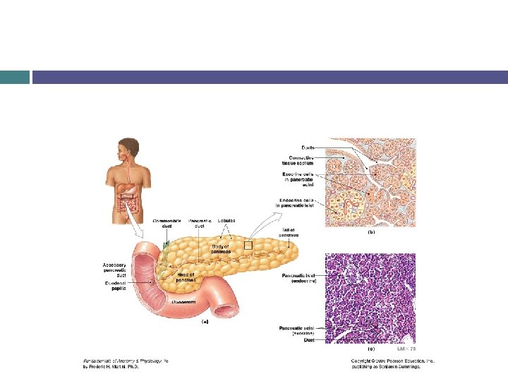
The Small Intestine and Associated Organs Pancreas Located posterior to the stomach Has a lumpy texture Secretes about 1000 ml of pancreatic juice/day into the small intestine Enzymes, water, and ions
The Small Intestine and Associated Organs Pancreatic Enzymes Pancreatic alpha-amylase – breaks down starches Similar to salivary amylase Pancreatic lipase – breaks down complex lipids Nucleases – break down nucleic acids Proteolytic enzymes – break down proteins Proteases – break down large proteins Peptidases – break down small proteins into amino acids
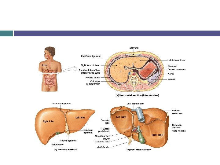
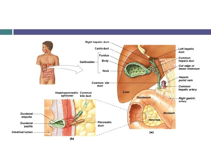
The Small Intestine and Associated Organs Liver Largest visceral organ, most versatile organ in the body Responsible for three general categories of functions Metabolic regulation Hematological regulation Bile production
The Small Intestine and Associated Organs Liver Metabolic regulation Carbohydrate metabolism Lipid metabolism If blood glucose levels fall, glycogen is broken down and glucose is released. The liver also can make glucose from amino acids. If blood glucose levels rise, the liver can remove glucose from the blood Can break down and release lipids as needed, or remove lipids from the blood stream as needed Amino acid metabolism Removes excess amino acids from blood to make proteins for use, or lipids for energy storage
The Small Intestine and Associated Organs Liver Metabolic Waste product removal Removes wastes from the blood Vitamin storage Converts iron reserves and stores as a protein-iron complex. Drug storage Fat-soluble vitamins (A, D, E, and K) along with B 12 are stored in the liver Mineral regulation (con’t) inactivation Removes and breaks down circulating drugs
The Small Intestine and Associated Organs Liver Hematological Liver regulation is largest blood reservoir in the body Removes old, dead, damaged RBCs, debris, and pathogens from the blood stream Removes circulating hormones Removes antibodies of immune system Removes toxins
The Small Intestine and Associated Organs Liver Makes bile Breaks down large droplets of lipids into smaller droplets (emulsification) This increases surface are for more efficient lipid digestion
The Small Intestine and Associated Organs Gall bladder Stores and concentrates bile to be released into the small intestine. A full gall bladder can contain 40 -70 ml of bile Gall stones form when the concentration of bile becomes to high
The Small Intestine and Associated Organs Intestinal Hormones Secretin – released when chyme reaches the duodenum Increases secretion of bile and buffers Reduces gastric motility (movement) and secretion Gastrin – secreted when large amounts of incompletely digested proteins are present Promotes enzymes gastric motility and secretion of acids and
The Small Intestine and Associated Organs Intestinal Absorption It takes about 5 hours for material to pass the length of the small intestine Absorption increased by villi
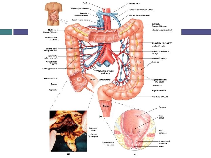
The Large Intestine Begins at the end of the small intestine and ends at the anus. Functions: Reabsorption of water and compaction of intestinal contents into feces Absorption of important vitamins from bacterial actions Storage of fecal matter prior to defecation About 5 ft long and is divided into 3 sections Cecum, colon, rectum
The Large Intestine The Cecum Material enters the cecum from the small intestine through the ileocecal valve The appendix is attached to the cecum Believed to have a function in the lympnatic system
The Large Intestine The Colon Four subdivisions Ascending Transverse Descending Sigmoid
The Large Intestine The Rectum Last 6 inches of digestive tract Expandable organ for the temporary storage of feces Anal canal – last portion of rectum Anus – the exit of the anal canal Anal sphincter – regulates the release of feces from the rectum
The Large Intestine Absorption in the Large Intestine About 1500 ml of material enters the large intestine each day Only about 200 ml of feces is ejected Composition 75% of feces water 5% bacteria Rest is indigestible materials, inorganic matter, remains of epithelial cells
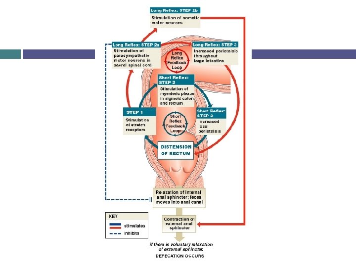
The Large Intestine Movement in the Large Intestine Movement from the cecum to the transverse colon is slow (hours) to allow for water absorption Peristaltic waves move material through the colon Mass movements-powerful peristatlic contractions As material moves from the sigmoid colon to the rectum, the defecation reflex is triggered Defecation reflex is responsible for moving feces toward the anus The anal sphincter must relax for feces to exit the body
Digestion and Absorption The nutrient molecules in food are too large to be absorbed and used by the body. Digestion breaks down the food into smaller particles, then disassembles the particles into absorbable and usable molecules Carbohydrases break down carbohydrates Proteases break down proteins Complex Lipases and time consuming break down lipids
Digestion and Absorption Carbohydrate and amino acid absorption takes place via facilitated diffusion. Lipid absorption takes place via diffusion The body then uses the absorbed molecules to make energy, or to synthesize carbohydrates, proteins or lipids.
Digestion and Absorption Water absorption takes place in the large intestine via osmosis. Ion absorption can take place via diffusion or active transport, depending on the concentrations and charges of the particular ions. Vitamin absorption takes place via diffusion.
Major Digestive Hormones
Digestive Enzymes