Digestive System Azami PHD Digestive System Anatomy Digestive


 • Three phases – Voluntary – Pharyngeal • Reflex – Esophageal • Reflex")


- Slides: 34
Digestive System Azami PHD
Digestive System Anatomy • Digestive tract – Alimentary tract or canal – GI tract • Accessory organs – Primarily glands • Regions – – – – Mouth or oral cavity Pharynx Esophagus Stomach Small intestine Large intestine Anus
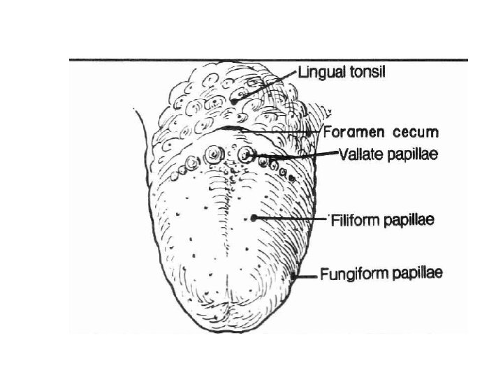
Oral Cavity • Mouth or oral cavity – Vestibule: Space between lips or cheeks and alveolar processes – Oral cavity proper • Lips (labia) and cheeks • Palate: Oral cavity roof – Hard and soft • Palatine tonsils • Tongue: Involved in speech, taste, mastication, swallowing
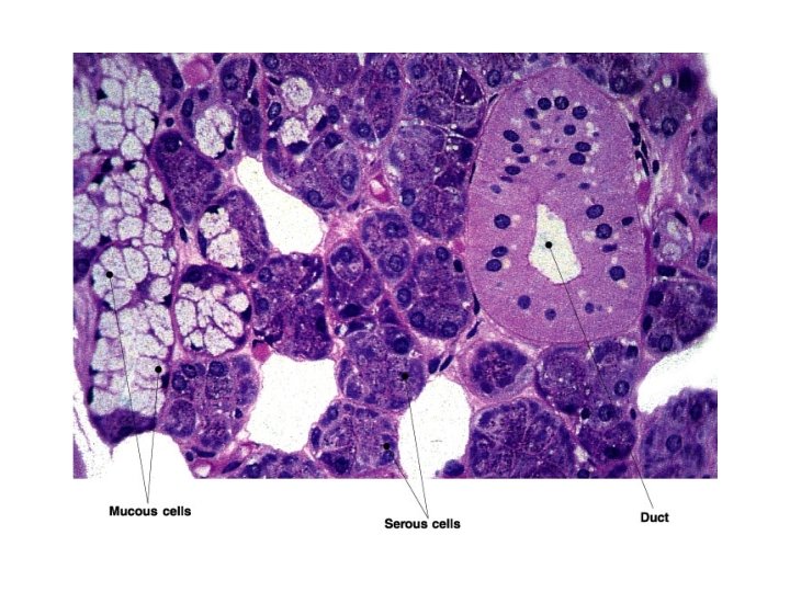
Salivary Glands • Produce saliva – Prevents bacterial infection – Lubrication – Contains salivary amylase • Breaks down Starch • Three pairs – Parotid: Largest – Submandibular – Sublingual: Smallest
Teeth • Two sets – Primary, deciduous, milk: Childhood – Permanent or secondary: Adult (32) • Types – Incisors, canine, premolar and molars
Tooth structure:
Pharynx and Esophagus • Pharynx – Nasopharynx – Oropharynx: Transmits food – Laryngopharynx: Transmits food • Esophagus – Transports food from pharynx to stomach – Passes through esophageal hiatus (opening) of diaphragm and ends at stomach
(Swallowing) • Three phases – Voluntary – Pharyngeal • Reflex – Esophageal • Reflex
Digestive Tract Histology
Digestive System Regulation • Nervous regulation – Involves enteric nervous system • Types of neurons: sensory, motor, interneurons – Coordinates peristalsis and regulates local reflexes • Chemical regulation – Production of hormones – Production of paracrine chemicals
Digestive Tract Anatomy
serous membranes
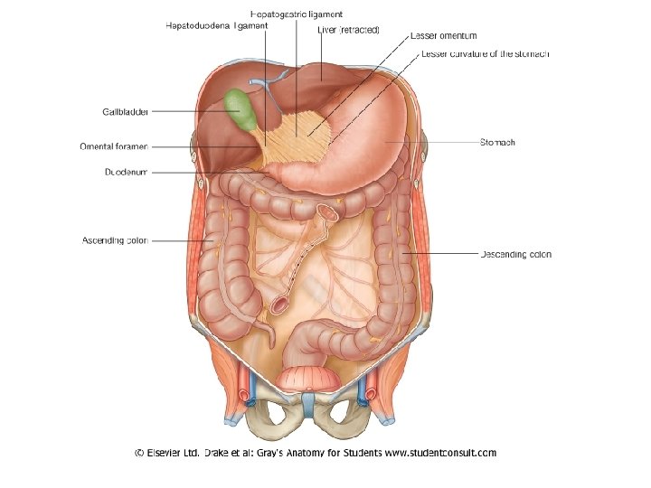
Peritoneum • Peritoneum – Visceral: Covers organs – Parietal: Covers interior Retroperitoneal surface of body wall – Retroperitoneal: Behind peritoneum as kidneys, pancreas, duodenum
• Mesenteries – Routes which vessels and nerves pass from body wall to organs
Mesenteries • Mesenteries – Routes which vessels and nerves pass from body wall to organs
Mesenteries
Mesenteries
Lesser omentum from ventral mesentery
Lesser omentum from ventral mesentery
greater omentum from dorsal mesentery
greater omentum from dorsal mesentery
greater omentum
Greater sac Lesser sac
Stomach Anatomy • Openings – Gastroesophageal : To esophagus – Pyloric: To duodenum • Regions – – Cardiac Fundus Body Pyloric
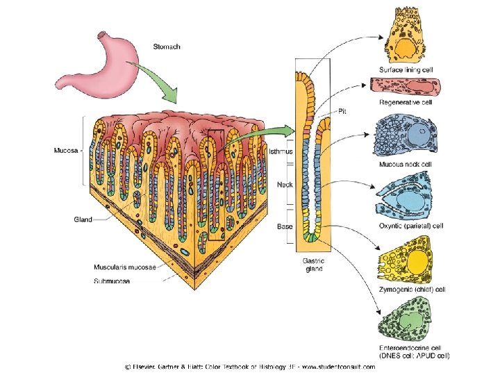
Stomach Histology: • Layers – Serosa or visceral peritoneum: Outermost – Muscularis: Three layers • Outer longitudinal • Middle circular • Inner oblique – Submucosa – Mucosa
Stomach Histology • Rugae: Folds in stomach when empty • Gastric pits: Openings for gastric glands – Contain cells
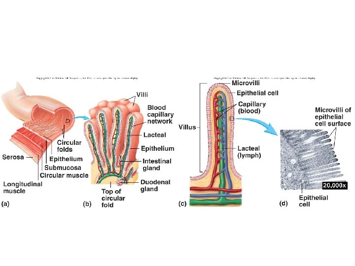
Small Intestine anatomy and Histology • Site of greatest amount of digestion and absorption • Divisions – Duodenum – Jejunum – Ileum: Peyer’s patches or lymph nodules • Modifications – Circular folds or plicae circulares, villi, microvilli • Cells of mucosa – Absorptive, goblet, granular, endocrine
Small Intestine Secretions • Mucus – Protects against digestive enzymes and stomach acids • Digestive enzymes – Disaccharidases: Break down disaccharides to monosaccharides – Peptidases: Hydrolyze peptide bonds – Nucleases: Break down nucleic acids • Duodenal glands – Stimulated by vagus nerve, secretin increases water and bicarbonate secretion from duodenal (Brunner's) glands
Duodenum