TRIAGE OF PLAYGROUND AND SPORTS INJURIES Jared Dixon
 INJURIES Jared Dixon MD, CAQSM 10 June 2019")


 -- Rapid trauma assessment •")


 • - Cases & Common Presentations • Ages 11")


 • - Cases & Common Presentations • Ages 10")
 • - Treatment • Same as for Osgood Schlatter")

 • - Cases & Common Presentations • Ages 8 -15")


 &")


")


 • - Common Presentations • Ages 11 -16 years")

 • - Common Presentations • 8 -15 years")


 • - Common presentation • Athletes with repetitive")


- Slides: 64
TRIAGE OF PLAYGROUND (AND SPORTS) INJURIES Jared Dixon MD, CAQSM 10 June 2019
GOALS • Review common injuries at school • Playground • Sports • Discuss: • Pathology • Exam • Triage
BACKGROUND • Playground Injuries • >200, 000 ER visits per year (ages 14 and younger) • 75% of nonfatal injuries occur on public playgrounds (most at school & daycare centers) • 1990 -2000, 147 children died from playground-related injuries • 82 strangulations • 31 falls • 70% occurred on home playgrounds • Girls 55%, Boys 45% • Common causes: • Falls (majority of injuries) • Slides • Other causes • Swing i. e. “wrecking ball” • Collisions
PREVENTION • Supervision • “Don’t slide with a long stick in your hands!” • “Don’t climb down from the top of the monkey bars with the toy gun strap around your neck!” • Playground design • Surfaces, layout, equipment installation/maintenance
PEDIATRIC SPORTS MEDICINE • - Estimated that over 30 -45 million children ages 6 -18 participate in athletics annually • - Nearly ¾ of US households have at least one child that participates in organized sports • - Sports participation is more accessible with increased variety • Increasing sports specialization • More year round and concurrent sports • - Drive for success, college scholarships, going professional • NCAA stats demonstrate that less than 0. 5 -1. 6% of high school athletes will earn partial scholarships to D 1 schools • 1% of college athletes go professional
PEDIATRIC SPORTS MEDICINE • - Over ½ of children under age 14 who seek medical care for injuries are due to overuse injuries • Most common injuries • Sprains, strains, bone or growth plate injuries, repetitive motion and overuse injuries, heat related illness • 62% of injuries occur during practice • - Over 1 in 10 will have an emergency room visit for a sports related injury
“ CHILD ATHLETES ARE NOT SMALL ADULT ATHLETES ” • • hyperelastic joints malleable bones epiphyses apophyses psychologic implications management by proxy all complaints must be thoroughly investigated
APOPHYSITIS
CONCUSSION • So a guy walks into a bar…
HEAD & NECK INJURY
WARNING SIGNS OF SPINAL CORD INJURY • Numbness • Weakness • Tingling • Paralysis
ASSESSING INJURIES • Scene Size-up: • Assess mechanism of injury • Initial assessment: • • • General impression C-Spine control if needed AVPU ABC’s Priority • Any problems – High Priority!
ASSESSMENT • Physical exam • Unstable patient (High Priority) -- Rapid trauma assessment • Stable patient -- exam focused on area of complaint
SIGNS AND SYMPTOMS OF A FRACTURE • Crepitus • False motion • Exposed fragments • Pain • Locked joint
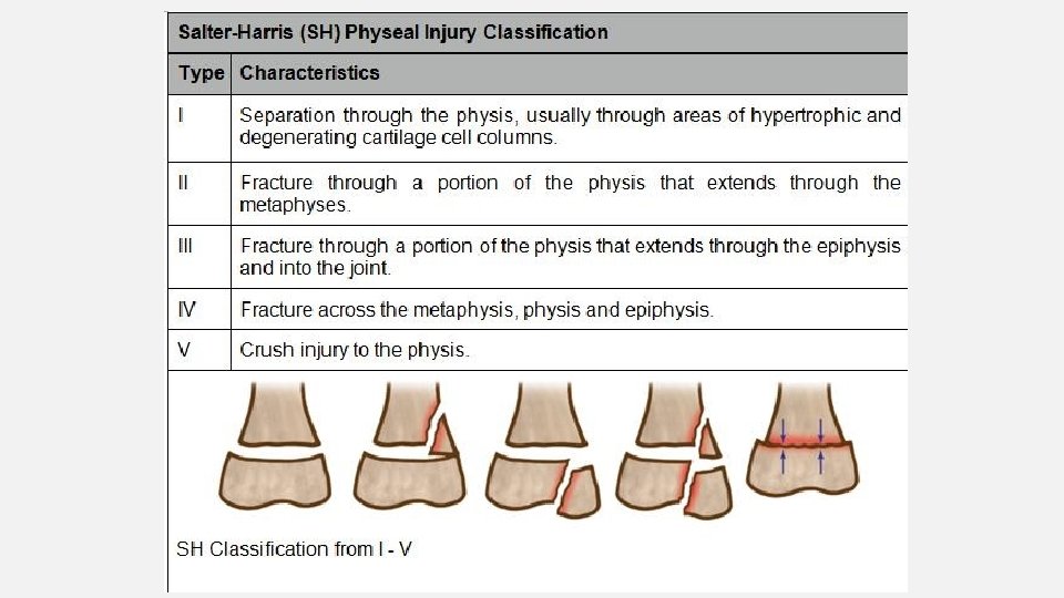
GREENSTICK FRACTURE Incomplete fracture: often occurs in children
DISLOCATION • A disruption in the joint, or separation of a joint • Caused by contact sports, high impact sports, and sports resulting in excessive stretching • Most common dislocated joints are in the hand, followed by the shoulder. Knees, hips, and elbows are less common. • Signs and symptoms: • Pain • Unable to use extremity involved • Numbness, tingling in effected extremity
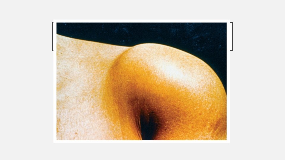
DISLOCATION OF THE PATELLA • Usually dislocates to lateral side. • Produces significant deformity. • Splint in position found. • Support with pillows.
Elbow Dislocation
ELBOW DISLOCATION
EVALUATING NEUROVASCULAR FUNCTION • Examination of the injured limb should include assessment of the following: • Pulse • Capillary refill • Sensation • Motor function
CLAVICLE FRACTURE
CLAVICLE AND SCAPULA INJURIES • Clavicle is one of the most fractured bones in the body. • Scapula is wellprotected. • Joint between clavicle and scapula is the acromioclavicular (A/C) joint. • Splint with a sling and swathe.
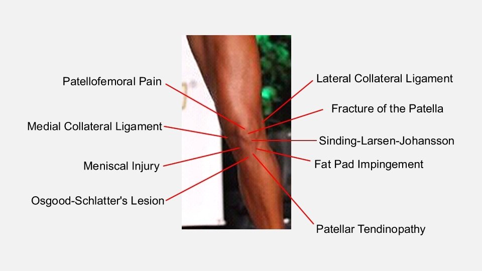
CASE #1 • 14 year old male with L knee pain x 1 year • Pain is located over anterior knee • Hurts more with running, jumping, squatting • Front of knee seems swollen at the area of pain
OSGOOD SCHLATTER (TIBIAL TUBERCLE APOPHYSITIS) • - Cases & Common Presentations • Ages 11 -15 years old • Males>females • Occurs with running, jumping or increase in physical activity • Worsens if hits/bangs/falls on tender area • - Physical exam • Tenderness on palpation of tibial tubercle • May have prominence/swollen appearance of tibial tubercle
OSGOOD SCHLATTER • - Imaging • Xrays demonstrate an open tibial tubercle • Xrays are not necessary • Help to exclude tibial tubercle avulsion, cyst, tumor, infection • - Treatment • Rest, activity modification • Ice • Patellar tendon strap • Increase flexibility of hamstrings & quadriceps • Closure of apophysis
CASE #2 • - 10 year old female with anterior knee pain x 2 weeks • - Pain occurs with running, kneeling, climbing • - Pain is located at inferior aspect of patella (superior to tender area in Osgood Schlatter)
SINDING LARSEN JOHANSSEN (PATELLAR APOPHYSITIS) • - Cases & Common Presentations • Ages 10 -13 years old • Pain present/worse with running, jumping, climbing, kneeling • - Physical Exam • Tenderness over inferior pole of the patella • - Imaging • Xrays not necessary • May demonstrate irregular calcification at inferior pole of the patella • -
SINDING LARSEN JOHANSSEN (PATELLAR APOPHYSITIS) • - Treatment • Same as for Osgood Schlatter • Rest/activity modification • Ice • Patellar tendon strap • Flexibility of hamstrings & quadriceps • Time to close growth plate • - Prevention? • Good flexibility • Gradual increase in activity
CASE #3 • 8 year old male soccer player with bilateral heel pain • Has been present for 2 years and is getting worse • Occurs with activity and patient will limp at the end of the game
SEVER’S DISEASE (CALCANEAL APOPHYSITIS) • - Cases & Common Presentations • Ages 8 -15 • Can be unilateral or bilateral • Usually occurs after physical activity but as worsens will occur during physical activity and at rest • May cause limping • Most common in running and high impact activities • Worse with cleats, flat feet • Pain at insertion of Achilles tendon and plantar fascia
SEVER’S DISEASE • - Physical Exam • Tenderness on palpation of medial & lateral aspect of calcaneus • + Calcaneal squeeze • May have tight calves, flat feet • - Imaging • Clinical diagnosis • Xrays demonstrate open physis • Often look irregular
SEVER’S DISEASE • - Treatment • Rest/activity modification • Ice • Heel cups • Cushion, 3/8” heel lift • Insert for arch support • May build up back to lift heel • Activity as tolerated, no limping allowed • - Prevention • Achilles flexibility • Arch support
CASE #4 • - 14 year old male football player who sprained his ankle during practice • - Wasn’t able to walk off the field • - Has bruising and swelling of ankle • - Pain with weightbearing • - Pain mainly located over lateral ankle and tenderness on palpation of distal fibula
SALTER HARRIS 1 FRACTURE OF DISTAL FIBULA • - Cases & Common Presentations • Usually inversion ankle injury • Swelling • May have pain with weightbearing • Ankle injury in skeletally immature patient • Most occur ages 8 -15 years old • Physis is the weakest link • Often missed and treated as ankle sprain • - Physical Exam • Tenderness on palpation of distal fibular physis (1 cm above distal tip of the fibula)
SALTER HARRIS 1 FRACTURE OF DISTAL FIBULA • - Imaging • Obtain WEIGHTBEARING ankle xrays (AP, lateral, and mortise views) • Xrays often normal • May demonstrate soft tissue swelling or widening of physis • Still treat for a SH 1 fracture if xrays normal • - Treatment • Tall walking boot & weightbearing as tolerated (use crutches if still has pain while in the boot) • Repeat exam in 3 -4 weeks • Refer displaced fractures to ortho
CASE #5 • - 16 year old male soccer player was kicking a soccer ball • - Felt and heard a pop from his hip • - Fell to the ground and had difficulty bearing weight • - Has bruising and swelling of his hip • - Tenderness on palpation of anterior hip • - Decreased strength & flexibility
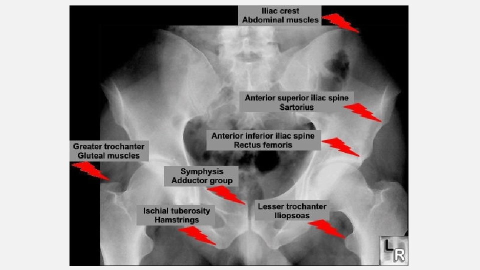
HIP AVULSION • - Cases & Common Presentations • Mechanism of injury is sudden forceful contraction of muscle • Kicking, sprinting, jumping • Most common at ASIS, AIIS, ischial tuberosity • Also can occur at iliac crest, lesser trochanter, pubic symphysis • Usually occurs between ages 14 -18 years old
HIP AVULSION • - Muscle attachments and mechanisms of injury • ASIS (Sartorius) & AIIS (Rectus femoris) • Kicking, coming out of starting blocks • Lesser trochanter (iliopsoas) • Sprinting, hip flexion • Ischial tuberosity (hamstring) • Hurdles, splits, high kick • Iliac crest (abdominal muscles) • Abrupt trunk rotation • Change of direction with running
HIP AVULSION • - Physical Exam • May have bruising & swelling • Tenderness on palpation over a growth plate • Pain with motion and manual resisted testing • Antalgic gait • - Imaging • Xray AP pelvis & frogleg lateral • - Treatment • If > 2 cm displacement refer to ortho • Acute: rest, crutches, ice, analgesics • Subacute: Physical therapy-> ROM, stretching, strengthening, then gradually guide back activities
HIP APOPHYSITIS • - Common Presentations • Gradual onset pain of pelvis/hip without specific trauma • Due to chronic traction at growth plate where tendon inserts • Skeletally immature • - Physical Exam • Tenderness on palpation at site of tendon insertion • - Imaging • Xray AP pelvis & frogleg lateral often normal • - Treatment • Rest x 4 weeks, physical therapy, gradual return to play
Ankle Injuries • Physeal plates • Ankle ligaments (sprains)
ANKLE SPRAIN VS FIBULAR FRACTURE
Acute ligmentous ankle injuries Work-up • assess site of maximal tenderness • distinguish between physeal injury and ligament injury • x-rays - AP, lateral, “mortise” view
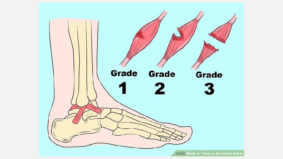
Acute ligamentous ankle injuries • Grade 1 Mild minimal swelling, pain, and disability • Grade 2 Moderate partial disruption of ligaments with difficulty with weight-bearing • Grade 3 Severe complete ligament disruption with extensive bleeding and disability
Acute ligamentous ankle injuries • 90% involve the lateral ligaments • 33% will require only 2 weeks of immobilization
Acute ligmentous injuries Management • • R. I. C. E. Grade 1 1 week off if necessary Grade 2 2 weeks on crutches with progressive weight-bearing Grade 3 7 -10 days of strict immobilization followed by 4 -8 weeks of “relative” immobilization
CASE #6 • - 12 year old left hand dominant baseball pitcher has 2 weeks of left shoulder pain • - Hurts when throwing, particularly if trying to throw hard • - Has been icing and taking ibuprofen but pain is still present • - Had pain at the end of last season that went away when the season finished
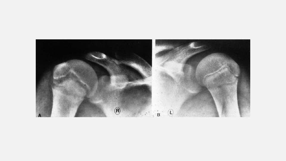
LITTLE LEAGUE SHOULDER (HUMERAL EPIPHYSITIS) • - Common Presentations • Ages 11 -16 years old • Mechanism of injury: Repetitive torsional stress • - Physical Exam • Tenderness over proximal humerus • Usually will have positive impingement signs • - Imaging • Xray Shoulder (AP, axillary, scapular Y views) may show widening of the proximal humeral epiphysis • Treatment: • Rest & Rehabilitation: Usually 3 or more months • Gradual return to throwing program
CASE #7 • - 12 year old right hand dominant baseball catcher with right elbow pain • - 2 months of elbow pain that is getting worse • - Initially was a pitcher but stopped due to pain and now catching but continues to have pain
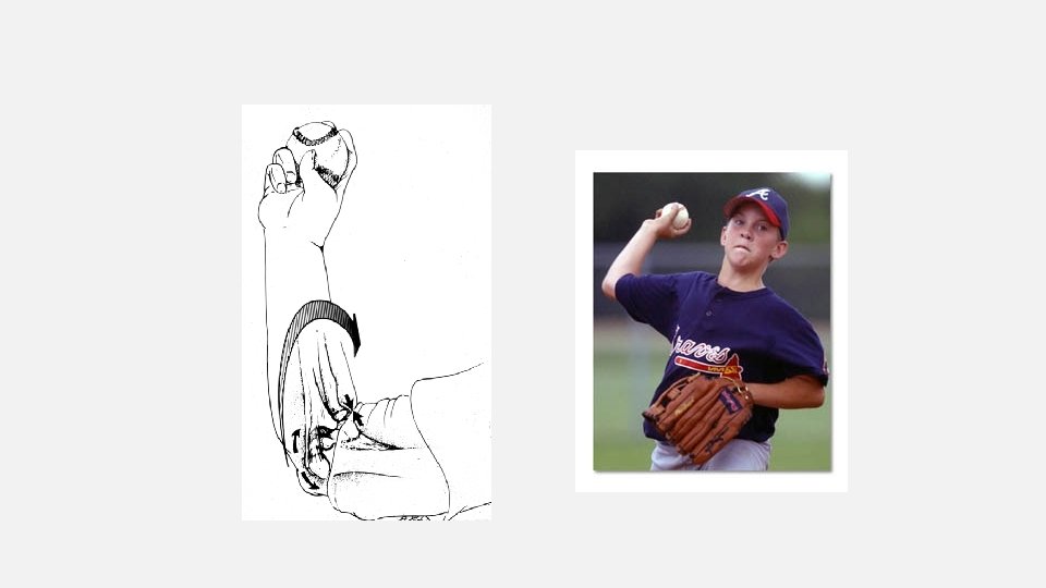
LITTLE LEAGUE ELBOW (MEDIAL CONDYLE APOPHYSITIS) • - Common Presentations • 8 -15 years old • Usually no trauma • May complain of weak & ineffective throws • Most common in pitchers, followed by catchers, 3 rd base, SS, outfield • Mechanism of injury= repetitive valgus stress on elbow from overhead throwing • - Physical Exam • Tenderness over medial epicondyle • Pain with resisted wrist flexion & forearm pronation
LITTLE LEAGUE ELBOW • - Imaging • Bilateral Elbow xrays (AP, lateral & oblique views) • May see widening of physis • - Treatment • Rest, ice, NSAIDs, immobilization (rarely) • Physical therapy: ROM, strength (elbow, shoulder, trunk, lower extremity)
UPPER EXTREMITY INJURY PREVENTION • - Prevention • Preseason strengthening and graded return to throwing program at least 6 -8 weeks prior to 1 st practice • Focus on scapular stabilizing, rotator cuff, hip, trunk, & lower extremity strengthening • Address deficits in the off season • Rest from overhead throwing at least 3 months out of the year • Follow pitch counts & rest days • Monitor all teams • Proper mechanics • Close attention to technique & monitored by coaches • No high velocity (>80 mph), curve balls or sliders until skeletally mature (~14 years old) • Stop if having pain & get evaluated promptly
CASE #8 • - 15 year old gymnast with right sided low back pain • - Bothers her with bending forward but worsens with backward bending • - Improves with rest
SPONDYLOLYSIS (STRESS FRACTURE OF PARS INTERARTICULARIS) • - Common presentation • Athletes with repetitive extension or rotation of spine • Gymnasts, dancers, figure skating, football linemen, rowing • Risk factors are family history and spina bifida • Most common at L 5 followed by L 4 • May be seen in higher lumbar vertebrae but much less frequent • - Physical Exam • Midline tenderness • Pain with lumbar extension • Positive stork test • Tight hamstrings
SPONDYLOLYSIS • - Imaging • Xrays AP and lateral lumbar spine • No obliques • MRI/CT lumbar spine • Determine what is best at your facility & be sure to talk with radiology
SPONDYLOLYSIS • - Treatment • Rest • Bracing controversial • Physical therapy • Avoid extension • Core strength, lower extremity flexibility • - Complications • Spondylolithesis: subluxation of upper vertebrae of lower vertebrae at site of bilateral spondylolysis • Chronic low back pain • Neurologic symptoms • Surgery for worsening spondylolithesis and chronic symptoms
QUESTIONS?