The Urinary System US Parts and Purposes Excretion
")


 is positioned in the cortex and medulla")


 monitors the blood pressure of the blood")


- Slides: 67
The Urinary System (US)
Parts and Purposes Excretion – the elimination of toxic and or excess materials from the body. This is accomplished by the kidneys, skin, rectum and lungs. The urinary system performs the excretion of substances from the blood.
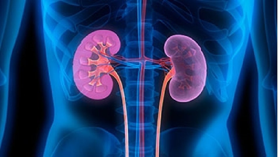
Kidney Structure A. External 1. The kidneys are in a position that is called retroperitoneal – outside the peritoneum lining.
2. They are covered by a tough layer called the renal capsule. The indention in the middle that the renal artery and vein use to enter and leave is called the renal hilus.
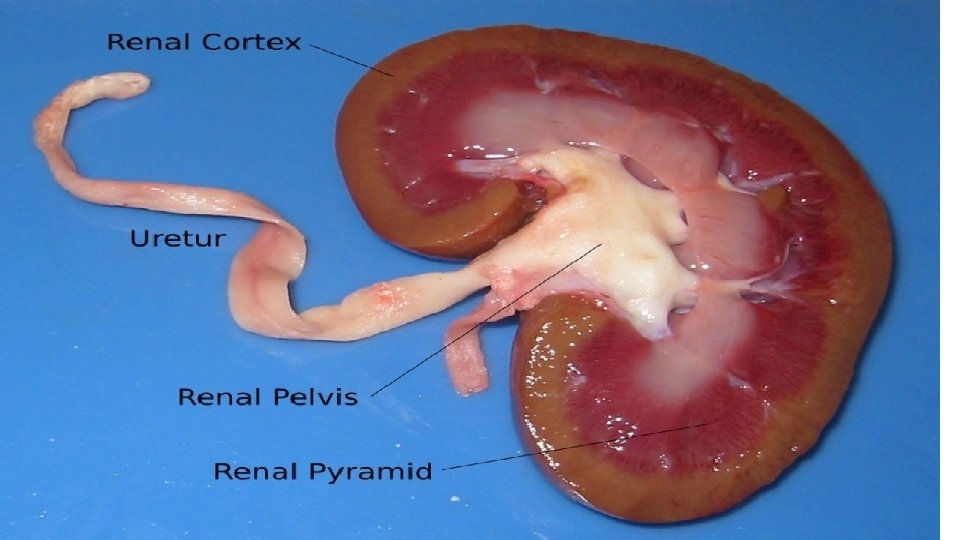
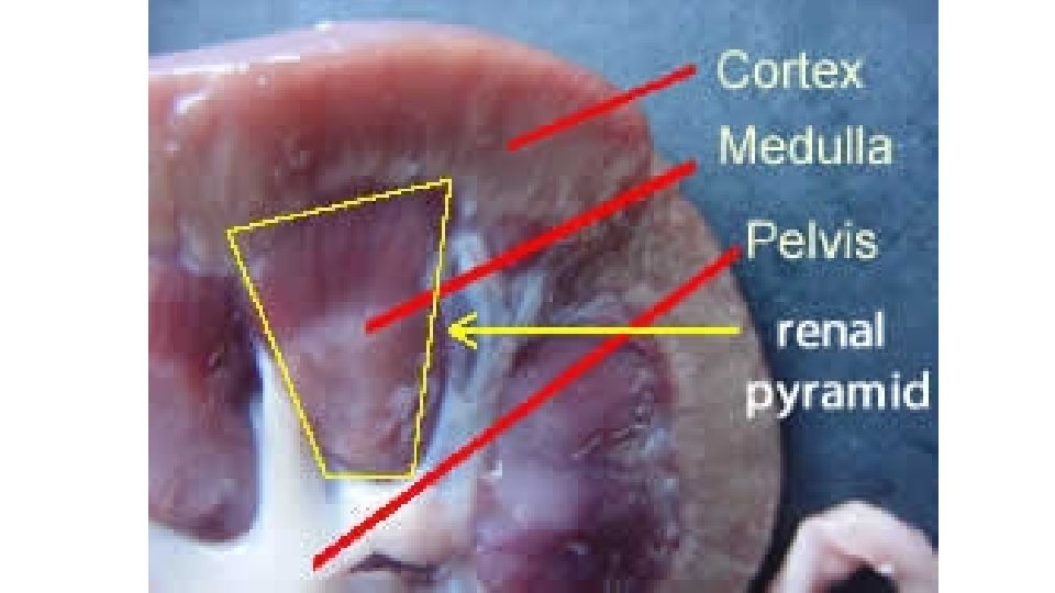
B. Internal 1. The inside can be divided from superficial to deep into the renal cortex, renal medulla and renal pelvis
2. The renal medulla can be divided into the renal pyramids and renal columns.
3. The nephron (filtering unit) is positioned in the cortex and medulla
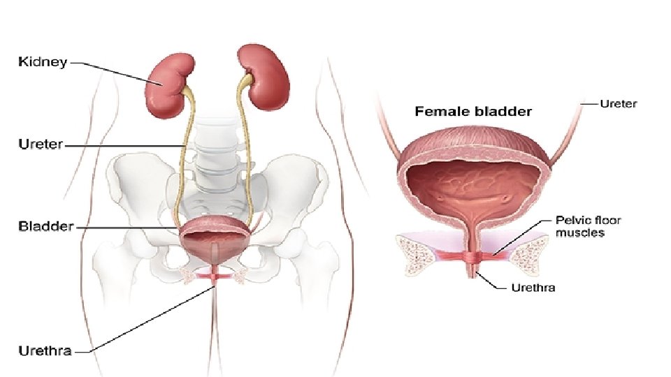
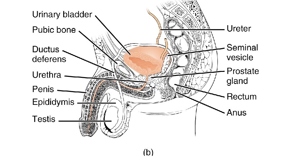
Ureter 1. Connects the kidney to the bladder
Ureter 1. Connects the kidney to the bladder 2. Made of mucus coat, smooth muscle and connective tissue
Ureter 1. Connects the kidney to the bladder 2. Made of mucus coat, smooth muscle and connective tissue 3. Peristaltic waves propel urine https: //www. youtube. com/watch? v=u. Ay 5 C 2 YYlbk
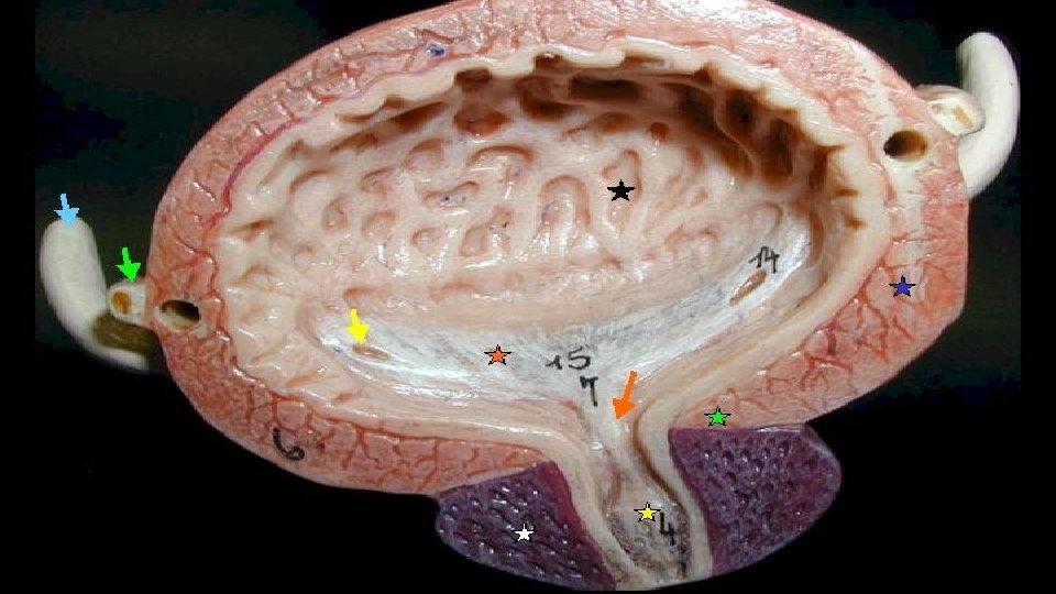
Bladder 1. A hollow distensible storage unit for urine
Bladder 1. A hollow distensible storage unit for urine 2. Has the same 4 layers as the alimentary canal
Bladder 1. A hollow distensible storage unit for urine 2. Has the same 4 layers as the alimentary canal 3. The trigone are the 3 holes of the bladder. 2 are for the ureters and one for the urethra
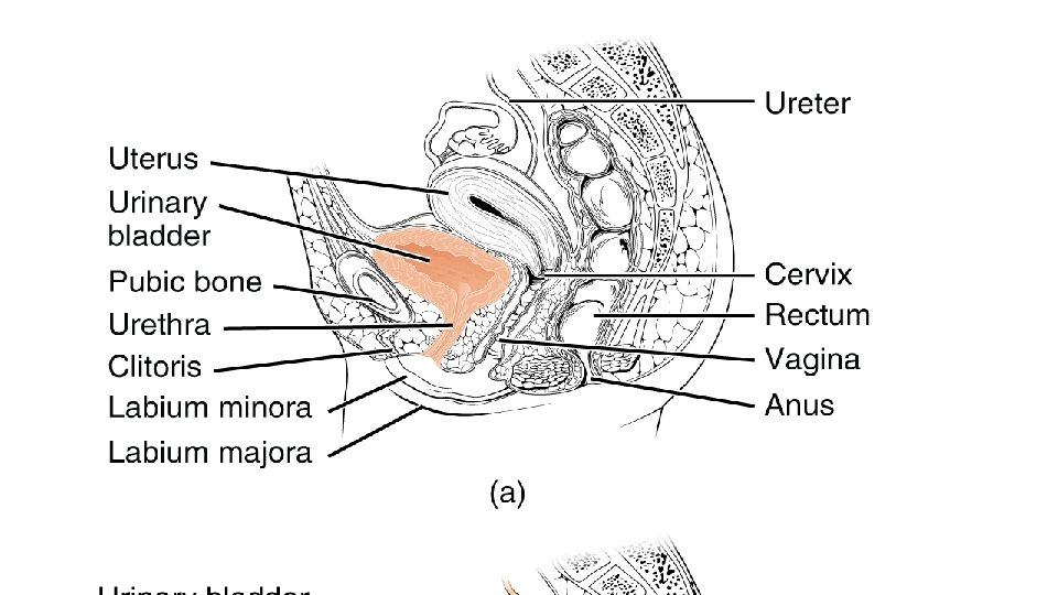
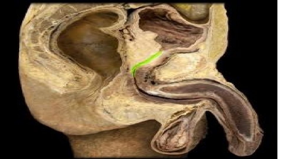
Urethra 1. Pathway from bladder to outside world 2. The structure is much the same as in the ureter
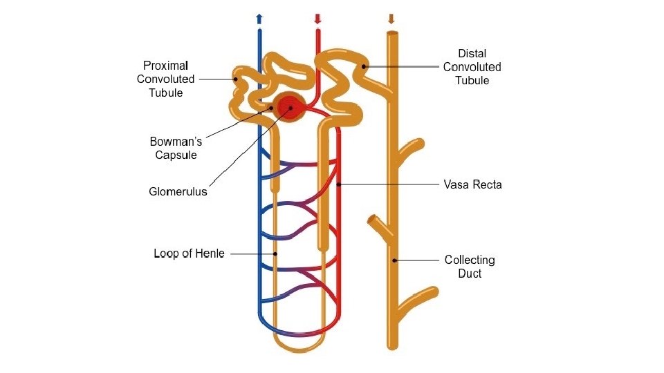
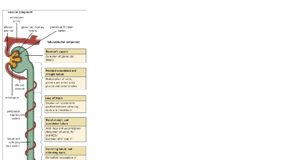
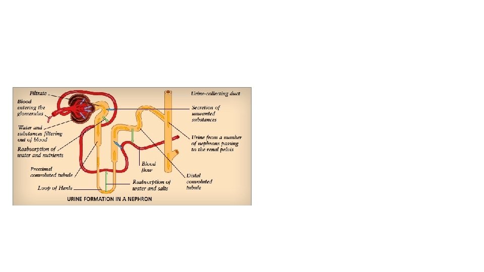
Urine formation There are 3 processes and 4 pathways involved in urine formation. The nephron is free to manipulate these 2 factors, in any way, to do its job of maintaining the volume, pressure and composition of the blood.
B. Once the nephron forms urine, it will leave the nephron via the collecting duct. Urine will then flow to the renal papilla minor calyx major calyx renal pelvis ureter bladder urethra
B. Once the nephron forms urine, it will leave the nephron via the collecting duct. Urine will then flow to the renal papilla minor calyx major calyx renal pelvis ureter bladder urethra
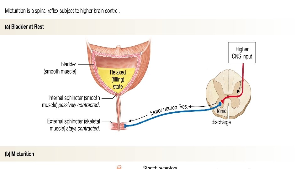
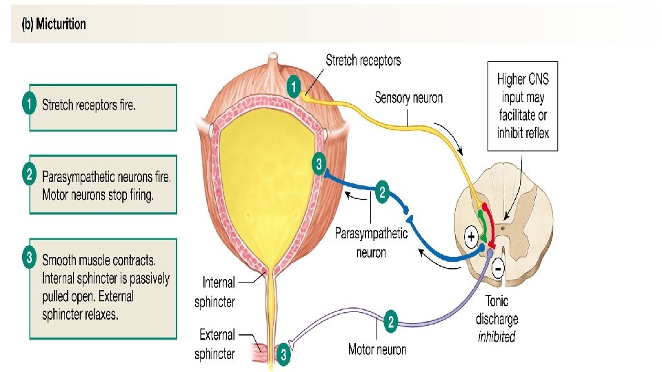
C. Micturition is the process of expelling urine from the bladder. The filling of the bladder signal stretch receptors that send messages to the micturition reflex center in the spinal cord. Parasympathetic impulses cause the detrusor muscle to contract the bladder. This occurs at around 150 ml of urine. The external urethral sphincter voluntarily relaxes to let the urine escape.
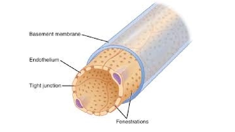
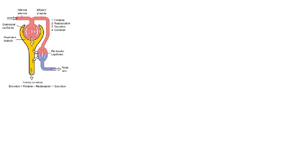
Pathway 1 - Filtration – Blood enters the renal artery and flows through the arcuate artery to the afferent arteriole and then the glomerulus. Filtration occurs at the glomerulus when substances are forced through the glomerular walls via fenestrae into the Bowman’s capsule of the nephron.
. The excessive pressure found in the glomerulus is caused by the fact that the blood vessel that exits the glomerulus, the efferent arteriole, has a smaller diameter than the blood vessel that enters it, afferent arteriole. Substances that can be filtered include water, salts, glucose, urea and H+. Filtration is the only process that the nephron cannot change. It is random and the substances that get filtered are filtered by chance.
Once in the Bowman’s capsule, the filtrate passes through the proximal convolute tubule descending loop of Henle ascending loop of Henle distal convoluted tubule collecting duct. This is the end of the nephron.
At this point the filtrate has become urine. Collecting ducts of several nephrons will drain into a minor calyx major calyx renal pelvis ureter
Pathway 2 – No filtration – Blood enters the renal artery and flows through the arcuate artery to the afferent arteriole and then the glomerulus. Substances following this pathway DO NOT get filtered into the nephron. They pass out the glomerulus via the efferent arteriole peritubular capillaries arcuate vein renal vein.
This pathway contains blood that is not cleaned or modified in any way by the nephron. Its purpose is to supply the nephron/kidney cells with oxygen and nutrients, while picking up CO 2 and waste. Normal blood functions. Substances in this pathway include water, salts, glucose, urea, RBC’s, proteins/amino acids and H+.
Pathway 3 – Reabsorption - Blood enters the renal artery and flows through the arcuate artery to the afferent arteriole and then the glomerulus. Filtration occurs at the glomerulus when substances are forced through the glomerular walls into the Bowman’s capsule of the nephron. Substances that can be filtered include water, salts, glucose, urea and H+. You filter about 180 liters in 24 hours. Do you urinate out that much? No. Substances that were filtered, but that the body needs back in the blood are pulled back into the peritubular capillaries (blood) through the process of reabsorption.
Looking at the list of filtered substances, you can see that glucose is filtered. Glucose is a vital substance to your body. It must be entirely reclaimed by reabsorption. Substances that are reabsorbed include water, salts, glucose, and urea.
Pathway 4 – Secretion Blood enters the renal artery and flows through the arcuate artery to the afferent arteriole and then the glomerulus. Substances following this pathway DO NOT get filtered into the nephron. They pass out the glomerulus via the efferent arteriole peritubular capillaries arcuate vein renal vein
Regretfully, there is still an excess amount of urea and H+ in the blood after their filtration at the glomerulus. So the urea and H+ that did not get filtered and are still in the blood, get secreted out of the peritubular capillaries/blood into the collecting duct of the nephron to join the substances in the urine that had been filtered into urine back at the glomerulus. This is how the nephron gets unwanted substances out of the blood if they did not get filtered out.
Use of the pathways and processes Blood volume What how does the nephron use these 3 processes and the 4 pathways to do its job? To accomplish the job of maintaining blood volume, the nephron will control water level in the blood. Say that to maintain homeostasis you need 8 water molecules in your blood. 12 water molecules enter the kidney. 6 water molecules would be filtered and 6 water molecules would pass by to remain in the blood. The 6 water molecules in the blood are not enough to maintain homeostasis. So, the nephron will reabsorb 2 more water molecules at the proximal convoluted tubule to bring the blood level up to the desired 8 water molecules. Out of the original 12 water molecules, only 4 water molecules would become urine and the remaining 8 (homeostasis) are in the blood.
What about if you were drinking more water – like 16 water molecules? 16 water molecules enter the kidney. 8 water molecules would be filtered and 8 water molecules would pass by to remain in the blood. The 8 water molecules in the blood are just enough to maintain homeostasis. So, the nephron will not reabsorb any more water molecules at the proximal convoluted tubule. Out of the original 16 water molecules, 8 water molecules would become urine and the remaining 8 (homeostasis) are in the blood.
What about if you were drinking less water – like 8 water molecules? 8 water molecules enter the kidney. 4 water molecules would be filtered and 4 water molecules would pass by to remain in the blood. The 4 water molecules in the blood are not enough to maintain homeostasis. So, the nephron will want to reabsorb all 4 water molecules in the nephron at the proximal convoluted tubule. But you can’t do that. Some water has to remain in the filtrate at all times. At most 3 water molecules can be reabsorbed. That leaves you with only 7 water molecules in the blood. You are now dehydrated. Out of the original 8 water molecules, only 1 water molecules would become urine.
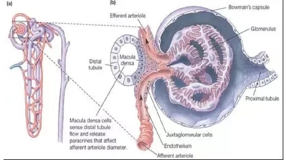
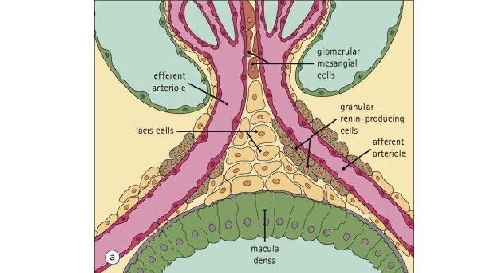
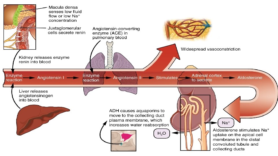
A sensor called the juxtaglomerular apparatus (JGA) monitors the blood pressure of the blood entering the glomerulus. The JGA is composed of some modified cells of the afferent arteriole and the macula densa of the ascending loop of Henle.
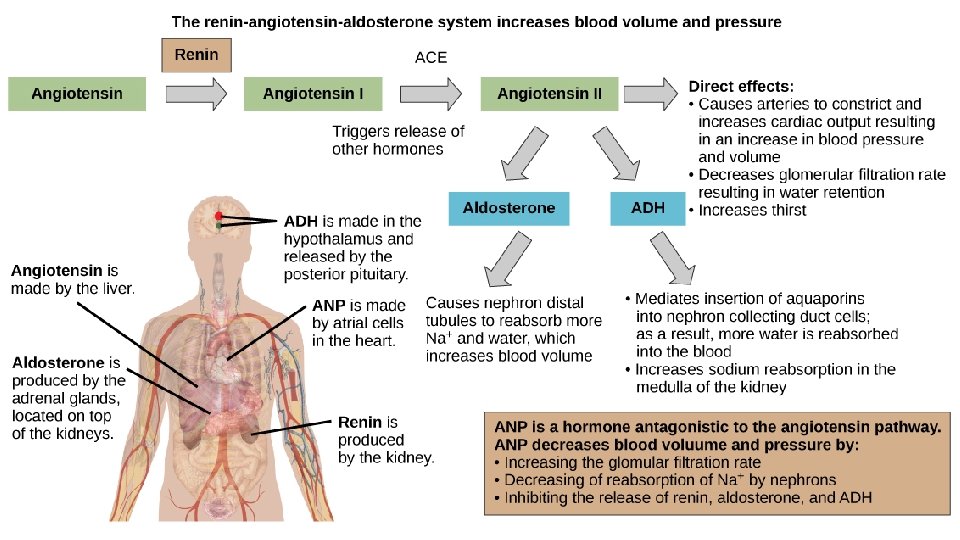
If the blood pressure is too high, the JGA will signal the brain. The brain will signal the nephron to NOT reabsorb water. High blood pressure indicates a high volume of water in the blood and no need to reabsorb water out of the nephron back into the blood.
If the blood pressure is too low, the JGA will signal the brain. The brain will signal the nephron to reabsorb water. Low blood pressure indicates a low volume of water in the blood and a need to reabsorb water out of the nephron back into the blood.
Blood composition To accomplish the job of maintaining blood composition, let’s pick blood p. H as an example. Most of the foods you eat are acidic releasing many H+ ions into your blood. As an example, to maintain homeostasis, you need 4 H+ ions in your blood. 12 H+ ions enter the kidney. 6 H+ ions would be filtered into urine and 6 H+ ions would pass by to remain in the blood. The 6 H+ ions in the blood are too many for homeostasis. The nephron will secrete 2 more H+ ions out of the blood in the peritubular capillaries into the collecting duct to bring the blood level down to the desired 4 H+ ions. Out of the original 12 H+ ions, 8 H+ ions would become urine and the remaining 4 (homeostasis) are in the blood
What about if you were eating more acidic food and created a blood level of 16 H+ ions ? Remember to maintain homeostasis, you need 4 H+ ions in your blood. 16 H+ ions enter the kidney. 8 H+ ions would be filtered into urine and 8 H+ ions would pass by to remain in the blood. The 8 H+ ions in the blood are too many for homeostasis. The nephron will secrete 4 more H+ ions out of the blood in the peritubular capillaries into the collecting duct to bring the blood level down to the desired 4 H+ ions. Out of the original 16 H+ ions, 12 H+ ions would become urine and the remaining 4 (homeostasis) are in the blood
Chemoreceptors in your blood vessels will determine your blood p. H and signal the brain. The brain will then signal the nephron as to how many H+ ions to secrete.
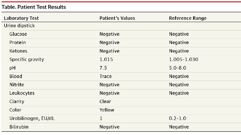
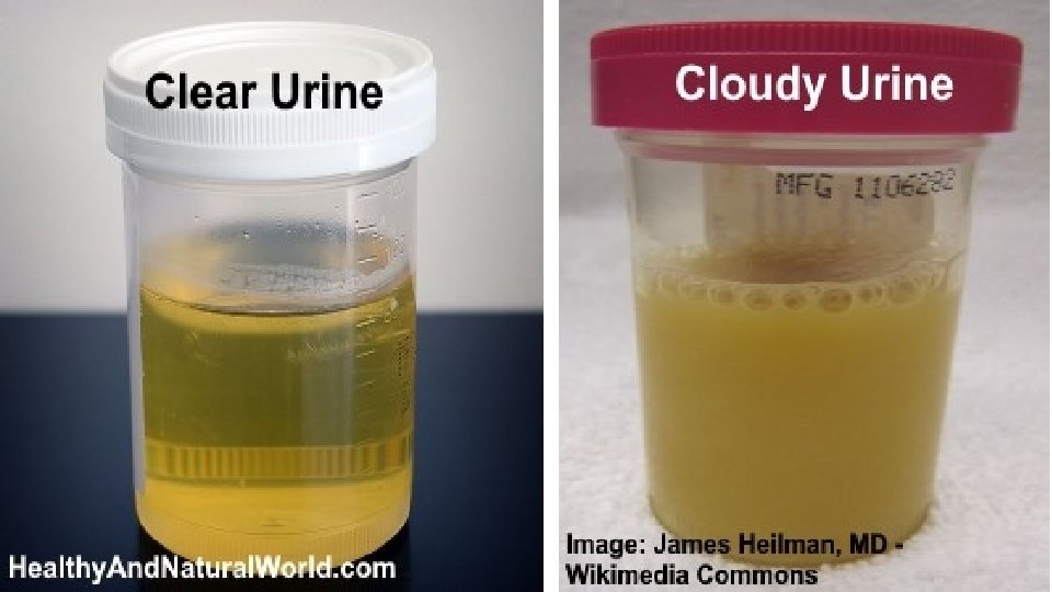
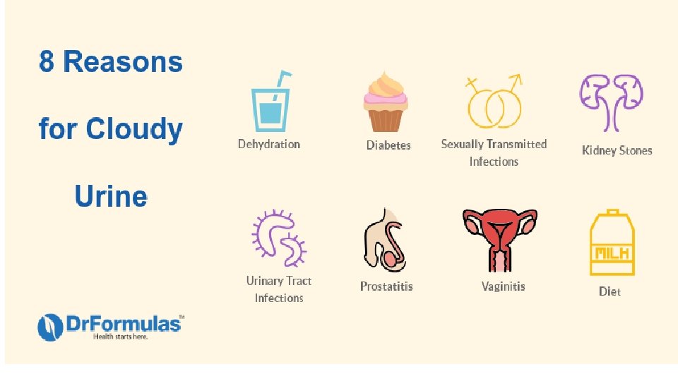
Urine Characteristics 1. Byproduct of kidney’s activities 2. Urinalysis – the testing of a urine sample – see reference sheet for abnormal urine examples
Urine Characteristics 1. Byproduct of kidney’s activities 2. Urinalysis – the testing of a urine sample – see reference sheet for abnormal urine examples 3. Daily volume 1500 ml 4. Transparent
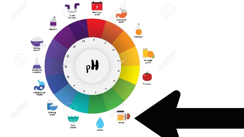
Urine Characteristics 1. Byproduct of kidney’s activities 2. Urinalysis – the testing of a urine sample – see reference sheet for abnormal urine examples 3. Daily volume 1500 ml 4. Transparent 5. Slight odor 6. p. H 6. 0
Urine Characteristics 1. Byproduct of kidney’s activities 2. Urinalysis – the testing of a urine sample – see reference sheet for abnormal urine examples 3. Daily volume 1500 ml 4. Transparent 5. Slight odor 6. p. H 6. 0 7. 95% water