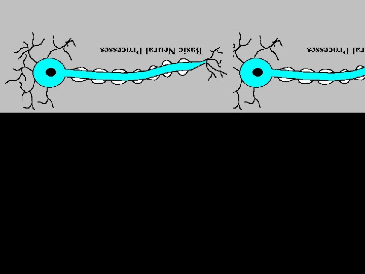
The Central Nervous System The Central Nervous System


 • Electrodes are placed on the scalp that amplify recordings")
 • A series of X-ray photographs")
 • A visual display of brain activity")
 • A technique that uses magnetic fields and")


")


- Slides: 107
The Central Nervous System
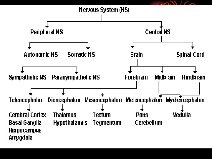
The Central Nervous System • The Central Nervous System processes all of the body’s information and includes the brain and the spinal cord
The Peripheral Nervous System • The Peripheral Nervous System includes all of the other nerves in the body • The Peripheral Nervous System is divided into two categories: • The Somatic Nervous System • The Autonomic Nervous System
The Somatic Nervous System • The Somatic Nervous System connects the brain to the muscles of the body. • controls all of our voluntary muscle movements. • It is connected to the motor cortex in the brain.
The Autonomic Nervous System • The Autonomic Nervous System controls the automatic functions of the body and connects the brain to the heart, lungs, internal organs, glands, etc.
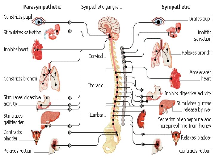
The Autonomic Nervous System • This system is also responsible for actions associated with stress and perceived threats. The autonomic system is further broken down into two systems: • Sympathetic Nervous System • Parasympathetic Nervous System
The Sympathetic Nervous System • The Sympathetic Nervous System mobilizes the body to respond to stress. It is our alert (fight) system. • Accelerates the heart, blood pressure, respiration) • Conserves energy for fight by slowing down digestion, etc.
The Parasympathetic Nervous System • The Parasympathetic Nervous System returns the body to a normal state after a stressful encounter.
THE BRAIN
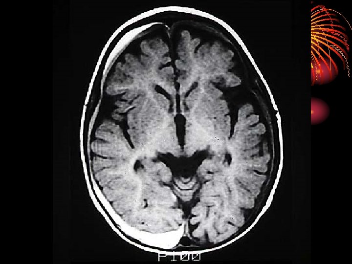
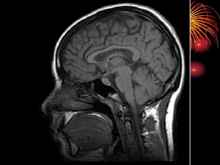
How Do We Study The Brain?
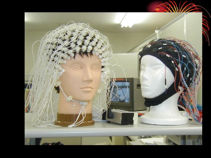
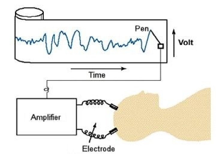
• Electroencephalogram (EEG) • Electrodes are placed on the scalp that amplify recordings of the waves of electrical activity across the brain’s surface
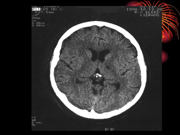
• Computed Tomography (CT or CAT Scan) • A series of X-ray photographs taken from different angles and combined by computer into a composite representation of the brain
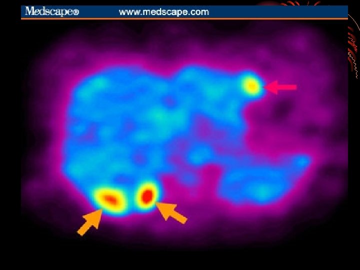
• Positron Emission Tomography (PET Scan) • A visual display of brain activity that detects where a radioactive form of glucose goes while the brain performs a given task
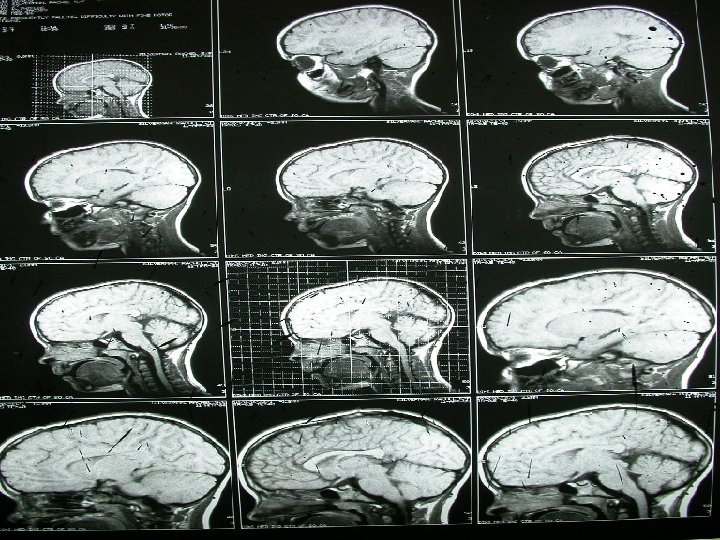
• Magnetic Resonance Imaging (MRI) • A technique that uses magnetic fields and radio waves to produce computergenerated images that allow us to see structures within the
• Accidents • Case study analysis of victims of suffer from a brain injury, resulting in variations in normal behavior • IE. Phineas Gage
• Lesions • Lesioning is the removal or destruction of part of the brain. • IE. Lobotomy
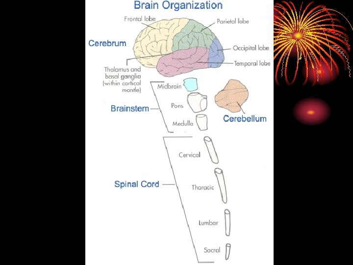
THE BRAIN AND ITS FUNCTIONS
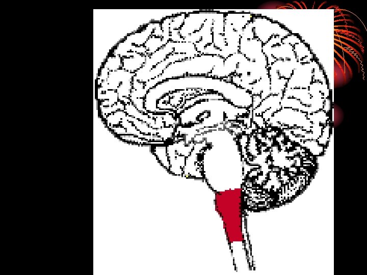
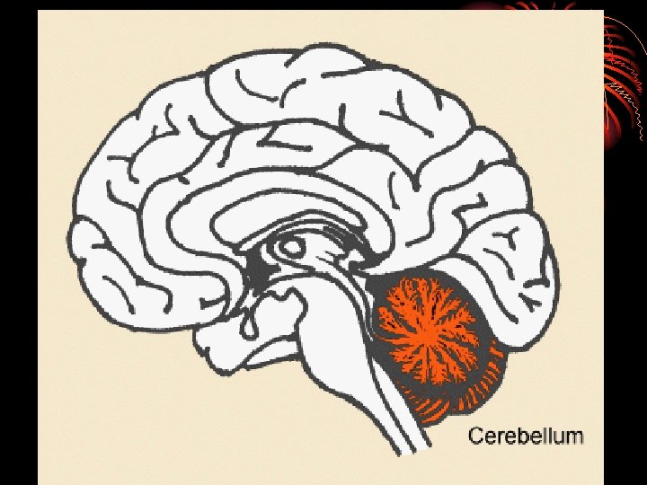
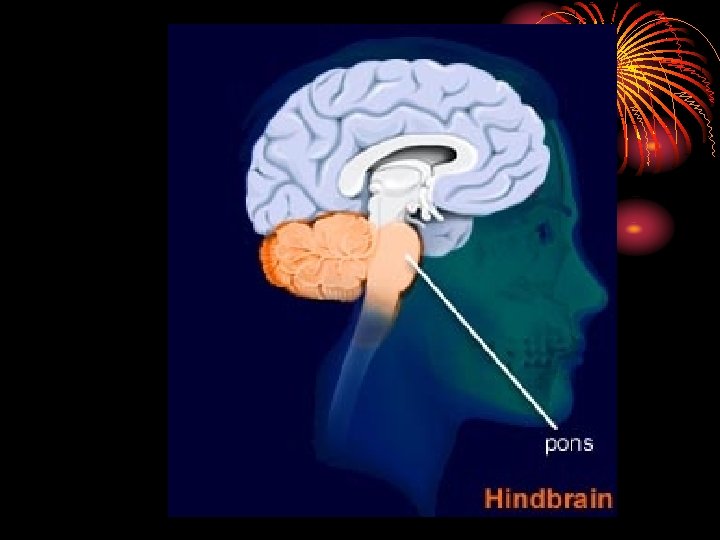
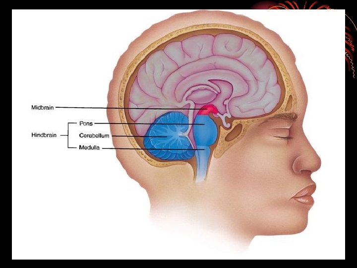
I. The Hindbrain
• The hindbrain is the oldest and innermost region of the brain
• The functions of the hindbrain are mostly done outside of our awareness, and occur without any conscious effort.
• The functions of the hindbrain control basic biological functions that keep the human body alive.
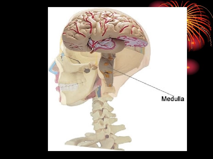
• There are three brain areas associated with the Hindbrain: • Medulla • Cerebellum • Pons
• The point at which the spinal cord enters the skull is called the MEDULLA • The MEDULLA controls heartbeat and breathing, blood pressure, and attention
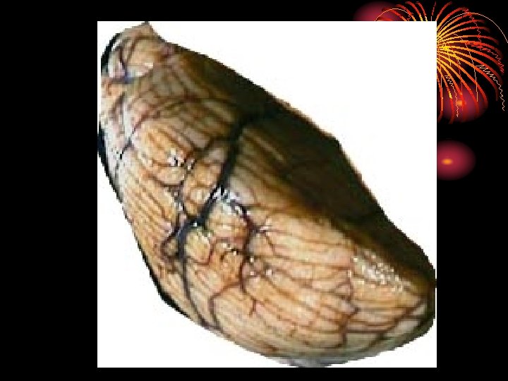
• Extending from the rear of the brainstem is the CEREBELLUM • The CEREBELLUM coordinates voluntary movements and balance (along with the BASIL GANGLIA)
• The PONS is responsible for helping to regulate breathing, to help with sleep and wake cycles, and controls facial expressions
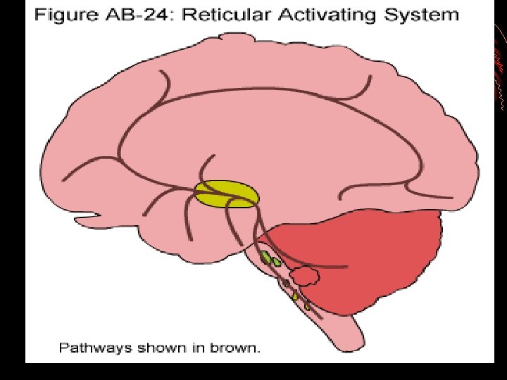
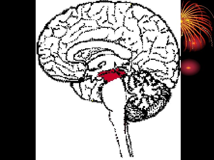
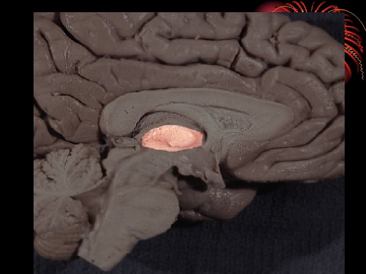
II. The Midbrain • The Midbrain is located between the hindbrain and the forebrain • This area is responsible for coordinating simple muscle movements with changes in sensory information
• The major area of the Midbrain is the RETICULAR FORMATION • The RETICULAR FORMATION extends from the spine to the thalamus, and is responsible for arousal/wakefulness and attentiveness
• The MIDBRAIN is also responsible for behaviors associated with hearing and sight • Pupil dilation and eyeball movement
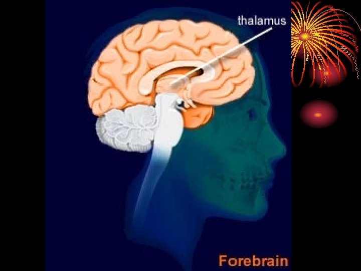
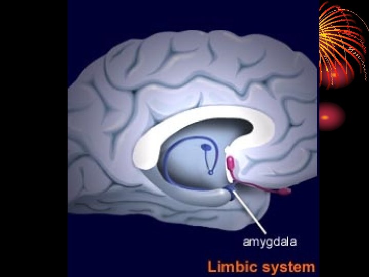
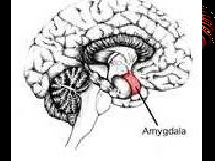
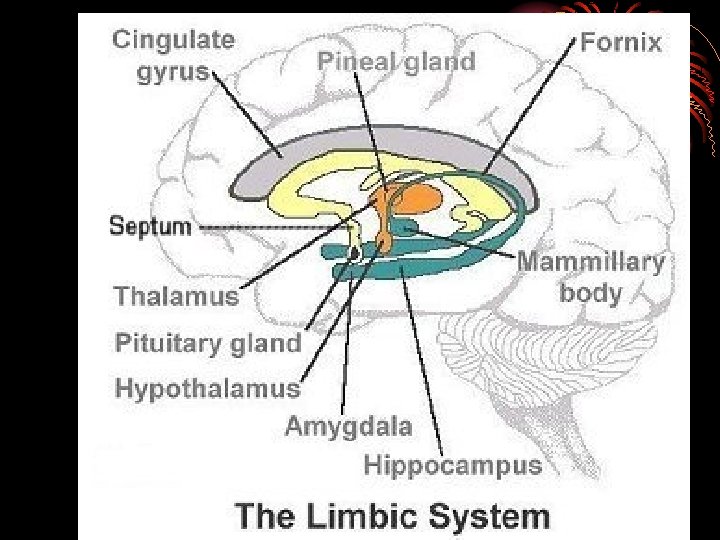
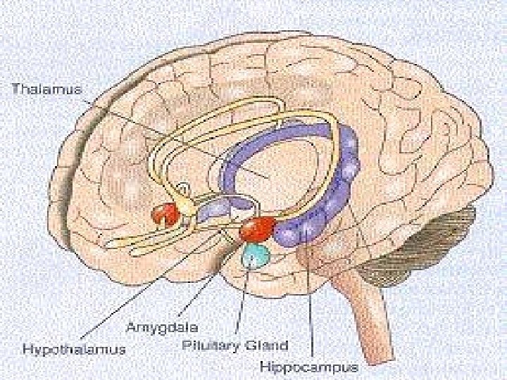
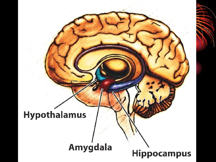
III. The Forebrain • Areas of the forebrain control thought and reason. • There are five main regions of the forebrain to study: • Thalamus • Hypothalamus • Amygdala • Hippocampus • The Cerebral Cortex
• On top of the hindbrain is the THALAMUS • The THALAMUS receives sensory input from all of the senses except smell, and routes it to the proper area of the brain for processing • The THALAMUS also helps to control the electrical currents in the brain
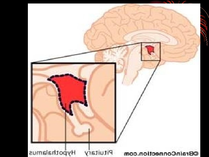
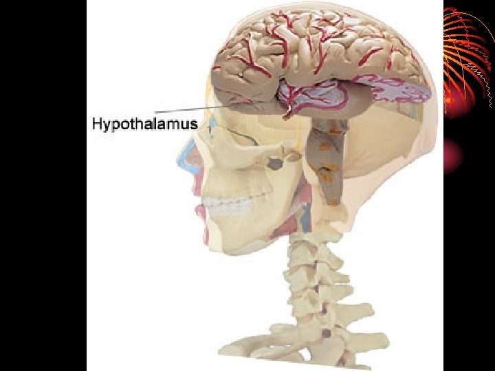
• The HYPOTHALAMUS is responsible for several maintenance activities, including eating, drinking, body temperature, and sexual arousal
• The HYPOTHALAMUS also relays communication between the brain and the endocrine system, via the pituitary gland, and then monitors the hormones released into the bloodstream
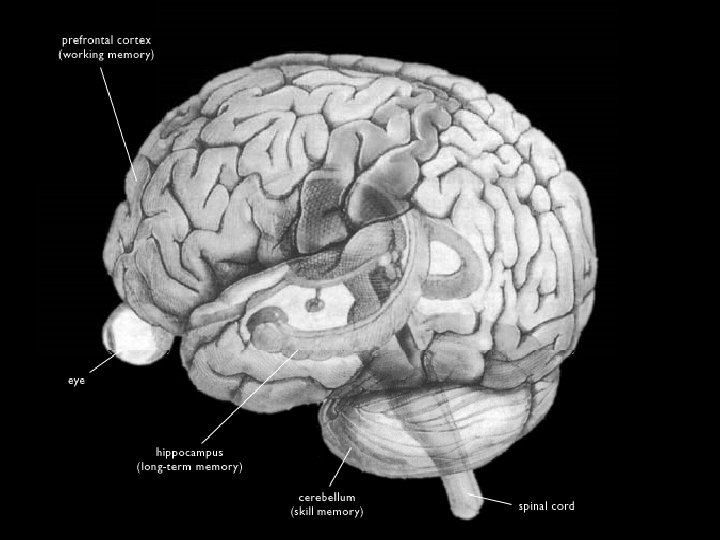
• The HIPPOCAMPUS is essential to memory processing
• The AMYGDALA is tied to emotions, especially those of aggression, rage, and fear
• Collectively, the thalamus, hypothalamus, hippocampus, and the amygdala are known as the Limbic System as well
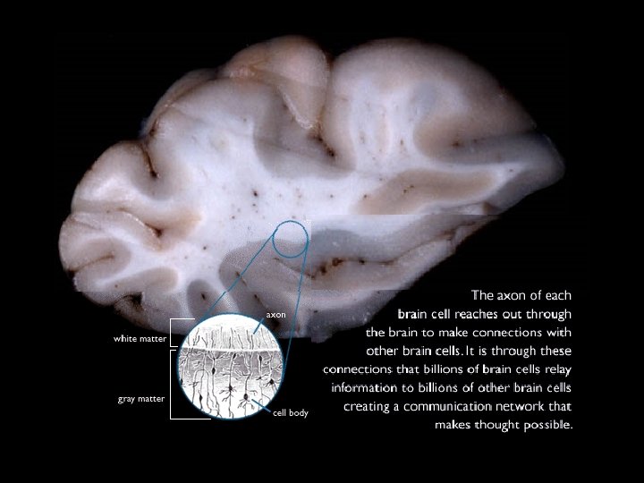
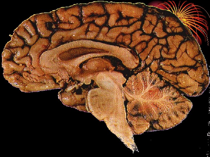
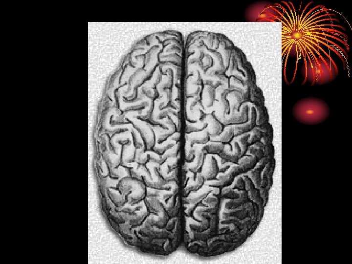
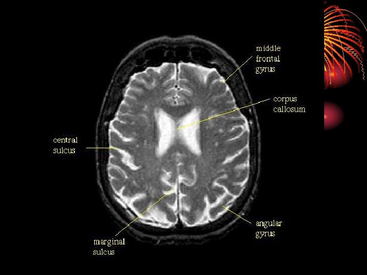
• The CEREBRAL CORTEX is the intricate, wrinkled covering of the brain (FISSURES) (actually a bump is a gyrus and a groove is a sulcus – but that’s a bit too much info…) • In addition to interneurons, it contains GLIAL CELLS, which guide neural connections, provide nutrients to myelin, and mop up neurotransmitters
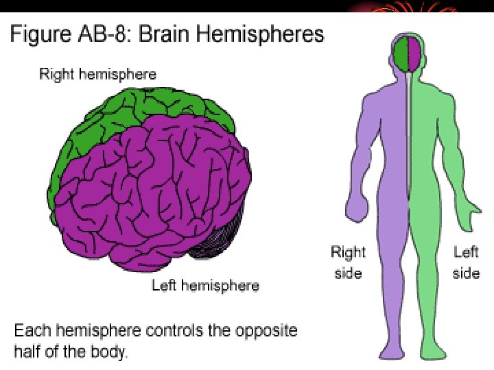
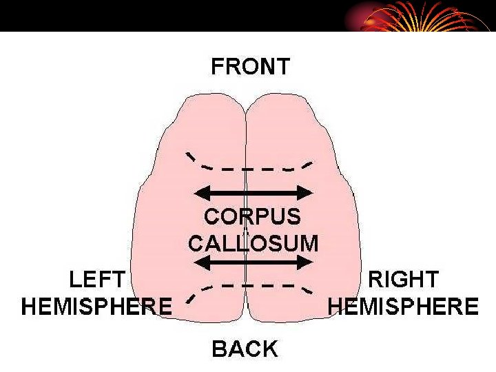
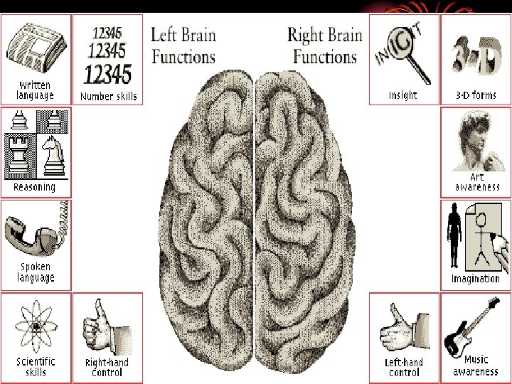
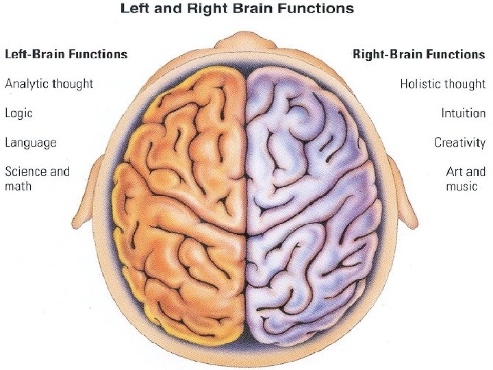
• The Cerebral Cortex is split into two halves, or Hemispheres: • Right Hemisphere • Left Hemisphere
• The two hemispheres of the brain are connected by the CORPUS CALLOSUM
Hemispheres of the Brain
• Each hemisphere of the brain has different functions. This is called hemispheric specialization.
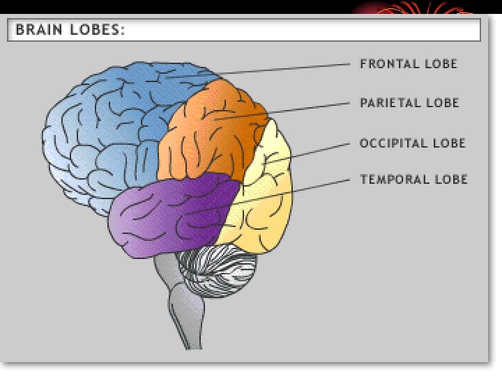
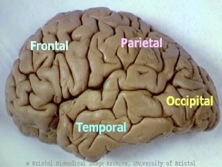
• The Cerebral Cortex is split into four LOBES, with half of each one on the left, and half of each one on the right: • The FRONTAL LOBE PARIETAL LOBE OCCIPITAL LOBE TEMPORAL LOBE
• The Frontal Lobes are the portions of the cortex lying just behind the forehead • Mostly involved in abstract thought, speaking, muscle movements, making plans, and judgments
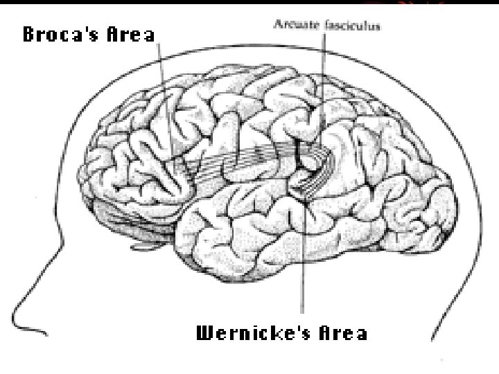
• In the left frontal lobe specifically, there is an association area responsible for language processing called Broca’s Area
• Broca’s Area controls language expression and the muscle’s involved with producing speech
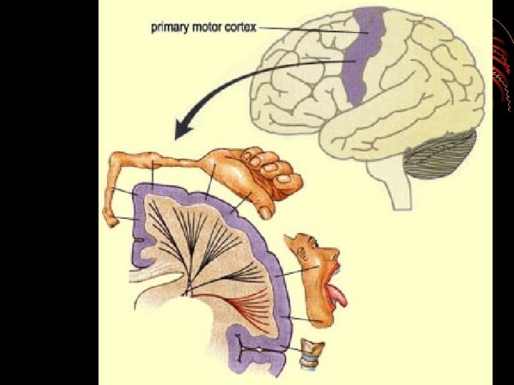
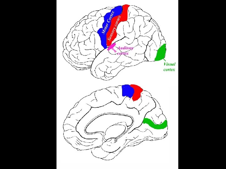
• Along the top of both front lobes runs the motor cortex
• The motor cortex receives messages from the rest of the brain and then sends messages back to the muscles of the body in order to control voluntary movements.
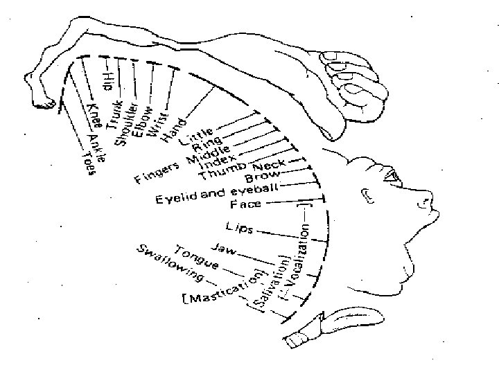
• The Parietal Lobes are the portion of the cortex lying at the top of the head includes the sensory (or somato-sensory) cortex
• The parietal lobes play important roles in integrating sensory touch information, and in the handling and manipulation of objects.
• The sensory cortex registers and processes touch sensations.
• Click the homunculus to understand the homunculus! http: //faculty. washington. edu/chudler/flash/hom. html
• The Temporal Lobes are the portions of the cerebral cortex roughly located above the ears • audio processing • comprehension, naming, verbal memory and other language functions.
• In the left temporal lobe specifically, there is an association area responsible for language processing called Wernike’s Area
• Wernicke’s Area interprets both written and spoken language.
• The Occipital Lobes are located at the back of the head • The occipital lobe is responsible for processing visual information.
Module 8: The Brain Plasticity
Plasticity • The ability of the brain tissue to take on new functions • Greatest in childhood • Important if parts of the brain are damaged or destroyed • Go to the next slide to see a video about brain plasticity! (may take a few seconds to load – be patient – click only once to load the video – then wait!