
Everything should be made as simple as possible

“Everything should be made as simple as possible, but not simpler” Albert Einstein
Stroke Alzheimer’s disease Multiple Sclerosis Meningitis Do you know …? Amyotrophic Lateral Sclerosis Parkinson’s Disease Cerebral Palsy Brain Tumor Fetal Alcohol Syndrome
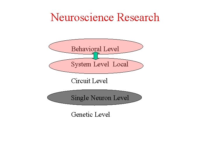
Neuroscience Research Behavioral Level System Level Local Circuit Level Single Neuron Level Genetic Level
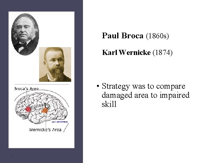
Paul Broca (1860 s) Karl Wernicke (1874) • Strategy was to compare damaged area to impaired skill 4
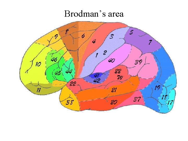
Brodman’s area
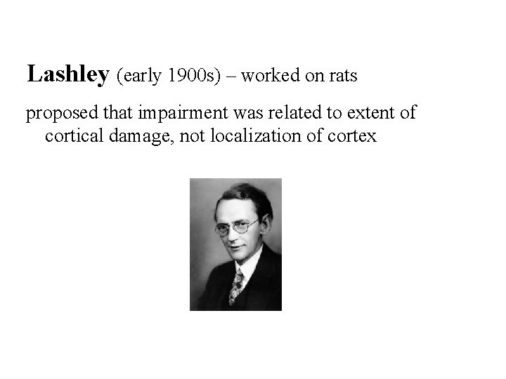
Lashley (early 1900 s) – worked on rats proposed that impairment was related to extent of cortical damage, not localization of cortex
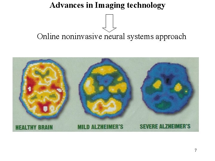
Advances in Imaging technology Online noninvasive neural systems approach 7
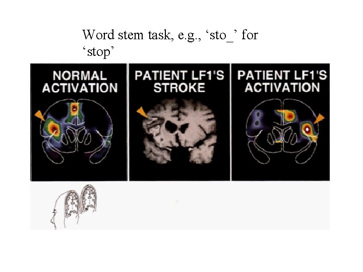
Word stem task, e. g. , ‘sto_’ for ‘stop’
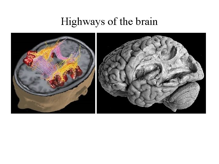
Highways of the brain
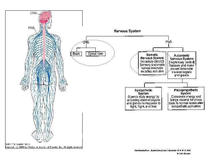
10 Downloaded from: Student. Consult (on 3 September 2010 04: 31 PM) © 2005 Elsevier
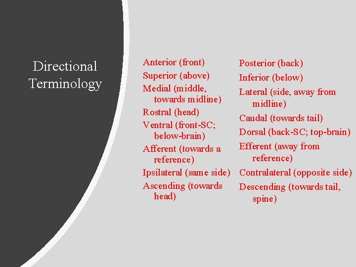
Directional Terminology Anterior (front) Superior (above) Medial (middle, towards midline) Rostral (head) Ventral (front-SC; below-brain) Afferent (towards a reference) Ipsilateral (same side) Ascending (towards head) Posterior (back) Inferior (below) Lateral (side, away from midline) Caudal (towards tail) Dorsal (back-SC; top-brain) Efferent (away from reference) Contralateral (opposite side) Descending (towards tail, spine)
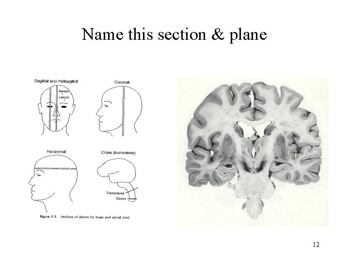
Name this section & plane 12
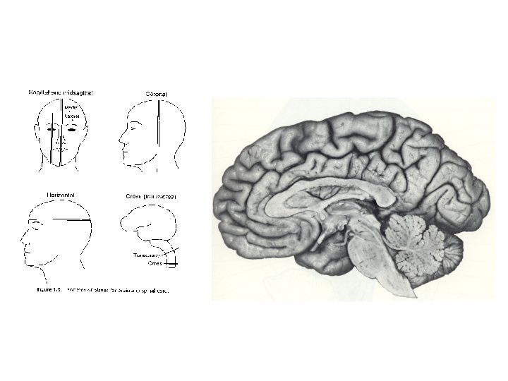
Name this section & plane 13
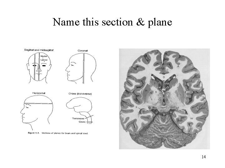
Name this section & plane 14
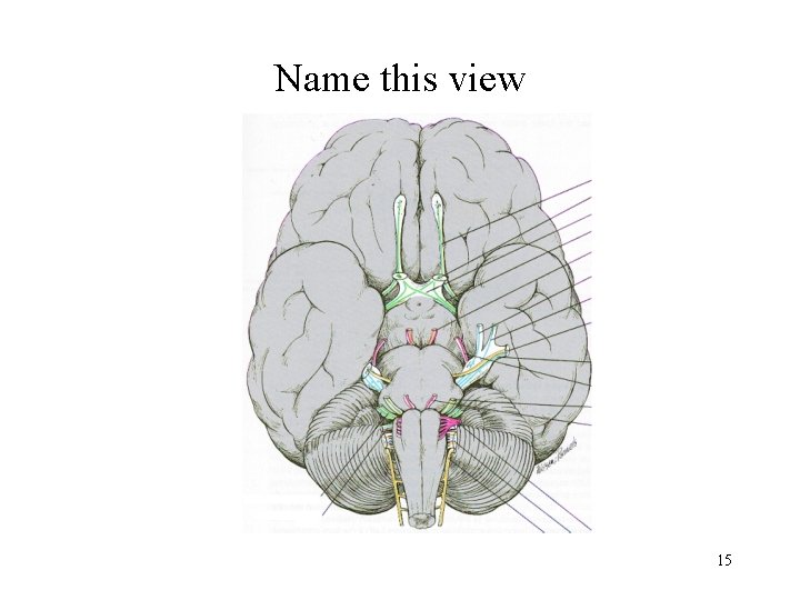
Name this view 15
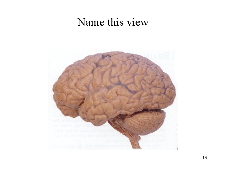
Name this view 16
17
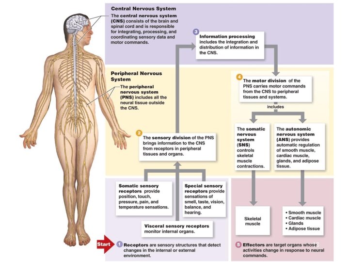
18
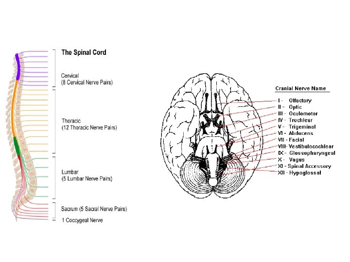
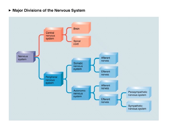
Somatic Nervous System 19
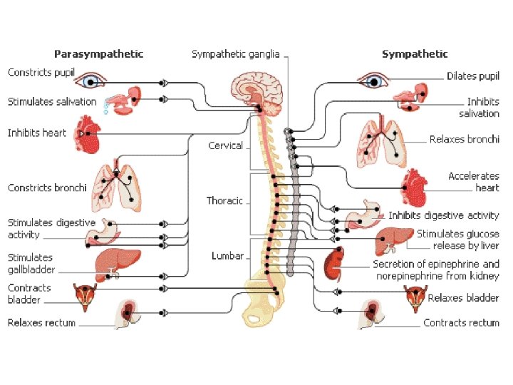
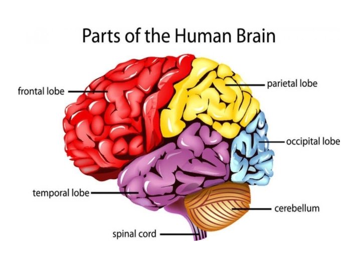
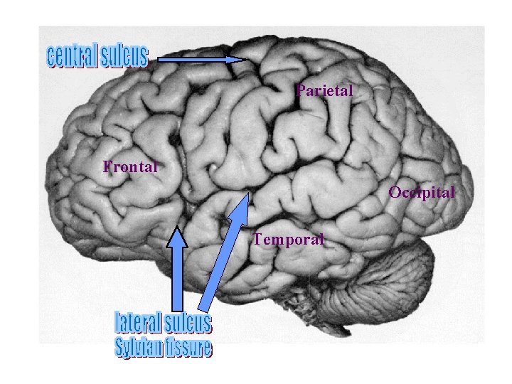
Parietal Frontal Occipital Temporal 22
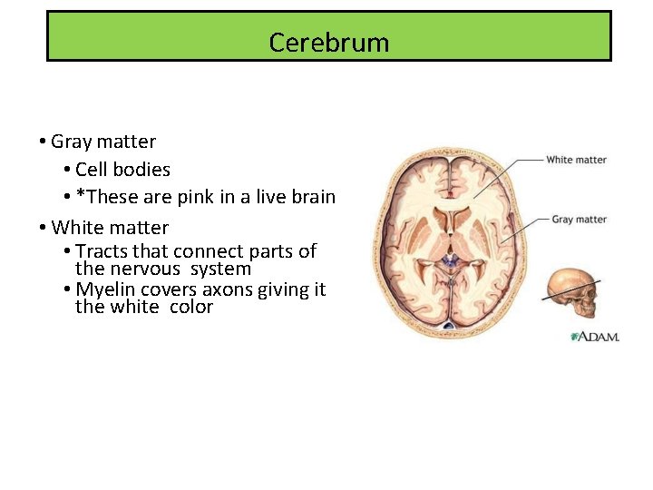
Cerebrum • Gray matter • Cell bodies • *These are pink in a live brain • White matter • Tracts that connect parts of the nervous system • Myelin covers axons giving it the white color
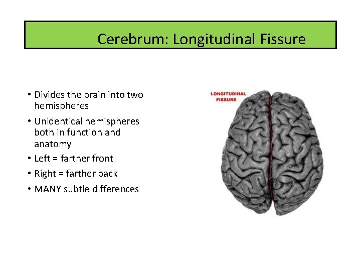
Cerebrum: Longitudinal Fissure • Divides the brain into two hemispheres • Unidentical hemispheres both in function and anatomy • Left = farther front • Right = farther back • MANY subtle differences
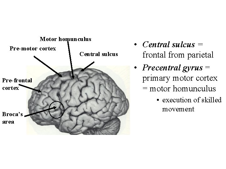
Frontal lobe Motor homunculus Pre-motor cortex Pre-frontal cortex Broca’s area Central sulcus • Central sulcus = frontal from parietal • Precentral gyrus = primary motor cortex = motor homunculus • execution of skilled movement 25
Cerebrum: Frontal Lobe • Anterior aspect of both hemispheres • Responsible for: • Judgment • Reasoning • Intellectual functioning • Personality • Abstract thinking • Memory • Motor control • “Executive Functioning”
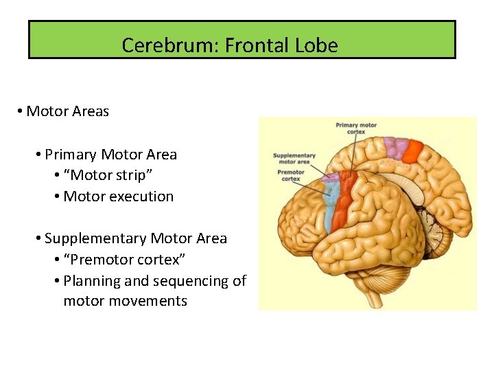
Cerebrum: Frontal Lobe • Motor Areas • Primary Motor Area • “Motor strip” • Motor execution • Supplementary Motor Area • “Premotor cortex” • Planning and sequencing of motor movements
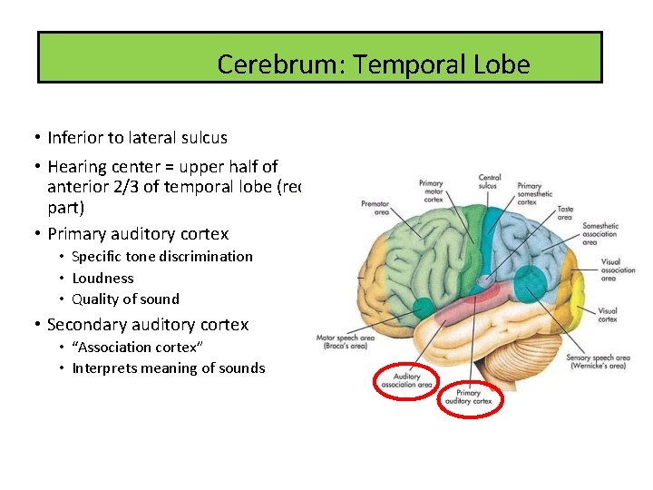
Cerebrum: Temporal Lobe • Inferior to lateral sulcus • Hearing center = upper half of anterior 2/3 of temporal lobe (red part) • Primary auditory cortex • Specific tone discrimination • Loudness • Quality of sound • Secondary auditory cortex • “Association cortex” • Interprets meaning of sounds
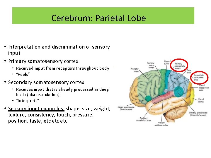
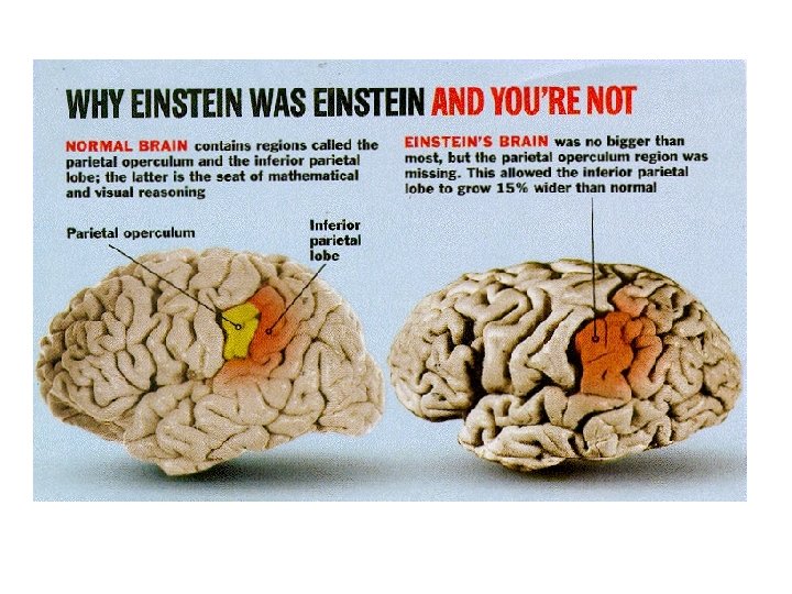
Cerebrum: Parietal Lobe • Interpretation and discrimination of sensory input • Primary somatosensory cortex • Received input from receptors throughout body • “Feels” • Secondary somatosensory cortex • Receives input that is already processed in deep brain (aka association) • “Interprets” • Sensory input examples: shape, size, weight, texture, consistency, touch, pressure, position, taste, etc etc
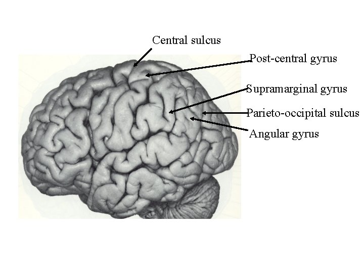
Central sulcus Post-central gyrus Supramarginal gyrus Parieto-occipital sulcus Angular gyrus 30
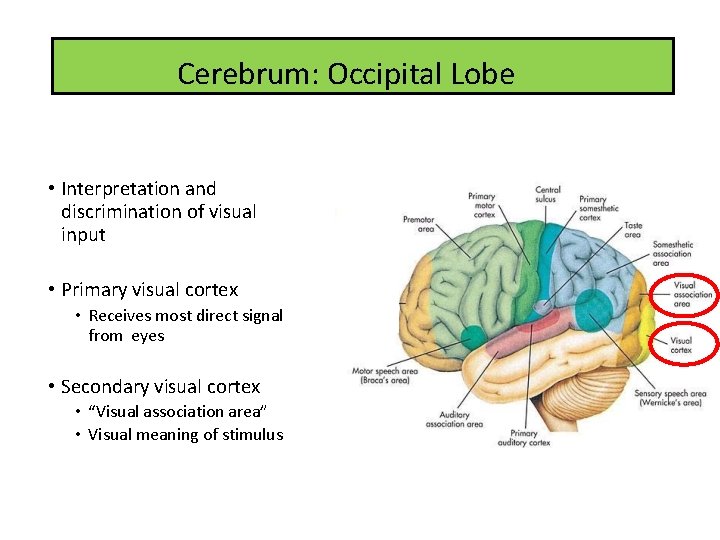
Cerebrum: Occipital Lobe • Interpretation and discrimination of visual input • Primary visual cortex • Receives most direct signal from eyes • Secondary visual cortex • “Visual association area” • Visual meaning of stimulus
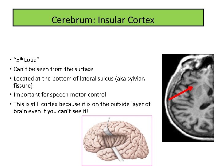
Cerebrum: Insular Cortex • “ 5 th Lobe” • Can’t be seen from the surface • Located at the bottom of lateral sulcus (aka sylvian fissure) • Important for speech motor control • This is still cortex because it is on the outside layer of brain even if you can’t see it!
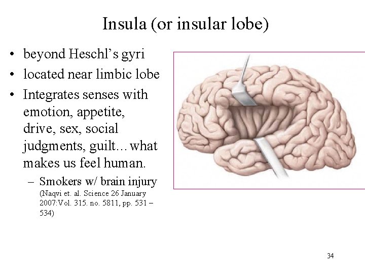
Insula (or insular lobe) • beyond Heschl’s gyri • located near limbic lobe • Integrates senses with emotion, appetite, drive, sex, social judgments, guilt…what makes us feel human. – Smokers w/ brain injury (Naqvi et. al. Science 26 January 2007: Vol. 315. no. 5811, pp. 531 – 534) 34
Primary cortical areas • Motor cortex - voluntary, skilled movement, contralateral • Somatosensory cortex – sensation, contralateral • Auditory cortex (Heschl’s gyrus, insula) input from both ears • Visual cortex (V 1) - visual perception • Olfactory cortex – ventro-posterior frontal & insula 35
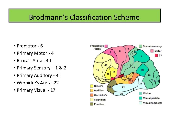
Brodmann’s Classification Scheme • Premotor - 6 • Primary Motor - 4 • Broca’s Area - 44 • Primary Sensory – 1 & 2 • Primary Auditory - 41 • Wernicke’s Area - 22 • Primary Visual - 17
White matter: Brain’s freeways • White matter is organized into tracts that connect regions of the nervous system • Three types: • Association Fibers • Transmit within one sweep of hemisphere (e. g. , arcuate fasciculus) • Commissural Fibers • Transmit from one cerebral hemisphere to the other (e. g. , corpus callosum) • Projection Fibers • Form ascending and descending pathways that connect the cerebral cortex to the lower CNS (e. g. , corona radiata) 37
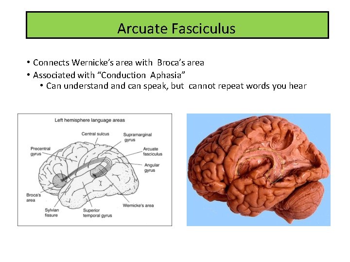
Arcuate Fasciculus • Connects Wernicke’s area with Broca’s area • Associated with “Conduction Aphasia” • Can understand can speak, but cannot repeat words you hear
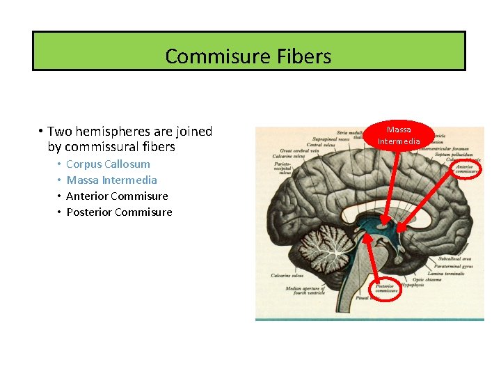
Commisure Fibers • Two hemispheres are joined by commissural fibers • • Corpus Callosum Massa Intermedia Anterior Commisure Posterior Commisure Massa Intermedia
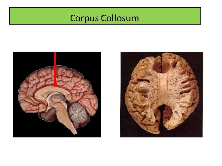
Corpus Collosum
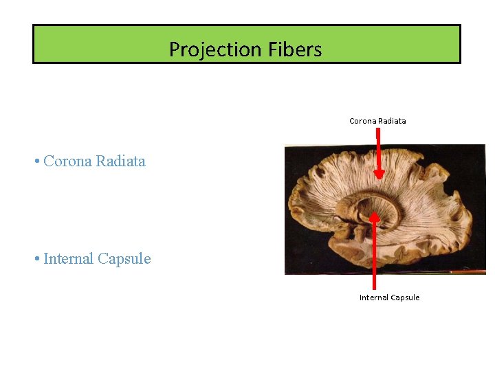
Projection Fibers • Send information to the primary cortexes (motor and sensory) Corona Radiata • Corona Radiata • “Fan-like” • An initial motor pathway or final sensory pathway • Carries information either to or from the cortex and brainstem • Internal Capsule • Convergence of descending axon fibers leaving cortex • OR where the corona radiata get smushed together Internal Capsule
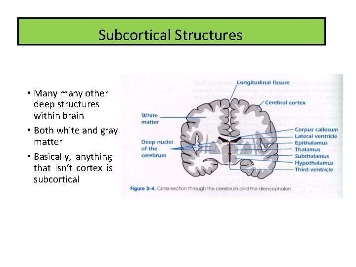
Subcortical Structures • Many many other deep structures within brain • Both white and gray matter • Basically, anything that isn’t cortex is subcortical
43
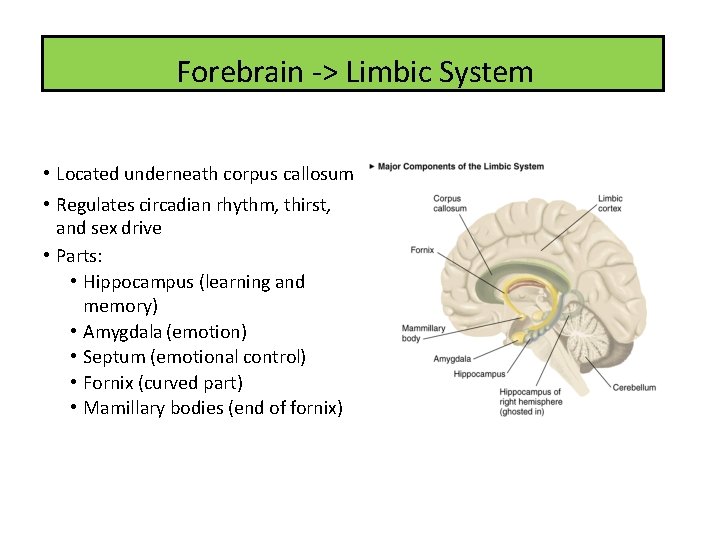
Forebrain -> Limbic System • Located underneath corpus callosum • Regulates circadian rhythm, thirst, and sex drive • Parts: • Hippocampus (learning and memory) • Amygdala (emotion) • Septum (emotional control) • Fornix (curved part) • Mamillary bodies (end of fornix)
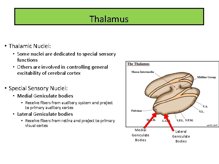
Thalamus • Thalamic Nuclei: • Some nuclei are dedicated to special sensory functions • Others are involved in controlling general excitability of cerebral cortex • Special Sensory Nuclei: • Medial Geniculate bodies • Receive fibers from auditory system and project to primary auditory cortex • Lateral Geniculate bodies • Receive fibers from retina and project to primary visual cortex Medial Geniculate Bodies Lateral Geniculate Bodies
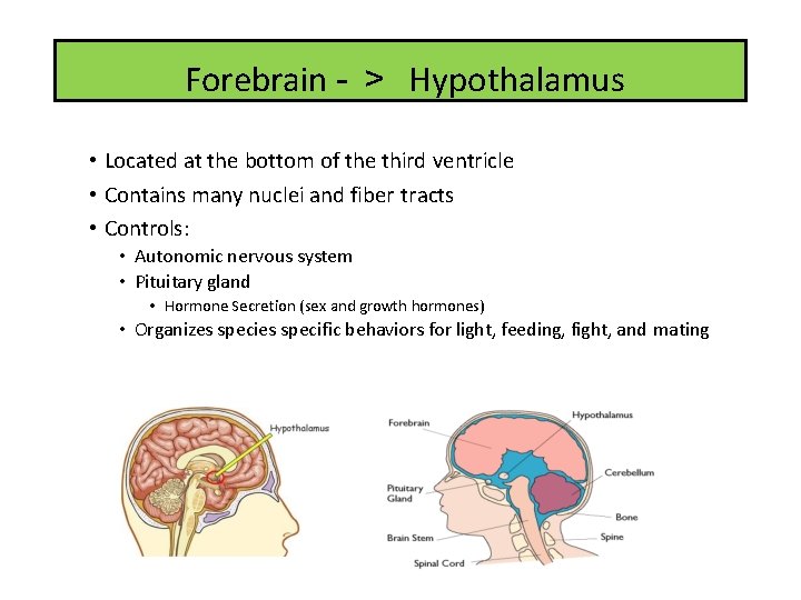
Forebrain - > Hypothalamus • Located at the bottom of the third ventricle • Contains many nuclei and fiber tracts • Controls: • Autonomic nervous system • Pituitary gland • Hormone Secretion (sex and growth hormones) • Organizes specific behaviors for light, feeding, fight, and mating
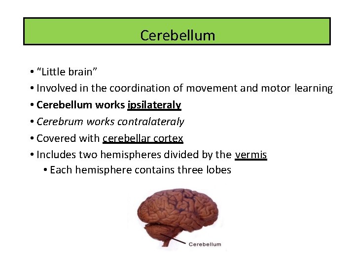
Cerebellum • “Little brain” • Involved in the coordination of movement and motor learning • Cerebellum works ipsilateraly • Cerebrum works contralateraly • Covered with cerebellar cortex • Includes two hemispheres divided by the vermis • Each hemisphere contains three lobes
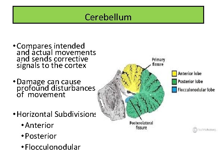
Cerebellum • Compares intended and actual movements and sends corrective signals to the cortex • Damage can cause profound disturbances of movement • Horizontal Subdivisions: • Anterior • Posterior • Flocculonodular
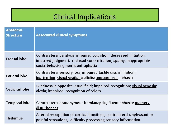
Clinical Implications Anatomic Structure Associated clinical symptoms Frontal lobe Contralateral paralysis; impaired cognition; decreased initiation; impaired judgment, reduced concentration, apathy, inappropriate social behaviors, nonfluent aphasia Parietal lobe Contralateral sensory loss; impaired tactile discrimination; inattention; visual spatial deficits; anosognosia; aphasia Occipital lobe Blindness in opposite visual field; impaired recognition; visual agnosia; alexia; impaired recognition of colors Temporal lobe Thalamus Contralateral homonymous hemianopsia; fluent aphasia; memory disturbances Altered recognition of cortical functions; contralateral unpleasant or painful sensations; difficulty processing sensory information
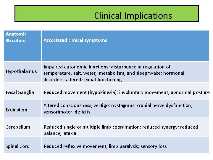
Clinical Implications Anatomic Structure Associated clinical symptoms Hypothalamus Impaired autonomic functions; disturbance in regulation of temperature, salt, water, metabolism, and sleep/wake; hormonal disorders; altered sexual functioning Basal Ganglia Reduced movement (hypokinesia); involuntary movement; abnormal posture Brainstem Altered consciousness; vertigo; nystagmus; cranial nerve dysfunction; sensorimotor deficits Cerebellum Reduced single or multiple limb coordination; reduced synergy; reduced balance; ataxia Spinal Cord Reduced reflexive movement; limb paralysis; sensory loss
- Slides: 50