Central Nervous System The central nervous system CNS
 appears at the beginning of the")


 the metencephalon, which")


 128 are entirely sensory; •")


 contains three groups")

 contains three groups")

, each basal plate")


- Slides: 46
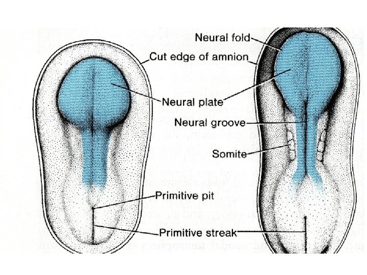
Central Nervous System The central nervous system (CNS) appears at the beginning of the third week as a slipper-shaped plate of thickened ectoderm, the neural plate, in the middorsal region in front of the primitive node. Its lateral edges soon elevate to form the neural folds (Fig. 17.
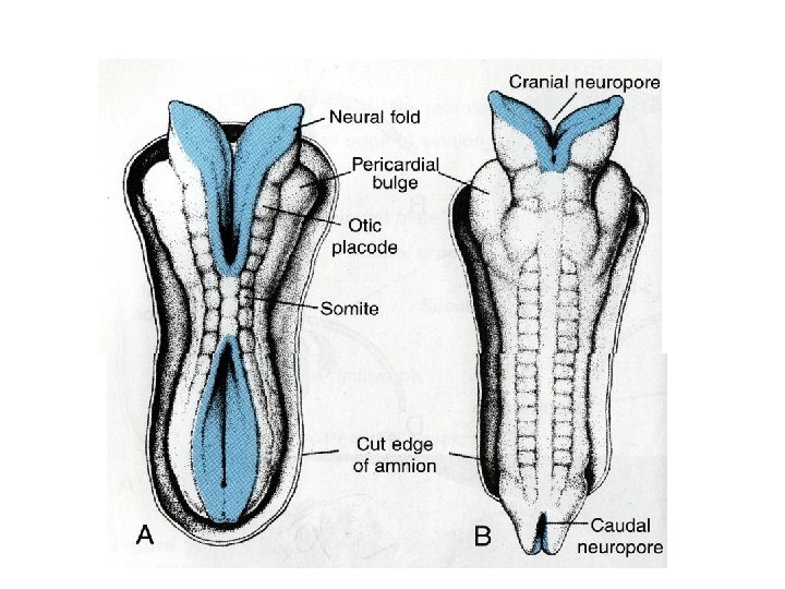
• With further development, the neural folds continue to elevate, approach each other in the midline, and finally fuse, forming the neural tube (Figs. 17. 2 and 17. 3). Fusion begins in the cervical region and proceeds in cephalic and caudal directions (Fig. 17. 3 A). Once fusion is initiated, the open ends of the neural tube form the cranial and caudal neuropores that communicate with the overlying amniotic cavity
• Closure of the cranial neuropore proceeds cranially from the initial closure site in the cervical region (Fig. 17. 3 A) and from a site in the forebrain that forms later. This latter site proceeds cranially, to close the rostral-most region of the neural tube, and caudally to meet advancing closure from the cervical site (Fig. 17. 3 B). Final closure of the cranial neuropore occurs at the 18 - to 20 -somite stage (25 th day); closure of the caudal neuropore occurs approximately 3 days later.
• The cephalic end of the neural tube shows three dilations, the primary brain vesicles: • (a) the prosencephalon, or forebrain; • (b) the mesencephalon, or midbrain; and • (c) the rhombencephalon, or hindbrain (Fig. 17. 4). Simultaneously, it forms two flexures: (a) the cervical flexure at the junction of the hindbrain and the spinal cord and • (b) the cephalic flexure in the midbrain region (Fig. 17. 4).
• When the embryo is 5 weeks old, the prosencephalon consists of two parts: • (a) the telencephalon, formed by a midportion and two lateral outpocketings, the primitive cerebral hemispheres; and • (b) the diencephalon, characterized by outgrowth of the optic vesicles (Fig. 17. 5). A deep furrow, the rhombencephalic isthmus, separates the mesencephalon from the rhombencephalon.
• The rhombencephalon also consists of two parts: • (a) the metencephalon, which later forms the pons and cerebellum and • (b) the myelencephalon. The boundary between these two portions is marked by the pontine flexure. It form medulla oblongata. (Fig. 17. 5
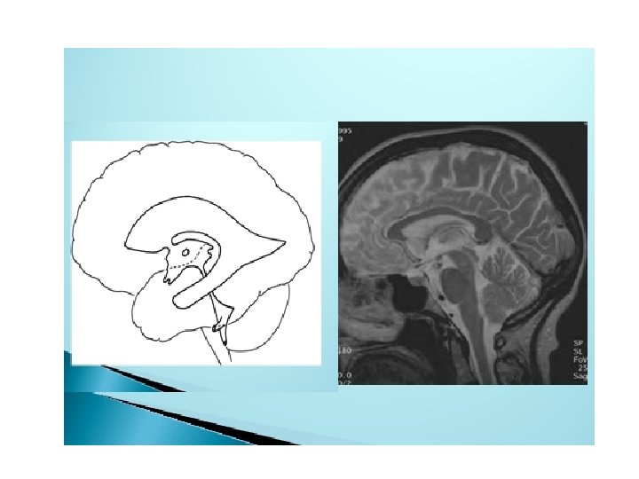
• The lumen of the spinal cord, the central canal, is continuous with that of the brain vesicles. The cavity of the rhombencephalon is the fourth ventricle, that of the diencephalon is the third ventricle, and those of the cerebral hemispheres are the lateral ventricles (Fig. 17. 5). The lumen of the mesencephalon connects the third and fourth ventricles. This lumen becomes very narrow and is then known as the aqueduct of Sylvius. The lateral ventricles communicate with the third ventricle through the interventricular foramina of Monro (Fig. 17. 5).
• SPINAL CORD • Neuroepithelial, Mantle, and Marginal Layers • The wall of a recently closed neural tube consists of neuroepithelial cells. These cells extend over the entire thickness of the wall and form a P. 295 thick pseudostratified epithelium (Fig. 17. 6). Junctional complexes at the lumen connect them. During the neural groove stage and immediately after closure of the tube, they divide rapidly, producing more and more neuroepithelial cells. Collectively, they constitute the neuroepithelial layer or neuroepithelium.
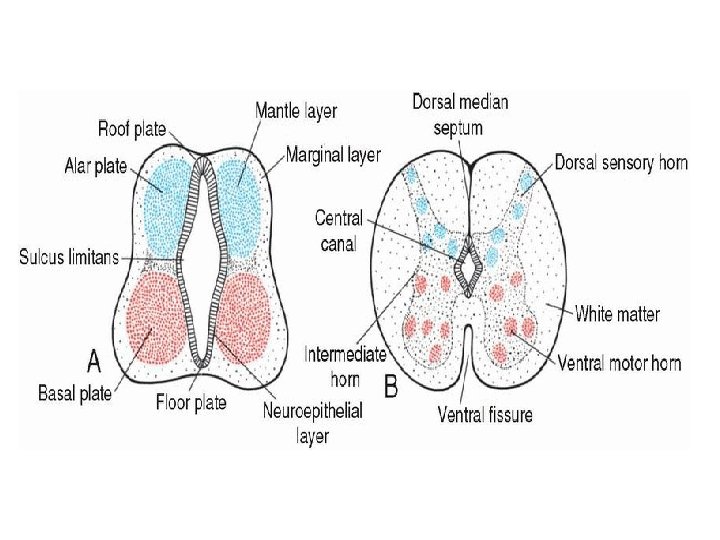
• Once the neural tube closes, neuroepithelial cells begin to give rise to another cell type characterized by a large round nucleus with pale nucleoplasm and a dark-staining nucleolus. These are the primitive nerve cells, or neuroblasts (Fig. 17. 7). They form the mantle layer, a zone around the neuroepithelial layer (Fig. 17. 8). The mantle layer later forms the gray matter of the spinal cord. • The outermost layer of the spinal cord, the marginal layer, contains nerve fibers emerging from neuroblasts in the mantle layer. As a result of myelination of nerve fibers, this layer takes on a white appearance and therefore is called the white matter of the spinal cord (Fig. 17. 8).
• Basal, Alar, Roof, and Floor Plates • As a result of continuous addition of neuroblasts to the mantle layer, each side of the neural tube shows a ventral and a dorsal thickening. The ventral thickenings, the basal plates, which contain ventral motor horn cells, form the motor areas of the spinal cord; the dorsal thickenings, the alar plates, form the sensory areas (Fig. 17. 8 A). A longitudinal groove, the sulcus limitans, marks the boundary between the two. The dorsal and ventral midline portions of the neural tube, known as the roof and floor plates, respectively, do not contain neuroblasts; they serve primarily as pathways for nerve fibers crossing from one side to the other.
• In addition to the ventral motor horn and the dorsal sensory horn, a group of neurons accumulates between the two areas and forms a small intermediate horn (Fig. 17. 8 B). This horn, containing neurons of the sympathetic portion of the autonomic nervous system (ANS), is present only at thoracic (T 1 -T 12) and upper lumbar levels (L 2 or L 3) of the spinal cord.
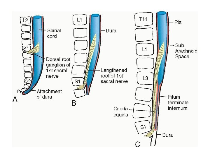
• Positional Changes of the Cord • In the third month of development, the spinal cord extends the entire length of the embryo, and spinal nerves pass through the intervertebral foramina at their level of origin (Fig. 17. 13 A). With increasing age, however, the vertebral column and dura lengthen more rapidly than the neural tube, and the terminal end of the spinal cord gradually shifts to a higher level. At birth, this end is at the level of the third lumbar vertebra (Fig. 17. 13 C). As a result of this disproportionate growth, spinal nerves run obliquely from their segment of origin in the spinal cord to the corresponding level of the vertebral column. The dura remains attached to the vertebral column at the coccygeal level
• In the adult, the spinal cord terminates at the level of L 2 to L 3, whereas the dural sac and subarachnoid space extend to S 2. At the end of the cord, a threadlike extension of pia mater passes caudally, goes through the dura, which provides a covering layer at S 2 and extends to the first coccygeal vertebra. This structure is called the filum terminale, and it marks the tract of regression of the spinal cord as well as providing support for the cord (the part covered by dura and extending from S 2, to the coccyx is also called the coccygeal ligament). Nerve fibers below the terminal end of the cord collectively constitute the cauda equina. When cerebrospinal fluid is tapped during a lumbar puncture, the needle is inserted at the lower lumbar level (L 4 -L 5), avoiding the lower end of the cord.
• CRANIAL NERVES • By the fourth week of development, nuclei for all 12 cranial nerves are present. All except the olfactory (I) and optic (II) nerves arise from the brain stem { pons , MO and midbrain}, and of these, only the oculomotor (III) arises outside the region of the hindbrain. In the hindbrain, proliferation centers in the neuroepithelium establish eight distinct segments, the rhombomeres. These rhombomeres give rise to motor nuclei of cranial nerves IV, V, VII, IX, X, XI, and XII (Figs. 17 and 17. 40).
• Three cranial nerves (I, II, and VIII) 128 are entirely sensory; • four (IV, VI, XI, and XII) {4 , 6, 11, 12} are entirely motor; • three (VII, IX, and X) { 7, 9, 10} have motor, sensory, and parasympathetic fibers; • and one (III) has only motor and parasympathetic components. In contrast, each spinal nerve has motor and sensory fibers. • V is motor and sensory
Hypophysis or Pituitary Gland The hypophysis, or pituitary gland, develops from two completely different parts: (a) an ectodermal outpocketing of the stomodeum (primitive oral cavity) immediately in front of the oropharyngeal membrane, known as Rathke's pouch (b) a downward extension of the diencephalon, the infundibulum (Fig. 17. 26). When the embryo is approximately 3 weeks old, Rathke's pouch appears as an evagination of the oral cavity and subsequently grows dorsally toward the infundibulum. By the end of the second month, it loses its connection with the oral cavity and is then in close contact with the infundibulum
• During further development, cells in the anterior wall of Rathke's pouch increase rapidly in number and form the anterior lobe of the hypophysis, or adenohypophysis (Fig. 17. 26 B). A small extension of this lobe, the pars tuberalis, grows along the stalk of the infundibulum and eventually surrounds it (Fig. 17. 26 C). The posterior wall of Rathke's pouch develops into the pars intermedia, which in humans seems to have little significance. • The infundibulum gives rise to the stalk and the pars nervosa, or posterior lobe of the hypophysis (neurohypophysis) (Fig. 17. 26 C). It is composed of neuroglial cells. In addition, it contains a number of nerve fibers from the hypothalamic area.
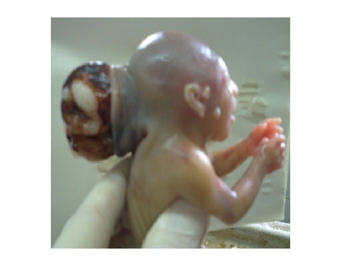
Clinical Correlates Hypophyseal Defects Occasionally, a small portion of Rathke's pouch persists in the roof of the pharynx as a pharyngeal hypophysis. Craniopharyngiomas arise from remnants of Rathke's pouch. They may form within the sella turcica or along the stalk of the pituitary but usually lie above the sella. They may cause hydrocephalus and pituitary dysfunction (e. g. , diabetes insipidus, growth failure
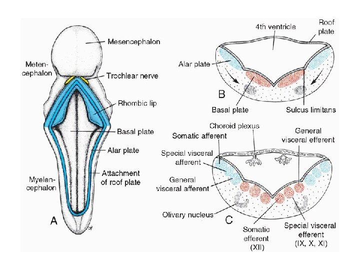
• BRAIN • Distinct basal and alar plates, representing motor and sensory areas, respectively, are found on each side of the midline in the rhombencephalon and mesencephalon. In the prosencephalon, however, the alar plates are accentuated and the basal plates regress. • Rhombencephalon: Hindbrain • The rhombencephalon consists of the myelencephalon, the most caudal of the brain vesicles, and the metencephalon, which extends from the pontine flexure to the rhombencephalic isthmus (Figs. 17. 5 and 17. 17).
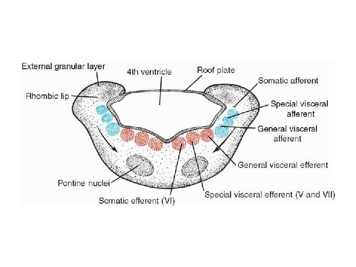
• Myelencephalon • The myelencephalon is a brain vesicle that gives rise to the mo. It differs from the spinal cord in that its lateral walls are everted (Fig. 17. 18). Alar and basal plates separated by the sulcus limitans can be clearly distinguished. The basal plate, similar to that of the spinal cord, contains motor nuclei. These nuclei are divided into three groups: • (a) a medial somatic efferent group, • (b) an intermediate special visceral efferent group, and • (c) a lateral general visceral efferent group
• The first group contains motor neurons, which form the cephalic continuation of the anterior horn cells. Since this somatic efferent group continues rostrally into the mesencephalon, it is called the somatic efferent motor column. In the myelencephalon, it includes neurons of the {12}hypoglossal nerve that supply the tongue musculature. In the metencephalon and the mesencephalon, the column contains neurons of the abducens (Fig. 17. 19), trochlear, and oculomotor nerves{3, 4, 6} (Fig. 17. 23), respectively. These nerves supply the eye musculature
• The special visceral efferent group extends into the metencephalon, forming the special visceral efferent motor column. Its motor neurons supply striated muscles of the pharyngeal arches. In the myelencephalon, the column is represented by neurons of the accessory, vagus, and glossopharyngeal {9, 10, 11} nerves. • The general visceral efferent group contains motor neurons that supply involuntary musculature of the respiratory tract, intestinal tract, and heart.
• The alar plate contains three groups of sensory relay nuclei (Fig. 17. 18 C). The most lateral of these, the somatic afferent (sensory) group, receives impulses from the ear and surface of the head by way of the vestibulocochlear and trigeminal nerves ( 5, 8). The intermediate, or special visceral afferent, group receives impulses from taste buds of the tongue and from the palate, oropharynx, and epiglottis ( 7 ). The medial, or general visceral afferent, group receives interoceptive information from the gastrointestinal tract and heart.
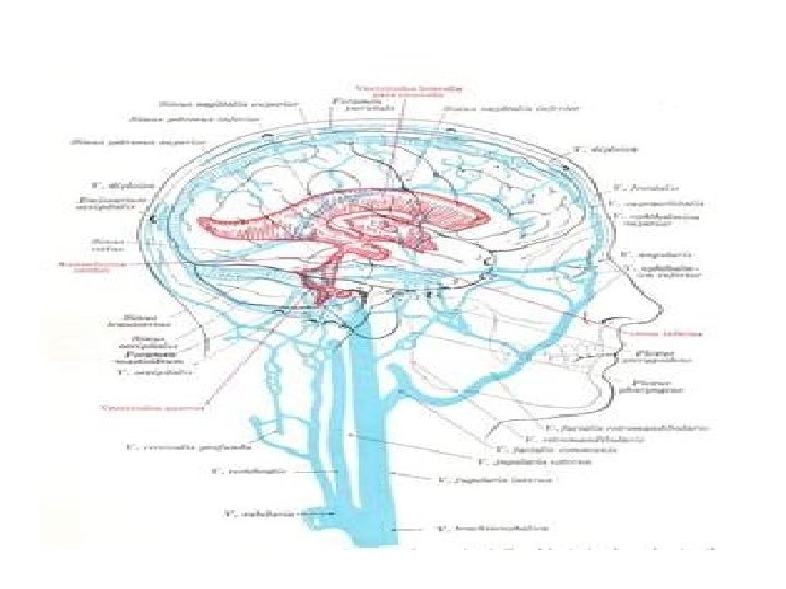
• The roof plate of the myelencephalon consists of a single layer of ependymal cells covered by vascular mesenchyme, the pia mater (Fig. 17. 18 C). The two combined are known as the tela choroidea. Because of active proliferation of the vascular mesenchyme, a number of sac-like invaginations project into the underlying ventricular cavity (Figs. 17. 18 C). These tuftlike invaginations form the choroid plexus, which produces cerebrospinal fluid
• Metencephalon • The metencephalon, similar to the myelencephalon, is characterized by basal and alar plates (Fig. 17. 19). Two new components form • (a) the cerebellum, a coordination center for posture and movement (Fig. 17. 20), and • (b) the pons, the pathway for nerve fibers between the spinal cord and the cerebral and cerebellar cortices
• Each basal plate of the metencephalon (Fig. 17. 19) contains three groups of motor neurons: (a) the medial somatic efferent group, which gives rise to the nucleus of the abducens (6) nerve; (b) the special visceral efferent group, containing nuclei of the trigeminal and facial nerves (5, 7), which innervate the musculature of the first and second pharyngeal arches; and (c) the general visceral efferent group, with axons that supply the submandibular and sublingual glands.
• The alar plates of the metencephalon contain three groups of sensory nuclei: • (a) a lateral somatic afferent group, which contains neurons of the trigeminal (5) nerve and a small portion of the vestibulocochlear(8) complex; • (b) the special visceral afferent group; and • (c) the general visceral afferent group (Fig. 17. 19
• Each basal plate of the metencephalon (Fig. 17. 19) contains three groups of motor neurons: (a) the medial somatic efferent group, which gives rise to the nucleus of the abducens nerve (6); • (b) the special visceral efferent group, containing nuclei of the trigeminal and facial nerves(5. 7), which innervate the musculature of the first and second pharyngeal arches; and • (c) the general visceral efferent group, with axons that supply the submandibular and sublingual glands.
• The marginal layer of the basal plates of the metencephalon expands as it makes a bridge for nerve fibers connecting the cerebral cortex and cerebellar cortex with the spinal cord. Hence, this portion of the metencephalon is known as the pons (bridge). In addition to nerve fibers, the pons contains the pontine nuclei, which originate in the alar plates of the metencephalon and myelencephalon mk
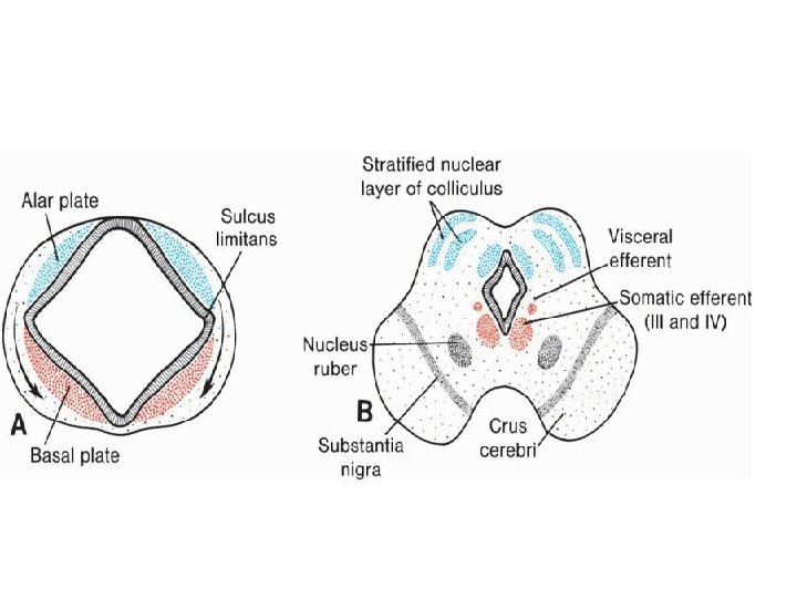
• Mesencephalon: Midbrain • In the mesencephalon (Fig. 17. 23), each basal plate contains two groups of motor nuclei: • (a) a medial somatic efferent group, represented by the oculomotor and trochlear • ( 3 , 4 )nerves, which innervate the eye musculature and • (b) a small general visceral efferent group, represented by the nucleus of Edinger. Westphal, which innervates the sphincter pupillary muscle (Fig. 17. 23 B
• Diencephalon • Roof Plate • The diencephalon, which develops from the median portion of the prosencephalon (Figs. 17. 5 and 17. 17), is thought to consist of a roof plate and two alar plates but to lack floor and basal plates (interestingly, sonic hedgehog, a ventral midline marker, is expressed in the floor of the diencephalon, suggesting that a floor plate does exist). The roof plate of the diencephalon consists of a single layer of ependymal cells covered by vascular mesenchyme. Together, these layers give rise to the choroid plexus of the third ventricle
• Alar Plate, Thalamus, and Hypothalamus • The alar plates form the lateral walls of the diencephalon. A groove, the hypothalamic sulcus, divides the plate into a dorsal and a ventral region, the thalamus and hypothalamus, respectively (Figs. 17. 24 and 17. 25).