ENTERIC NERVOUS SYSTEM The enteric nervous system ENS

 or intrinsic nervous system is one of the main")
 an outer plexus")


- Slides: 23
ENTERIC NERVOUS SYSTEM
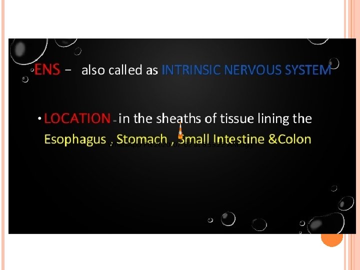
The enteric nervous system (ENS) or intrinsic nervous system is one of the main divisions of the autonomic nervous system (ANS) and consists of a mesh-like system of neurons that governs the function of the gastrointestinal tract. It is capable of acting independently of the sympathetic and parasympathetic nervous systems, although it may be influenced by them. The ENS is also called the second brain It is derived from neural crest cells
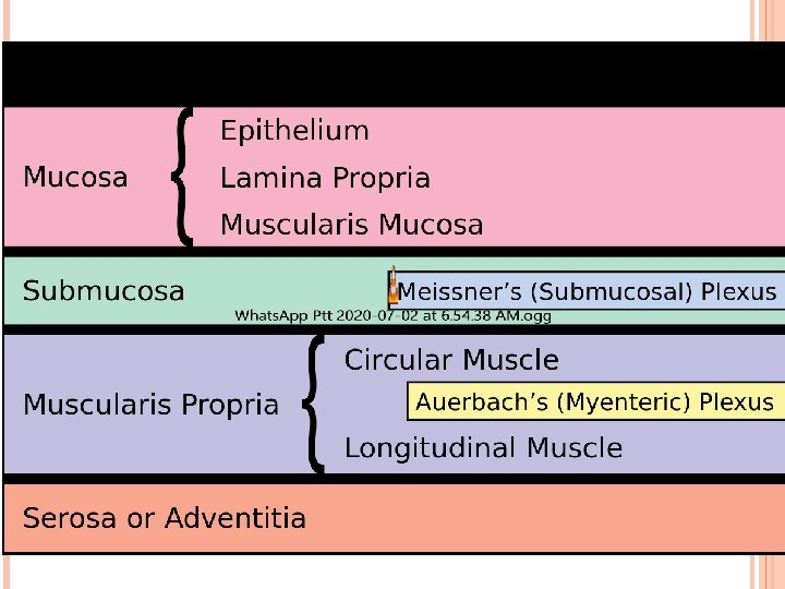
The enteric nervous system is composed mainly of two plexuses, (1) an outer plexus lying between the longitudinal and circular muscle layers, called the myenteric plexus or Auerbach’s plexus, (2) an inner plexus, called the submucosal plexus or Meissner’s plexus, that lies in the submucosa
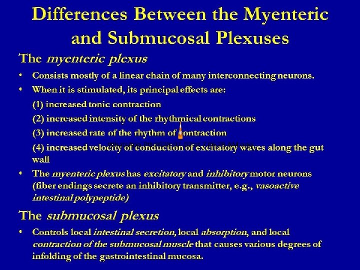
The myenteric plexus controls mainly the gastrointestinal movements, and the submucosal plexus controls mainly gastrointestinal secretion and local blood flow. Although the enteric nervous system can function on its own, independently of these extrinsic nerves, stimulation by the parasympathetic and sympathetic systems can greatly enhance or inhibit gastrointestinal functions Also there are sensory nerve endings that originate in the gastrointestinal epithelium or gut wall and send afferent fibers to both plexuses of the enteric system, as well as (1) to the prevertebral ganglia of the sympathetic nervous system, (2) to the spinal cord, and (3) in the vagus nerves all the way to the brain stem.
Differences Between the Myenteric and Submucosal Plexuses
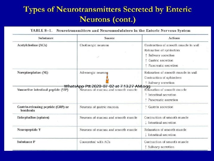
Types of Neurotransmitters Secreted by Enteric Neurons
Autonomic Control of the Gastrointestinal Tract Parasympathetic Innervation.
The parasympathetic sup-ply to the gut is divided into cranial and sacral divisions. Except for a few parasympathetic fibers to the mouth and pharyngeal regions of the alimentary tract, the CRANIAL PARASYMPATHETIC NERVE FIBERS are almost entirely in the vagus nerves. These fibers provide extensive innervation to the oesophagus, stomach, and pancreas and somewhat less to the intestines down through the first half of the large intestine. The SACRAL PARASYMPATHETICS originate in the second, third, and fourth sacral segments of the spinal cord and pass through the pelvic nerves to the distal half of the large intestine and all the way to the anus. The sigmoidal, rectal, and anal regions are considerably better supplied with parasympathetic fibers than are the other intestinal areas. These fibers function especially to execute the defecation reflexes. The POSTGANGLIONIC NEURONS of the gastrointestinal parasympathetic system are located mainly in the myenteric and submucosal plexuses. Stimulation of these parasympathetic nerves causes general increase in activity of the entire enteric nervous system. This in turn enhances activity of most gastrointestinal functions.
Sympathetic Innervation. The sympathetic fibers to the gastrointestinal tract originate in the spinal cord between segments T-5 and L-2. The sympathetics innervate essentially all of the gastrointestinal tract, rather than being more extensive nearest the oral cavity and anus as is true of the parasympathetics. The sympathetic nerve endings secrete mainly norepinephrine but also small amounts of epinephrine.
In general, stimulation of the sympathetic nervous system inhibits activity of the gastrointestinal tract, causing many effects opposite to those of the parasympathetic system. It exerts its effects in two ways: (1) to a slight extent by direct effect of secreted norepinephrine to inhibit intestinal tract smooth muscle (except the mucosal muscle, which it excites) and (2) to a major extent by an inhibitory effect of norepinephrine on the neurons of the entire enteric nervous system. Strong stimulation of the sympathetic system can inhibit motor movements of the gut so greatly that this literally can block movement of food through the gastrointestinal tract.
Afferent Sensory Nerve Fibers from the Gut Many afferent sensory nerve fibers innervate the gut. Some of them have their cell bodies in the enteric nervous system itself and some in the dorsal root ganglia of the spinal cord. These sensory nerves can be stimulated by (1) irritation of the gut mucosa, (2) excessive distention of the gut, or (3) presence of specific chemical substances in the gut. Signals trans-mitted through the fibers can then cause excitation or, under other conditions , inhibition of intestinal movements or intestinal secretion.
Gastrointestinal Reflexes The anatomical arrangement of the enteric nervous system and its connections with the sympathetic and parasympathetic systems support three types of gastrointestinal reflexes that are essential to gastrointestinal control.
1. Reflexes that are integrated entirely within the gut wall enteric nervous system. These include reflexes that control much gastrointestinal secretion, peristalsis, mixing contractions, local inhibitory effects, and so forth. 2. Reflexes from the gut to the prevertebral sympathetic ganglia and then back to the gastrointestinal tract. These reflexes transmit signals long distances to other areas of the gastrointestinal tract, such as signals from the stomach to cause evacuation of the colon (the gastrocolic reflex), signals from the colon and small intestine to inhibit stomach motility and stomach secretion (the enterogastric reflexes), and reflexes from the colon to inhibit emptying of ileal contents into the colon (the colonoileal reflex).
3. Reflexes from the gut to the spinal cord or brain stem and then back to the gastrointestinal tract. These include especially (1) reflexes from the stomach and duodenum to the brain stem and back to the stomach—by way of the vagus nerves—to control gastric motor and secretory activity; (2) pain reflexes that cause general inhibition of the entire gastrointestinal tract; and (3) defecation reflexes that travel from the colon and rectum to the spinal cord and back again to produce the powerful colonic, rectal, and abdominal contractions required for defecation (the defecation reflexes).
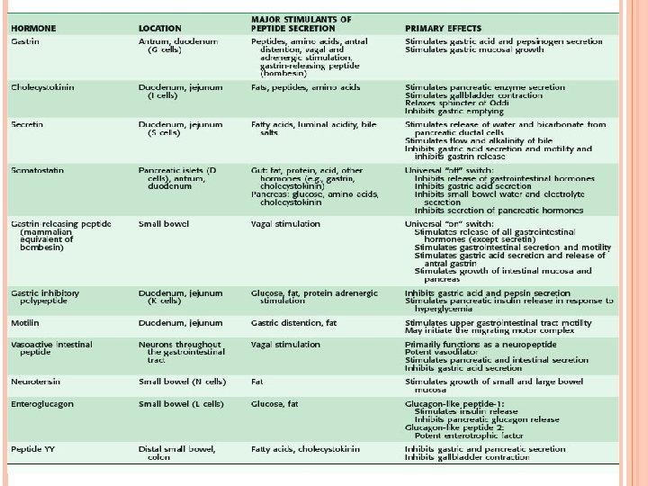
Hormonal Control of Gastrointestinal Motility Most of the hormones affect motility in some parts of the gastrointestinal tract. Although the motility effects are usually less important than the secretory effects of the hormones, some of the more important of them are the following. Gastrin is secreted by the “G” cells of the antrum of the stomach in response to stimuli associated with ingestion of a meal, such as distention of the stomach, the products of proteins, and gastrin releasing peptide, which is released by the nerves of the gastric mucosa during vagal stimulation. The primary actions of gastrin are (1) stimulation of gastric acid secretion and(2) stimulation of growth of the gastric mucosa.
Cholecystokinin is secreted by “I” cells in the mucosa of the duodenum and jejunum mainly in response to digestive products of fat, fatty acids, and monoglycerides in the intestinal contents. Actions : This hormone strongly contracts the gallbladder, expelling bile into the small intestine where the bile in turn plays important roles in emulsifying fatty substances, allowing them to be digested and absorbed. Cholecystokinin also inhibits stomach contraction moderately. Therefore, at the same time that this hormone causes emptying of the gallbladder, it also slows the emptying of food from the stomach to give adequate time for digestion of the fats in the upper intestinal tract.
Secretin was the first gastrointestinal hormone discovered and is secreted by the “S” cells in the mucosa of the duodenum in response to acidic gastric juice emptying into the duodenum from the pylorus of the stomach. Action : Secretin has a mild effect on motility of the gastrointestinal tract and acts to promote pancreatic secretion of bicarbonate which in turn helps to neutralize the acid in the small intestine. Gastric inhibitory peptide is secreted by the mucosa of the upper small intestine, mainly in response to fatty acids and amino acids but to a lesser extent in response to carbohydrate. Action : It has a mild effect in decreasing motor activity of the stomach and therefore slows emptying of gastric contents into the duodenum when the upper small intestine is already overloaded with food products.
Motilin is secreted by the upper duodenum during fasting, and the only known function of this hormone is to increase gastrointestinal motility. Motilin is released cyclically and stimulates waves of gastrointestinal motility called interdigestive myoelectric complexes that move through the stomach and small intestine every 90 minutes in a fasted person. Motilin secretion is inhibited after ingestion by mechanisms that are not fully understood.