
Nervous System Speech Language Chapter 11 Perry C
- Slides: 76
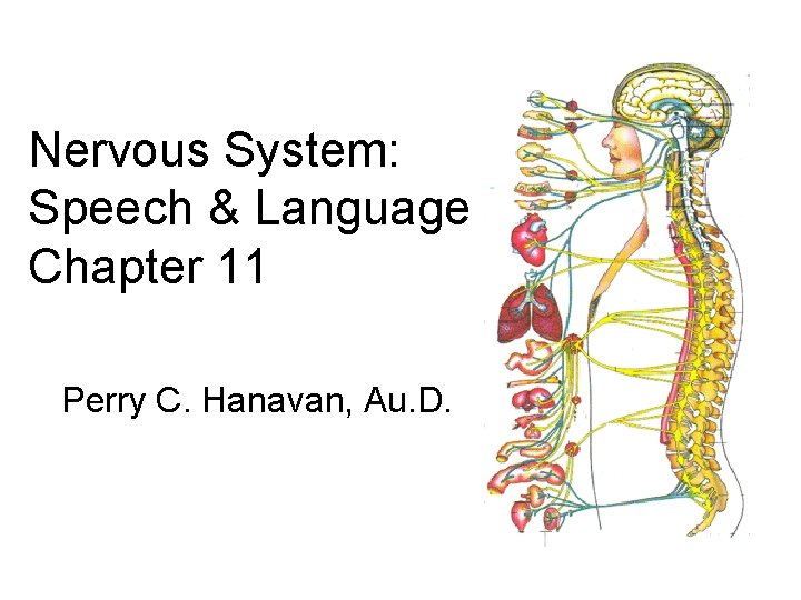
Nervous System: Speech & Language Chapter 11 Perry C. Hanavan, Au. D.
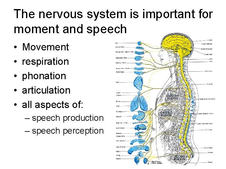
The nervous system is important for moment and speech • • • Movement respiration phonation articulation all aspects of: – speech production – speech perception
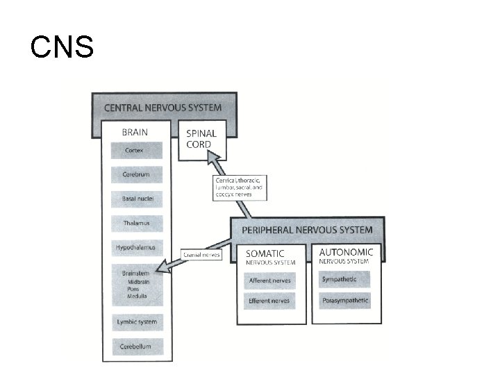
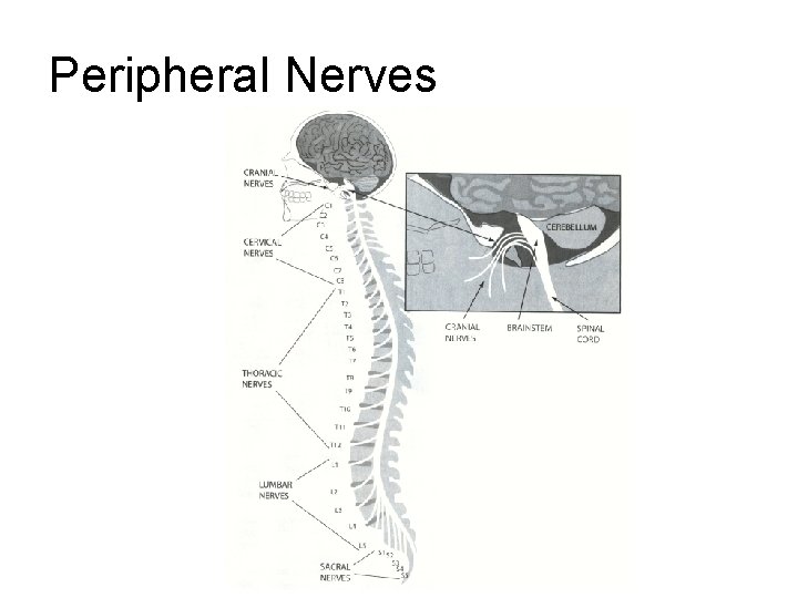
The nervous system divisions • CNS – brain and spinal cord – glial cells • astrocytes – oligodendrocytes • forms myelin • PNS – cranial nerves and spinal nerves – Schwann cells • forms myelin
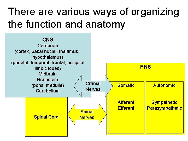
There are various ways of organizing the function and anatomy CNS Cerebrum (cortex, basal nuclei, thalamus, hypothalamus) (parietal, temporal, frontal, occipital limbic lobes) Midbrain Brainstem Cranial (pons, medulla) Nerves Cerebellum Spinal Cord Spinal Nerves PNS Somatic Autonomic Afferent Efferent Sympathetic Parasympathetic
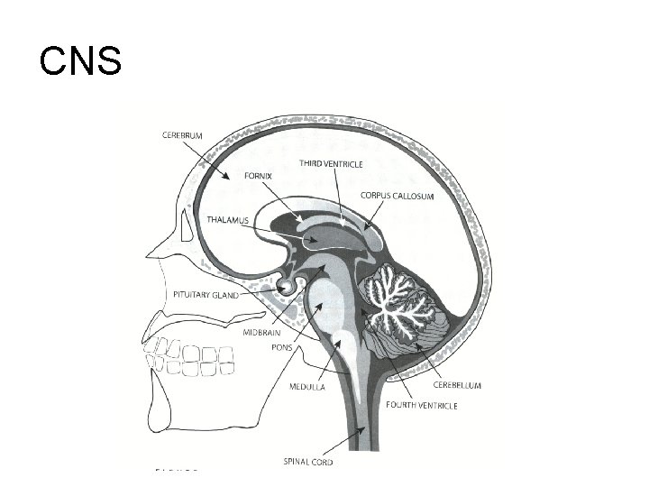
CNS
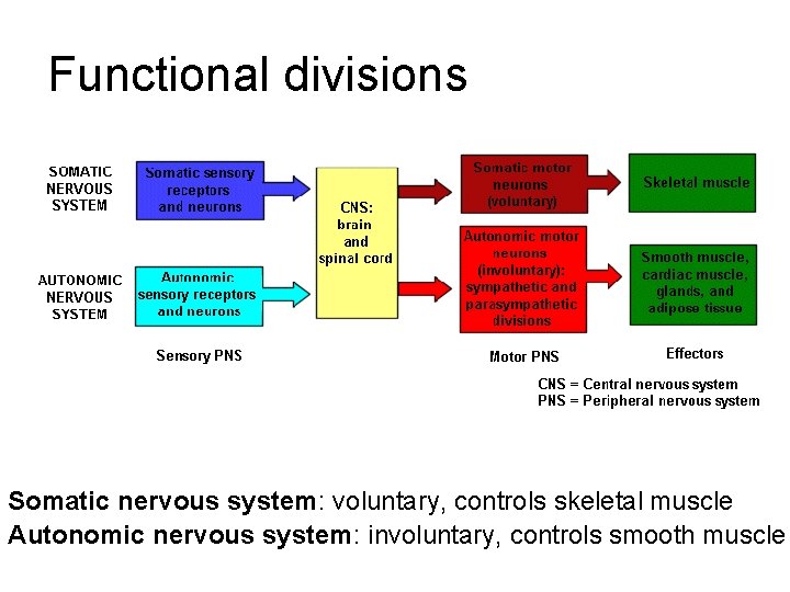
Functional divisions Somatic nervous system: voluntary, controls skeletal muscle Autonomic nervous system: involuntary, controls smooth muscle
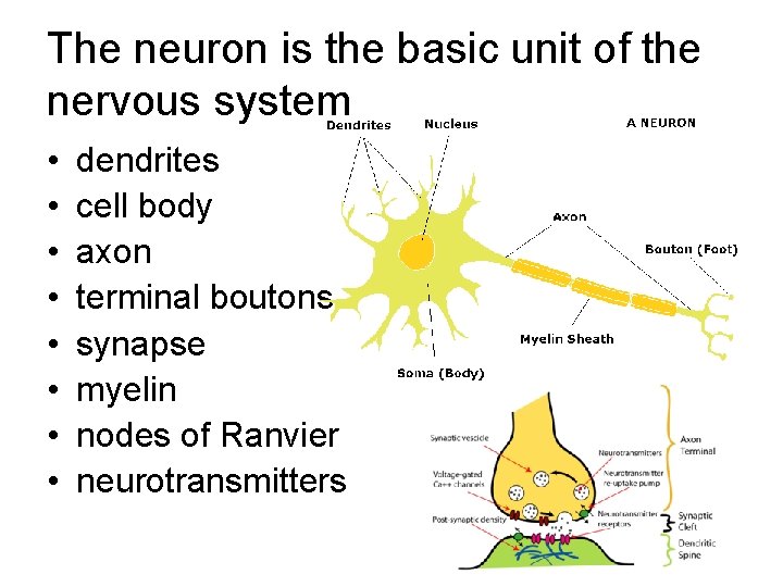
The neuron is the basic unit of the nervous system • • dendrites cell body axon terminal boutons synapse myelin nodes of Ranvier neurotransmitters
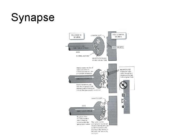
Synapse
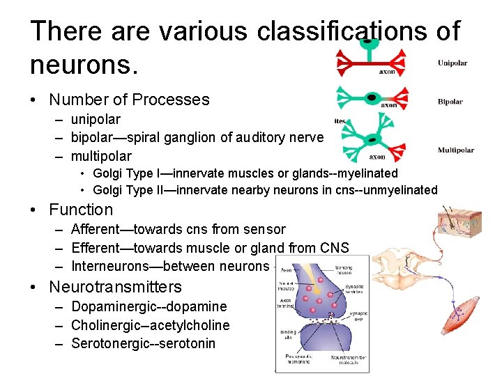
There are various classifications of neurons. • Number of Processes – unipolar – bipolar—spiral ganglion of auditory nerve – multipolar • Golgi Type I—innervate muscles or glands--myelinated • Golgi Type II—innervate nearby neurons in cns--unmyelinated • Function – Afferent—towards cns from sensor – Efferent—towards muscle or gland from CNS – Interneurons—between neurons • Neurotransmitters – Dopaminergic--dopamine – Cholinergic--acetylcholine – Serotonergic--serotonin
Question Afferent is? A. From CNS B. Towards CNS C. Between interneurons D. All the above E. None of the aboe
Question Spiral ganglion is? A. Unipolar B. Bipolar C. Tripolar D. Quadrapolar E. Multipolar
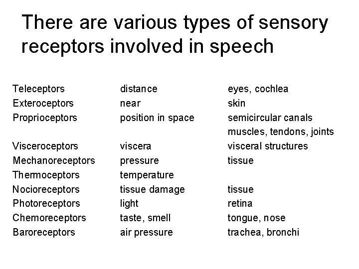
There are various types of sensory receptors involved in speech Teleceptors Exteroceptors Proprioceptors distance near position in space Visceroceptors Mechanoreceptors Thermoceptors Nocioreceptors Photoreceptors Chemoreceptors Baroreceptors viscera pressure temperature tissue damage light taste, smell air pressure eyes, cochlea skin semicircular canals muscles, tendons, joints visceral structures tissue retina tongue, nose trachea, bronchi
Question Proprioceptors sense? A. Air pressure B. Distance C. Pressure (touch) D. Position in space E. Temperature
CNS
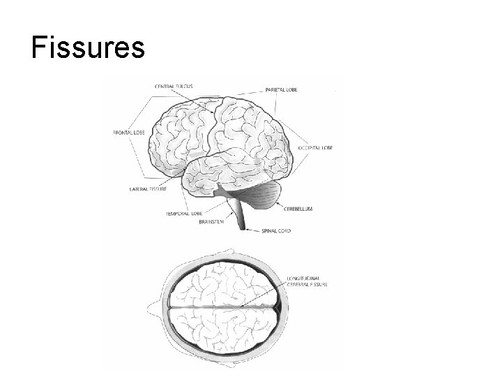
Fissures
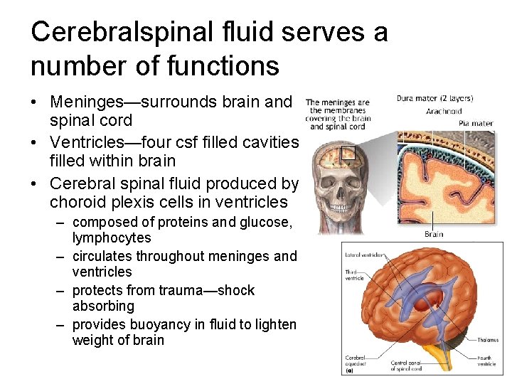
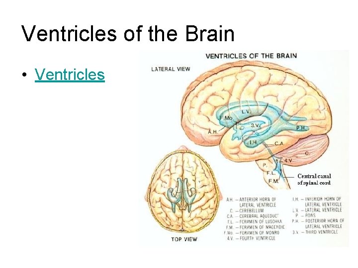
Cerebralspinal fluid serves a number of functions • Meninges—surrounds brain and spinal cord • Ventricles—four csf filled cavities filled within brain • Cerebral spinal fluid produced by choroid plexis cells in ventricles – composed of proteins and glucose, lymphocytes – circulates throughout meninges and ventricles – protects from trauma—shock absorbing – provides buoyancy in fluid to lighten weight of brain
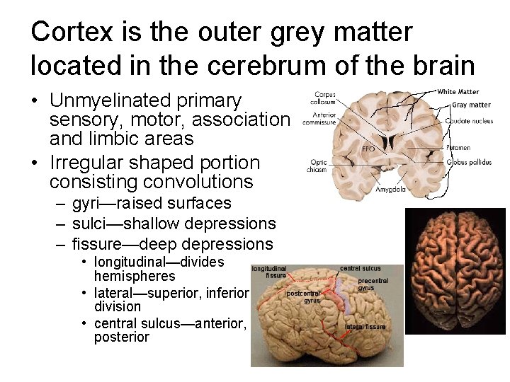
Cortex is the outer grey matter located in the cerebrum of the brain • Unmyelinated primary sensory, motor, association and limbic areas • Irregular shaped portion consisting convolutions – gyri—raised surfaces – sulci—shallow depressions – fissure—deep depressions • longitudinal—divides hemispheres • lateral—superior, inferior division • central sulcus—anterior, posterior
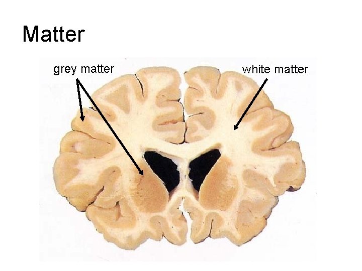
Matter grey matter white matter
Question Which lobe of the brain is primarily involved in speech production? A. Temporal B. Frontal C. Parietal D. Occipital E. Limbic
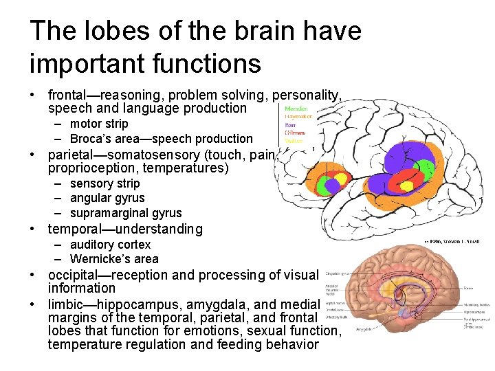
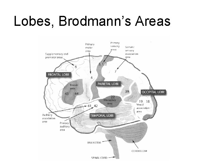
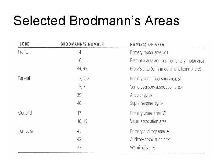
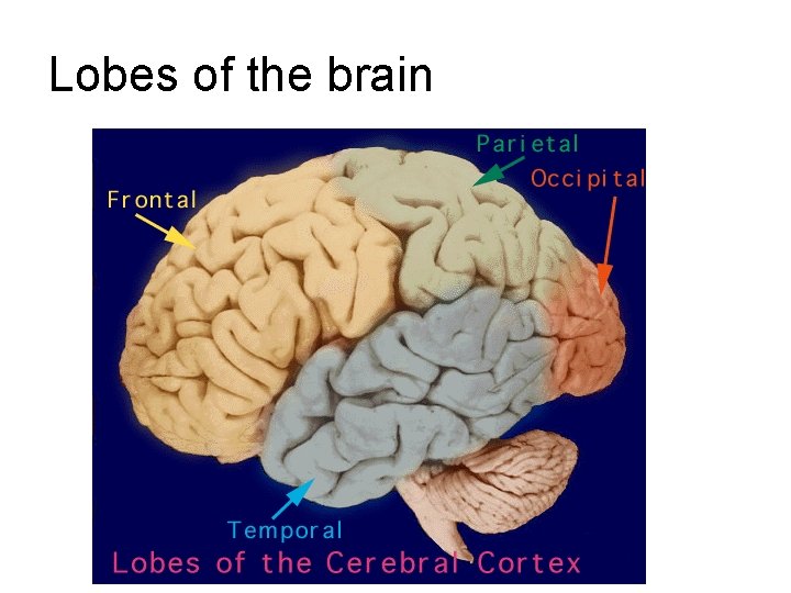
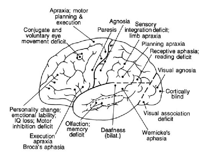
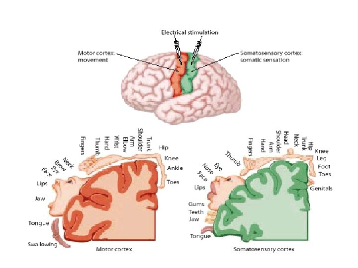
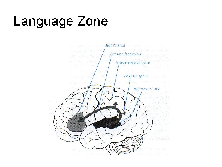
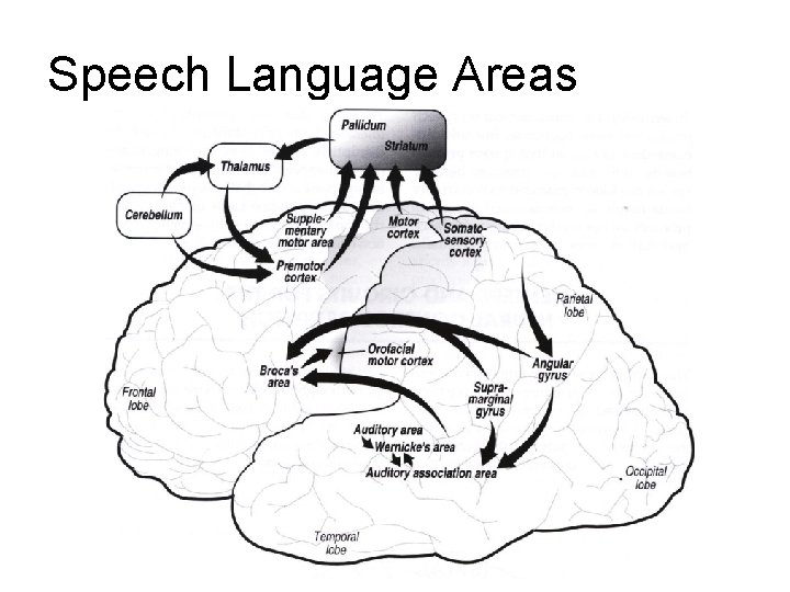
The lobes of the brain have important functions • frontal—reasoning, problem solving, personality, speech and language production – motor strip – Broca’s area—speech production • parietal—somatosensory (touch, pain, proprioception, temperatures) – sensory strip – angular gyrus – supramarginal gyrus • temporal—understanding – auditory cortex – Wernicke’s area • occipital—reception and processing of visual information • limbic—hippocampus, amygdala, and medial margins of the temporal, parietal, and frontal lobes that function for emotions, sexual function, temperature regulation and feeding behavior
Lobes, Brodmann’s Areas
Selected Brodmann’s Areas
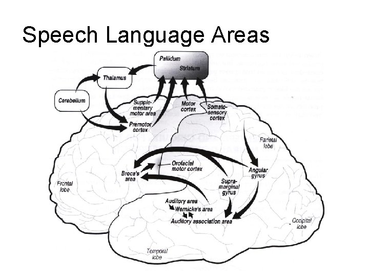
Speech Language Areas
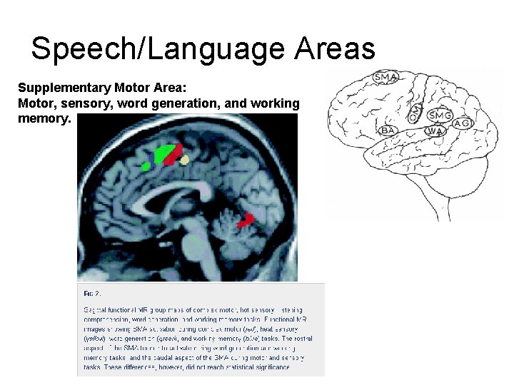
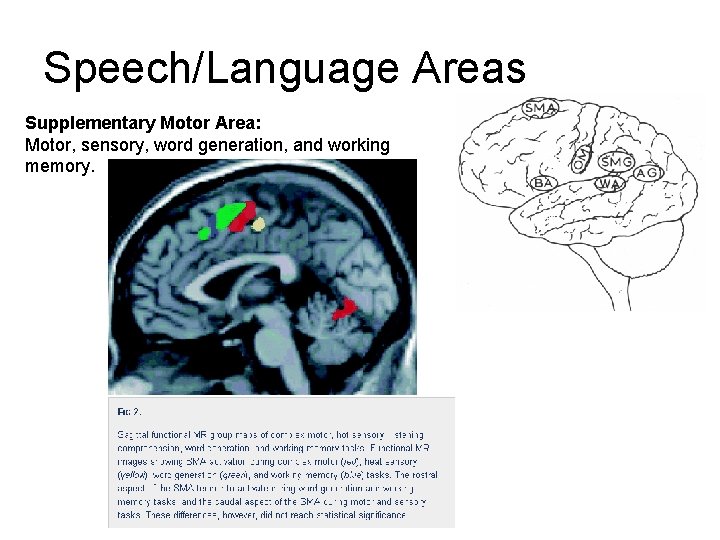
Speech/Language Areas Supplementary Motor Area: Motor, sensory, word generation, and working memory.
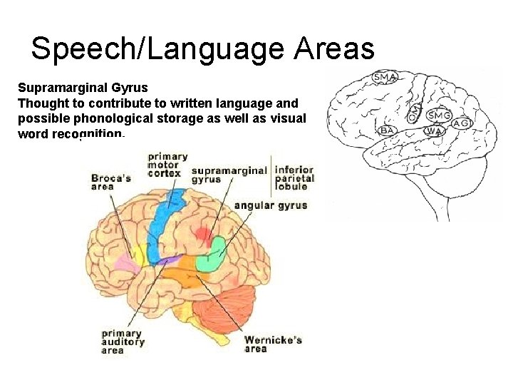
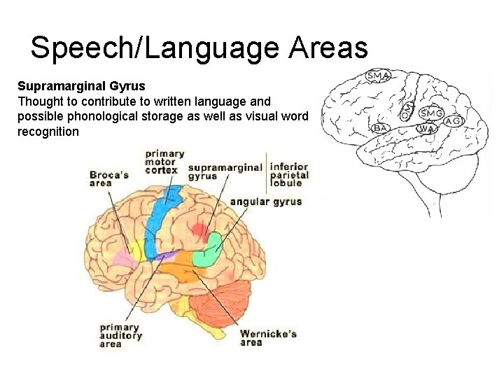
Speech/Language Areas Supramarginal Gyrus Thought to contribute to written language and possible phonological storage as well as visual word recognition.
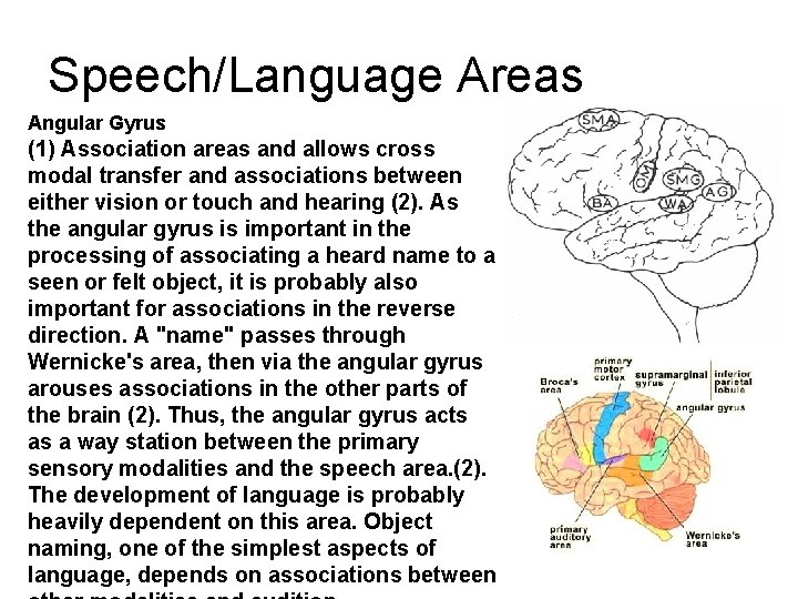
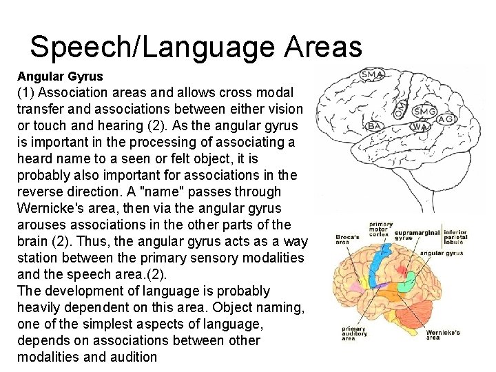
Speech/Language Areas Angular Gyrus (1) Association areas and allows cross modal transfer and associations between either vision or touch and hearing (2). As the angular gyrus is important in the processing of associating a heard name to a seen or felt object, it is probably also important for associations in the reverse direction. A "name" passes through Wernicke's area, then via the angular gyrus arouses associations in the other parts of the brain (2). Thus, the angular gyrus acts as a way station between the primary sensory modalities and the speech area. (2). The development of language is probably heavily dependent on this area. Object naming, one of the simplest aspects of language, depends on associations between
Lobes of the brain
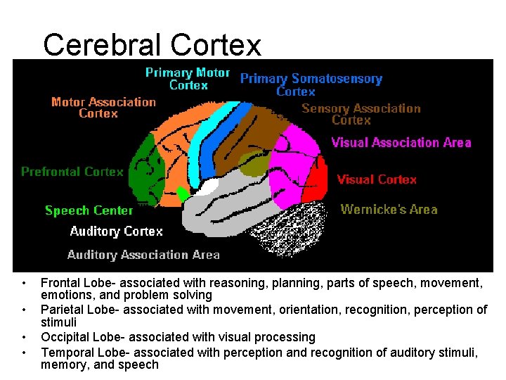
Cerebral Cortex • • Frontal Lobe- associated with reasoning, planning, parts of speech, movement, emotions, and problem solving Parietal Lobe- associated with movement, orientation, recognition, perception of stimuli Occipital Lobe- associated with visual processing Temporal Lobe- associated with perception and recognition of auditory stimuli, memory, and speech
Question The sensory strip is located in the? A. Frontal lobe B. Temporal lobe C. Occipital lobe D. Parietal lobe E. Limbic lobe
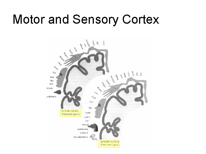
Motor and Sensory Cortex
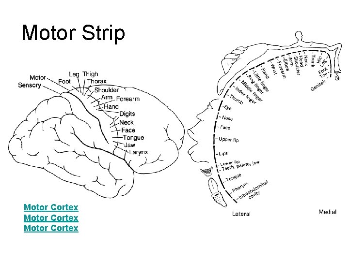
Motor Strip Motor Cortex
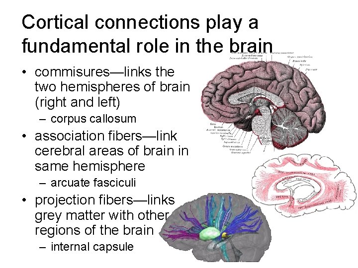
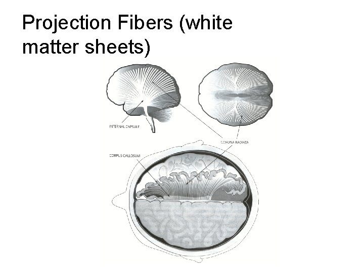
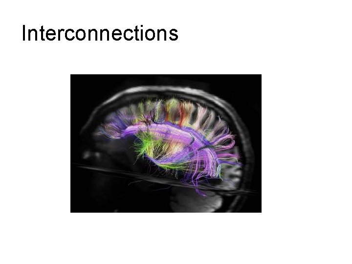
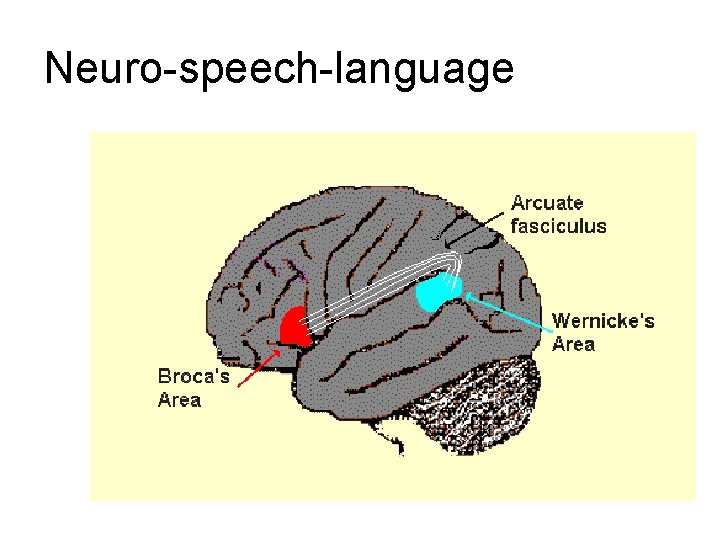
Cortical connections play a fundamental role in the brain • commisures—links the two hemispheres of brain (right and left) – corpus callosum • association fibers—link cerebral areas of brain in same hemisphere – arcuate fasciculi • projection fibers—links grey matter with other regions of the brain – internal capsule
Projection Fibers (white matter sheets)
Interconnections
Neuro-speech-language
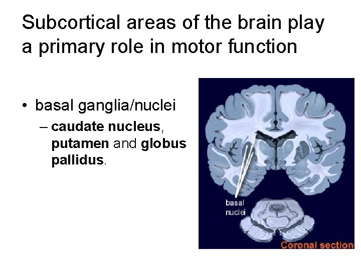
Subcortical areas of the brain play a primary role in motor function • basal ganglia/nuclei – caudate nucleus, putamen and globus pallidus.
Ventricles of the Brain • Ventricles
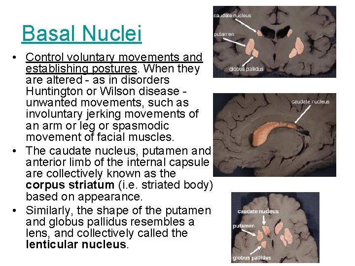
Basal Nuclei • Control voluntary movements and establishing postures. When they are altered - as in disorders Huntington or Wilson disease - unwanted movements, such as involuntary jerking movements of an arm or leg or spasmodic movement of facial muscles. • The caudate nucleus, putamen and anterior limb of the internal capsule are collectively known as the corpus striatum (i. e. striated body) based on appearance. • Similarly, the shape of the putamen and globus pallidus resembles a lens, and collectively called the lenticular nucleus.
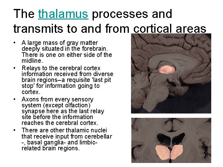
The thalamus processes and transmits to and from cortical areas • A large mass of gray matter deeply situated in the forebrain. There is one on either side of the midline. • Relays to the cerebral cortex information received from diverse brain regions--a requisite 'last pit stop' for information going to cortex. • Axons from every sensory system (except olfaction) synapse here as the last relay site before the information reaches the cerebral cortex. • There are other thalamic nuclei that receive input from cerebellar -, basal ganglia- and limbicrelated brain regions.
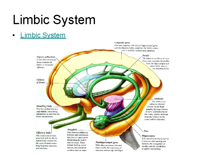
Limbic System • Limbic System
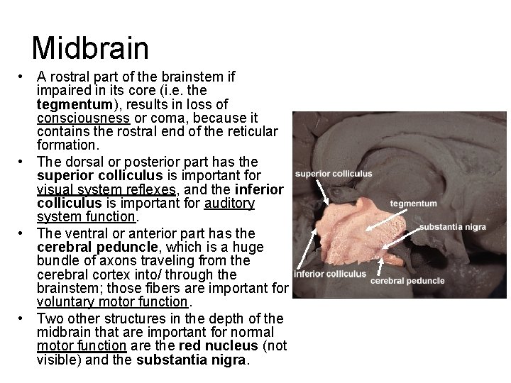
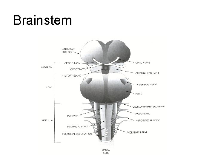
Midbrain • A rostral part of the brainstem if impaired in its core (i. e. the tegmentum), results in loss of consciousness or coma, because it contains the rostral end of the reticular formation. • The dorsal or posterior part has the superior colliculus is important for visual system reflexes, and the inferior colliculus is important for auditory system function. • The ventral or anterior part has the cerebral peduncle, which is a huge bundle of axons traveling from the cerebral cortex into/ through the brainstem; those fibers are important for voluntary motor function. • Two other structures in the depth of the midbrain that are important for normal motor function are the red nucleus (not visible) and the substantia nigra.
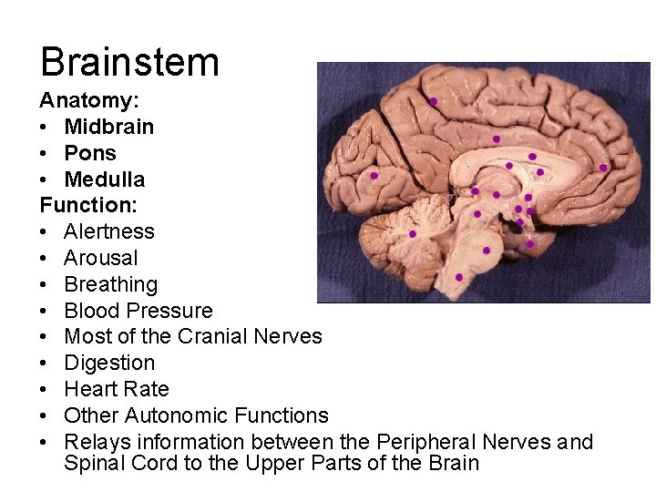
Brainstem Anatomy: • Midbrain • Pons • Medulla Function: • Alertness • Arousal • Breathing • Blood Pressure • Most of the Cranial Nerves • Digestion • Heart Rate • Other Autonomic Functions • Relays information between the Peripheral Nerves and Spinal Cord to the Upper Parts of the Brain
Brainstem
Cranial Nerves
Peripheral Nerves
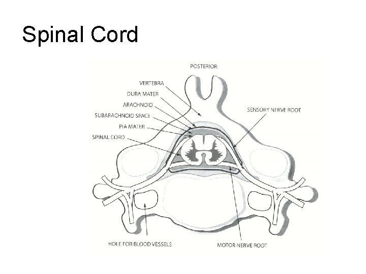
Spinal Cord
Spinal Cord
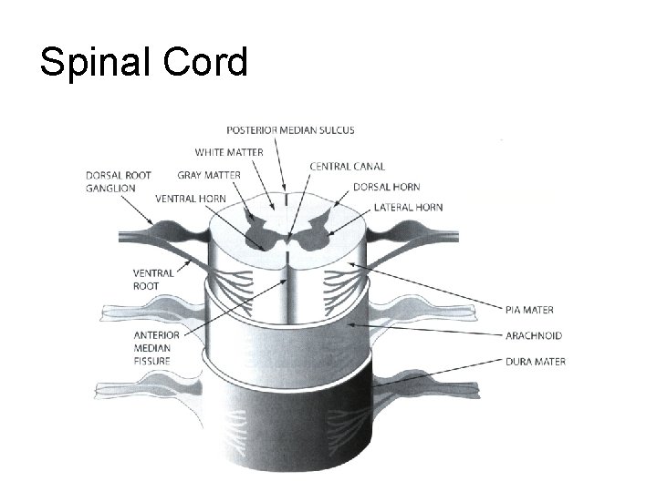
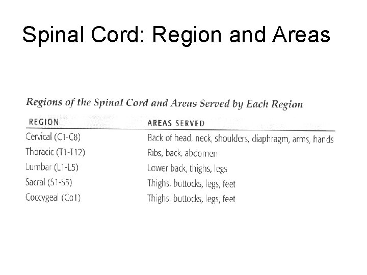
Spinal Cord: Region and Areas
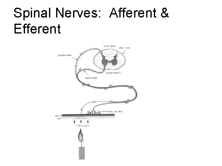
Spinal Nerves: Afferent & Efferent
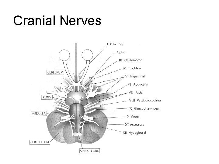
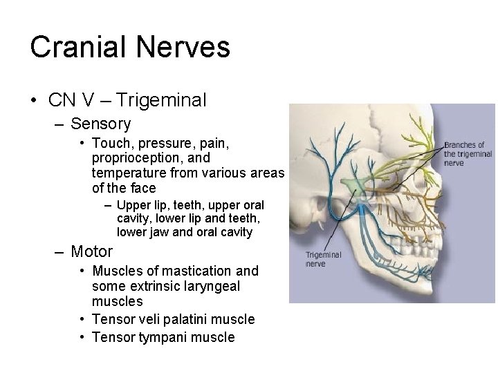
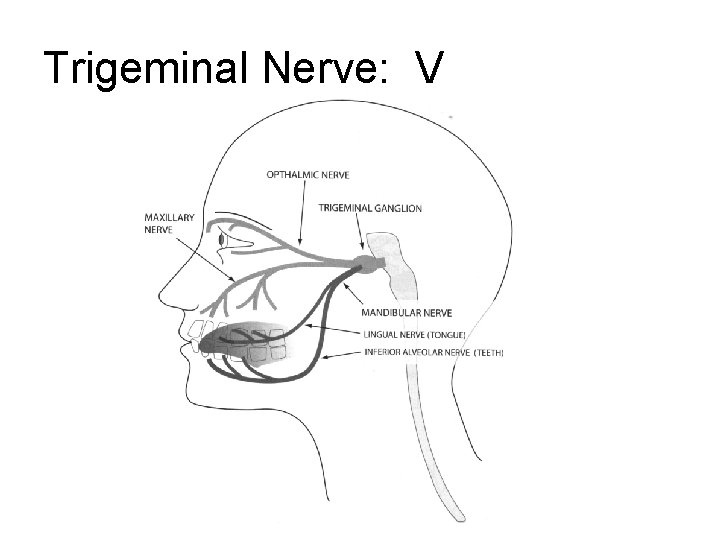
Cranial Nerves • CN V – Trigeminal – Sensory • Touch, pressure, pain, proprioception, and temperature from various areas of the face – Upper lip, teeth, upper oral cavity, lower lip and teeth, lower jaw and oral cavity – Motor • Muscles of mastication and some extrinsic laryngeal muscles • Tensor veli palatini muscle • Tensor tympani muscle
Trigeminal Nerve: V
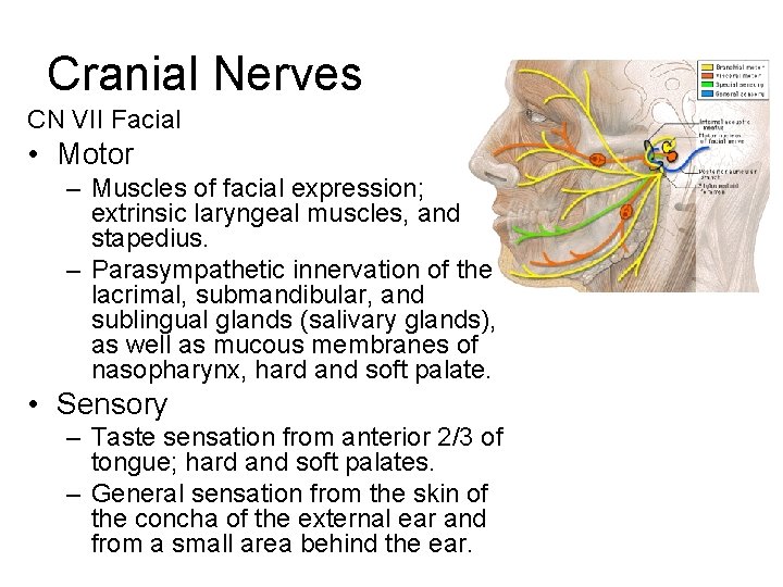
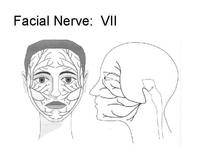
Cranial Nerves CN VII Facial • Motor – Muscles of facial expression; extrinsic laryngeal muscles, and stapedius. – Parasympathetic innervation of the lacrimal, submandibular, and sublingual glands (salivary glands), as well as mucous membranes of nasopharynx, hard and soft palate. • Sensory – Taste sensation from anterior 2/3 of tongue; hard and soft palates. – General sensation from the skin of the concha of the external ear and from a small area behind the ear.
Facial Nerve: VII
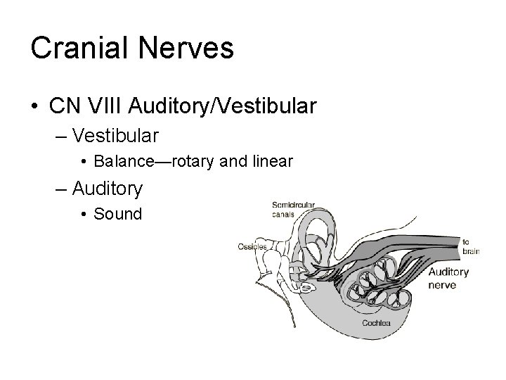
Cranial Nerves • CN VIII Auditory/Vestibular – Vestibular • Balance—rotary and linear – Auditory • Sound
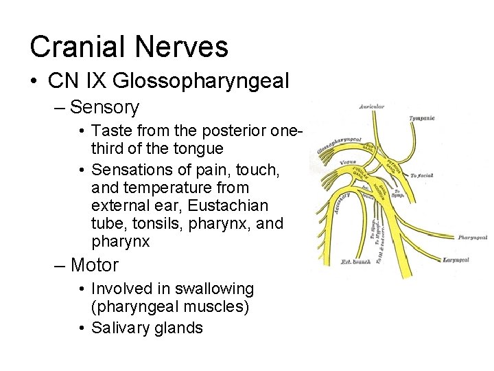
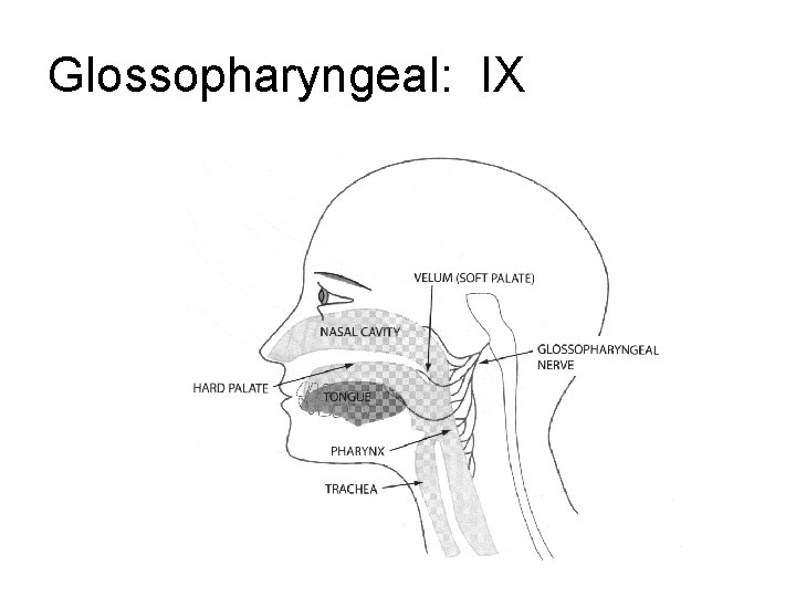
Cranial Nerves • CN IX Glossopharyngeal – Sensory • Taste from the posterior onethird of the tongue • Sensations of pain, touch, and temperature from external ear, Eustachian tube, tonsils, pharynx, and pharynx – Motor • Involved in swallowing (pharyngeal muscles) • Salivary glands
Glossopharyngeal: IX
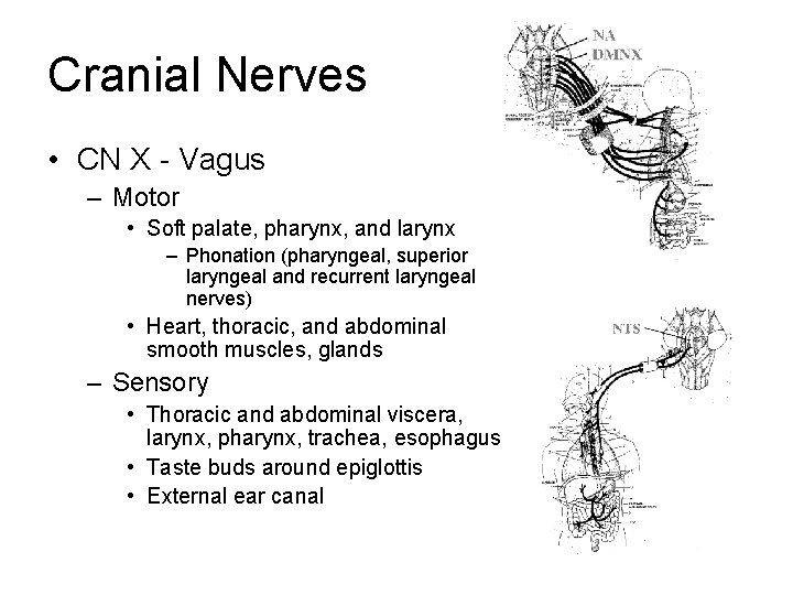
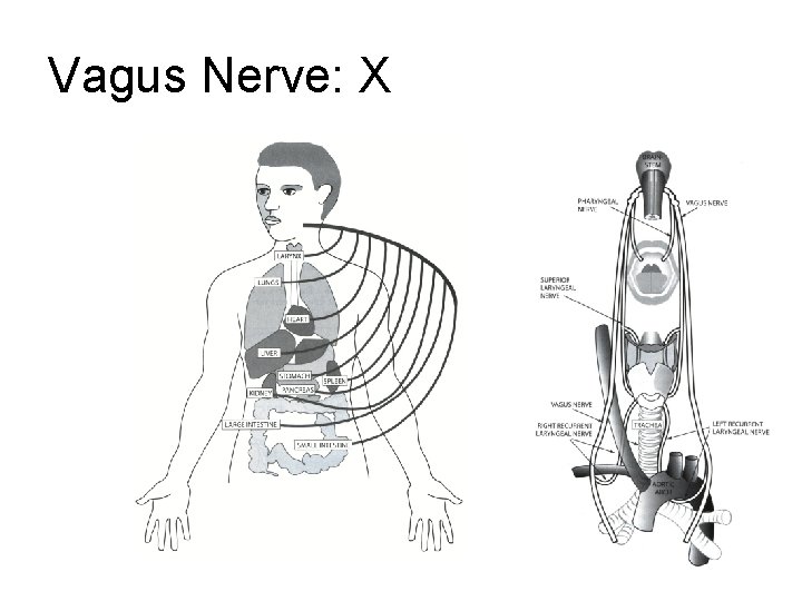
Cranial Nerves • CN X - Vagus – Motor • Soft palate, pharynx, and larynx – Phonation (pharyngeal, superior laryngeal and recurrent laryngeal nerves) • Heart, thoracic, and abdominal smooth muscles, glands – Sensory • Thoracic and abdominal viscera, larynx, pharynx, trachea, esophagus • Taste buds around epiglottis • External ear canal
Vagus Nerve: X
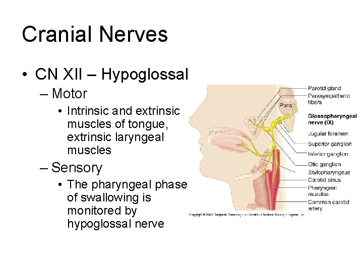
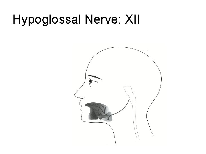
Cranial Nerves • CN XII – Hypoglossal – Motor • Intrinsic and extrinsic muscles of tongue, extrinsic laryngeal muscles – Sensory • The pharyngeal phase of swallowing is monitored by hypoglossal nerve
Hypoglossal Nerve: XII
Language Zone
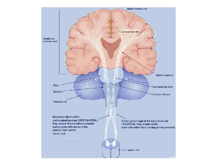
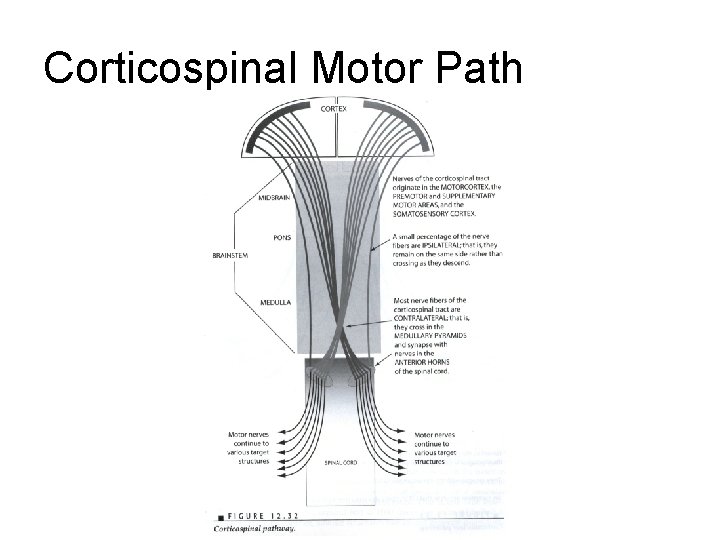
Corticospinal Motor Path
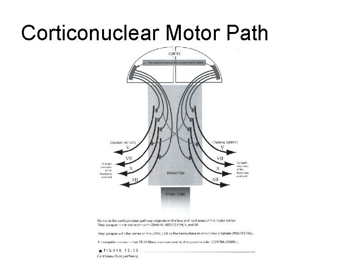
Corticonuclear Motor Path
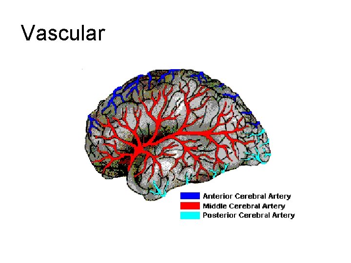
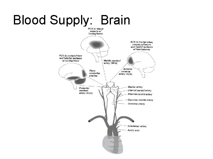
Vascular
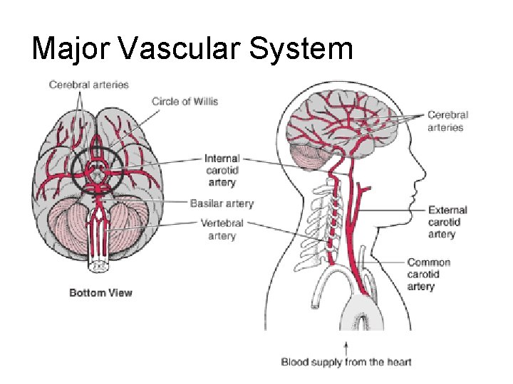
Major Vascular System
Blood Supply: Brain
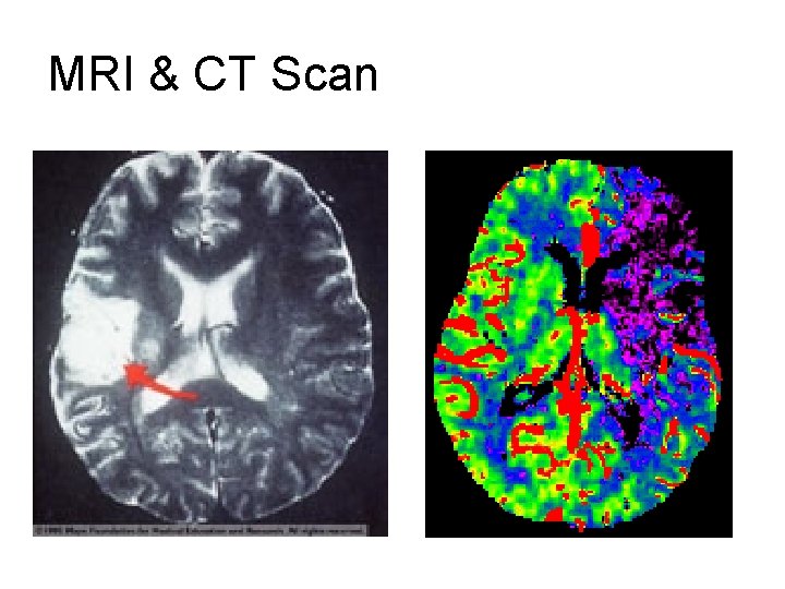
MRI & CT Scan
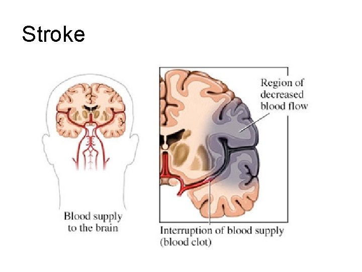
Stroke
Speech Language Areas
Speech/Language Areas Supplementary Motor Area: Motor, sensory, word generation, and working memory.
Speech/Language Areas Supramarginal Gyrus Thought to contribute to written language and possible phonological storage as well as visual word recognition.
Speech/Language Areas Angular Gyrus (1) Association areas and allows cross modal transfer and associations between either vision or touch and hearing (2). As the angular gyrus is important in the processing of associating a heard name to a seen or felt object, it is probably also important for associations in the reverse direction. A "name" passes through Wernicke's area, then via the angular gyrus arouses associations in the other parts of the brain (2). Thus, the angular gyrus acts as a way station between the primary sensory modalities and the speech area. (2). The development of language is probably heavily dependent on this area. Object naming, one of the simplest aspects of language, depends on associations between other modalities and audition