Susunan saraf autonom Anatomi fisiologi dan Neurotransmister Oleh




- Slides: 55
Susunan saraf autonom Anatomi fisiologi dan Neurotransmister Oleh: Slamet Sumarno
Pokok bahasan 1. 2. 3. 4. 5. 6. Anatomi susunan saraf otonom Faal susunan saraf ototnom Tranmisi neurohumeral Tranmisi kolinergik Tranmisi adrenergik Respons berbagai organ efektor terhadap rangsangan saraf otonom 7. Cara kerja obat otonom: …. .
Anatomi fisiologi Thoracolumbal SSA terdiri dua komponen Ortosympatic Adrenargic Kraniosacral Parasympatic Kolinergic Epineprin (adrenalin) Pre glanglion Asetilkholin Post ganglion Noreepinefrin (noradrenalin) Epineprin Beta (dilatasi bronkus) Naikkan insulin Alfa Vasokontriksi Glikogenolisis(hepar) Penurunan insulin noreepinefrin Pre & post ganglion asetilkolin Sifat Muskarinik (nikorinik) Motor end plate Otot lurik Otot polos Kelenjar Eksokrin SAnode & AV node
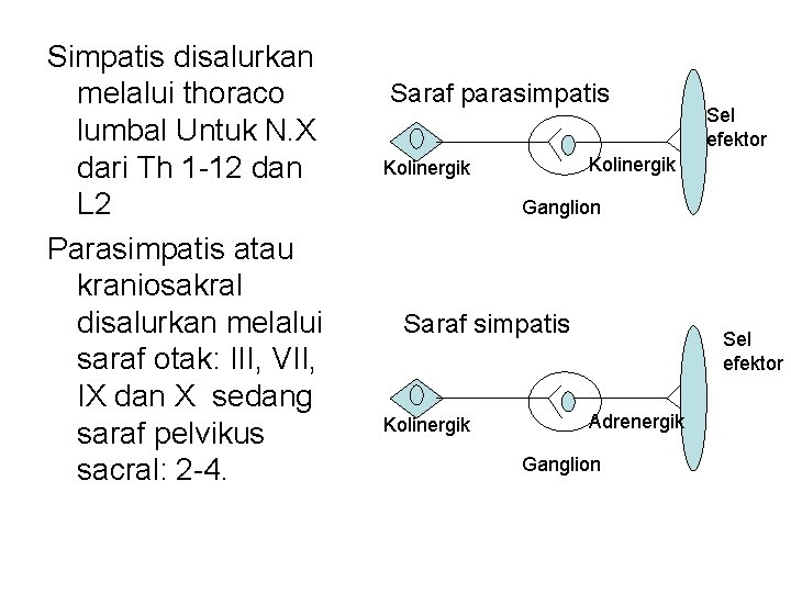
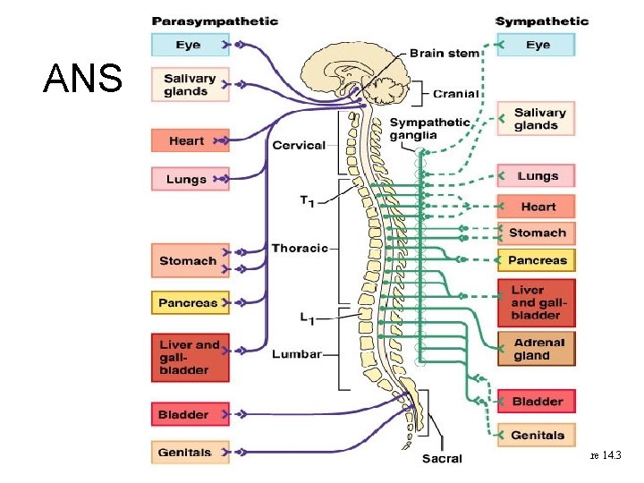
Simpatis disalurkan melalui thoraco lumbal Untuk N. X dari Th 1 -12 dan L 2 Parasimpatis atau kraniosakral disalurkan melalui saraf otak: III, VII, IX dan X sedang saraf pelvikus sacral: 2 -4. Saraf parasimpatis Sel efektor Kolinergik Ganglion Saraf simpatis Kolinergik Sel efektor Adrenergik Ganglion
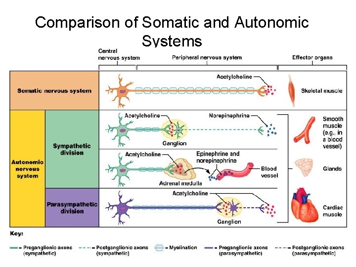
Comparison of Somatic and Autonomic Systems Figure 14. 2
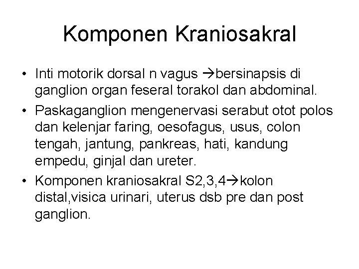
Komponen Kraniosakral • Inti motorik dorsal n vagus bersinapsis di ganglion organ feseral torakol dan abdominal. • Paskaganglion mengenervasi serabut otot polos dan kelenjar faring, oesofagus, usus, colon tengah, jantung, pankreas, hati, kandung empedu, ginjal dan ureter. • Komponen kraniosakral S 2, 3, 4 kolon distal, visica urinari, uterus dsb pre dan post ganglion.
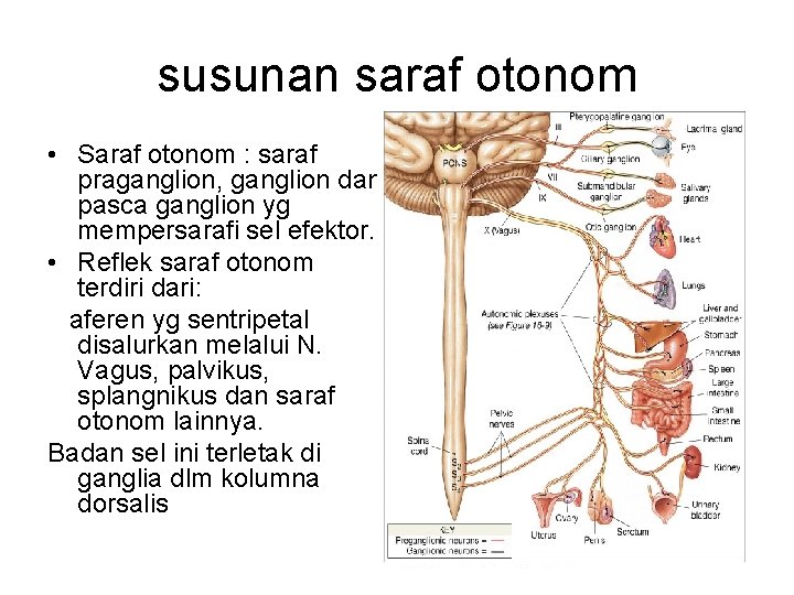
susunan saraf otonom • Saraf otonom : saraf praganglion, ganglion dan pasca ganglion yg mempersarafi sel efektor. • Reflek saraf otonom terdiri dari: aferen yg sentripetal disalurkan melalui N. Vagus, palvikus, splangnikus dan saraf otonom lainnya. Badan sel ini terletak di ganglia dlm kolumna dorsalis
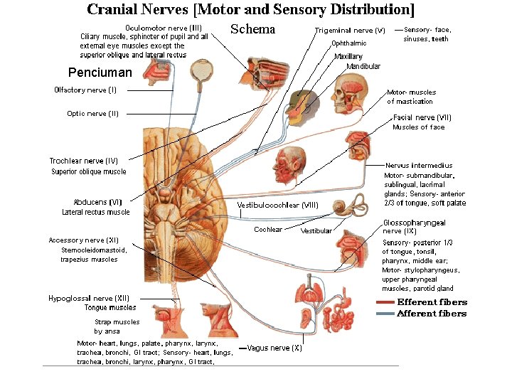
Penciuman
• Perbedaan tak jelas antara afreren otonom dan somatik shg tak dikenal obat kusus yg pengaruhi saraf aferen otonom. • Saraf aferen pra, ganglion dan pasca berakir di sel efektor otonom. • Saraf otonom berhubungan dgn saraf somatik saling mempengaruhi. • Terdapat beberapa pusat saraf otonom: 1) medula oblongata pengatur pernafasan dan tekanan darah. 2) Hipotalamus dan hipofisis mengatur suhu tubuh, keseimbangan air, metabolisme karbohidrat dan lemak, pusat tidur dan dianggap sebagai pusat saraf otonom, yg lebih tinggi adalah: korpus striatum dan kortek serebelum.
• Saraf praganglion simpatis dan parasipatis pascaganglion parasimpatis bersifat kolinergik berarti ujungnya melepas asetilkolin sebagai neurotranmister. • Pascaganglion simpatis bersifat adrenergik berarti ujung sarafnya melepas norepineprin • Saraf aferen yg berasal preresptor dan kemoreseptor dlm sinus karotikus, badan karotikus dan aorta diteruskan Via N IX dan X menuju medula oblongata. • Sistem ini berhubungan dengan reflek pertahanan tekanan darah dan frekuensi jantung dan pernafasan.
5 perbedaan pokok otonom dan somatik 1. 2. 3. 4. 5. Saraf otonom mengenervasi seluruh tubuh kecuali otot rangka. Sinaps saraf otonom yg paling distal terletak dlm ganglia diluar susunan saraf pusat. Sinaps saraf somatis dlm ssp. Saraf otonom membentuk pleksus diluar ssp, saraf somatis tidak membentuk pleksus. Saraf somatis diselubungi mielin, sedang otonom pasca ganglion tidak. Saraf otonom mengenervasi sel efektor yg bersifat otonom (sel efektor masih bisa bekerja tanpa persarafan), sebaliknya somatis putus maka otot rangka paralises dan kemudian atropi.
fungsi saraf otonom. • Secara umum dikatakan sistem simpatis dan parasimpatis berfungsi antagonis berarti simpatis memacu dan parasimpatis menghambat fungsi. • Organ tubuh umumnya dipersarafi simpatis dan parasimpatis dan tonus yg terlihat merupakan keseimbangan ke 2 nya. • Inhibisi salah satu sitem obat shg aktivitas organ didominasi simpatis, ada yg tidak antagonis kadang 2 efeknya sama seperti kelenjar liur dirangsang simpatis dan para simpatis tetapi simpatis liurnya pekat dan parasimpatis liurnya encer. Atau saling melengkapi, misal fungsi seksual : ereksi fungsi parasimpatis sedang eyagulasi fungsi simpatis.
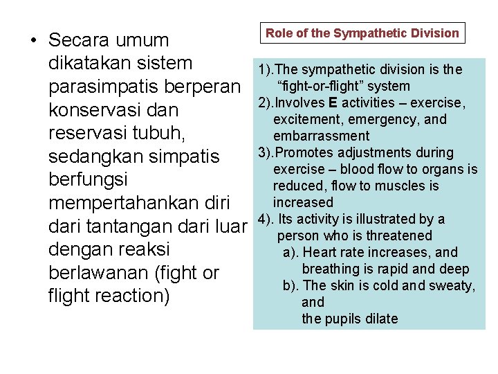
• Secara umum dikatakan sistem parasimpatis berperan konservasi dan reservasi tubuh, sedangkan simpatis berfungsi mempertahankan diri dari tantangan dari luar dengan reaksi berlawanan (fight or flight reaction) Role of the Sympathetic Division 1). The sympathetic division is the “fight-or-flight” system 2). Involves E activities – exercise, excitement, emergency, and embarrassment 3). Promotes adjustments during exercise – blood flow to organs is reduced, flow to muscles is increased 4). Its activity is illustrated by a person who is threatened a). Heart rate increases, and breathing is rapid and deep b). The skin is cold and sweaty, and the pupils dilate
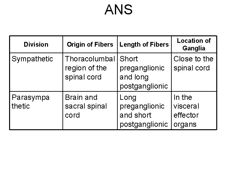
ANS Division Sympathetic Parasympa thetic Origin of Fibers Length of Fibers Thoracolumbal Short region of the preganglionic spinal cord and long postganglionic Brain and Long sacral spinal preganglionic cord and short postganglionic Location of Ganglia Close to the spinal cord In the visceral effector organs
ANS Figure 14. 3
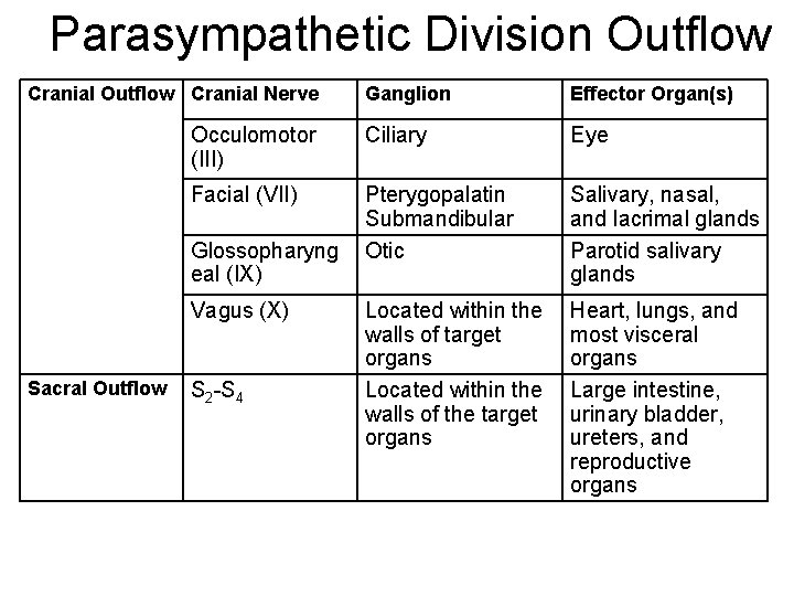
Parasympathetic Division Outflow Cranial Nerve Ganglion Effector Organ(s) Occulomotor (III) Ciliary Eye Facial (VII) Pterygopalatin Submandibular Otic Salivary, nasal, and lacrimal glands Parotid salivary glands Located within the walls of target organs Located within the walls of the target organs Heart, lungs, and most visceral organs Large intestine, urinary bladder, ureters, and reproductive organs Glossopharyng eal (IX) Vagus (X) Sacral Outflow S 2 -S 4
Parasym pathetic Division Outflow Figure 14. 4
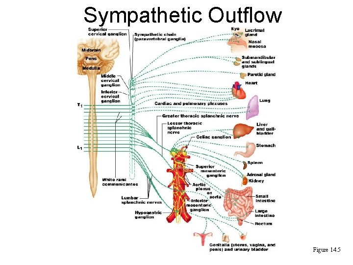
• • • Sympathetic Outflow (Pelimpahan saraf simpatik). Arises from spinal cord segments T 1 through L 2 Sympathetic neurons produce the lateral horns of the spinal cord Preganglionic fibers pass through the white rami communicantes and synapse in the chain (paravertebral) ganglia Fibers from T 5 -L 2 form splanchnic nerves and synapse with collateral ganglia Postganglionic fibers innervate the numerous organs of the body
Sympathetic Outflow Figure 14. 5
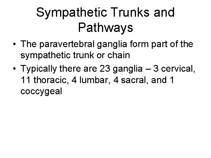
Sympathetic Trunks and Pathways • The paravertebral ganglia form part of the sympathetic trunk or chain • Typically there are 23 ganglia – 3 cervical, 11 thoracic, 4 lumbar, 4 sacral, and 1 coccygeal
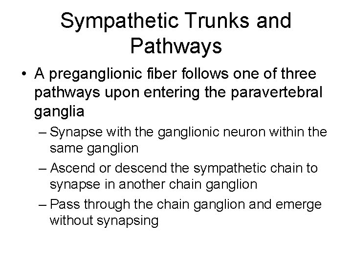
Sympathetic Trunks and Pathways • A preganglionic fiber follows one of three pathways upon entering the paravertebral ganglia – Synapse with the ganglionic neuron within the same ganglion – Ascend or descend the sympathetic chain to synapse in another chain ganglion – Pass through the chain ganglion and emerge without synapsing
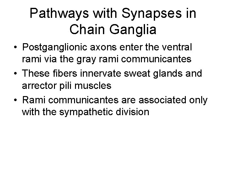
Pathways with Synapses in Chain Ganglia • Postganglionic axons enter the ventral rami via the gray rami communicantes • These fibers innervate sweat glands and arrector pili muscles • Rami communicantes are associated only with the sympathetic division
Pathways to the Head • Preganglionic fibers emerge from T 1 -T 4 and synapse in the superior cervical ganglion • These fibers: – Serve the skin and blood vessels of the head – Stimulate dilator muscles of the iris – Inhibit nasal and salivary glands
Pathways to the Thorax • Preganglionic fibers emerge from T 1 -T 6 and synapse in the cervical chain ganglia • Postganglionic fibers emerge from the middle and inferior cervical ganglia and enter nerves C 4 C 8 • These fibers innervate the heart via the cardiac plexus, as well as innervating the thyroid and the skin • Other T 1 -T 6 preganglionic fibers synapse in the nearest chain ganglia • Postganglionic fibers directly serve the heart, aorta, lungs, and esophagus
Pathways with Synapses in Collateral Ganglia • These fibers (T 5 -L 2) leave the sympathetic chain without synapsing • They form thoracic, lumbar, and sacral splanchnic nerves • Their ganglia include the celiac, the superior and inferior mesenterics, and the hypogastric
Pathways to the Abdomen • Sympathetic nerves innervating the abdomen have preganglionic fibers from T 5 -L 2 • They travel through the thoracic splanchnic nerves and synapse at the celiac and superior mesenteric ganglia • Postganglionic fibers serve the stomach, intestines, liver, spleen, and kidneys
Pathways to the Pelvis • Preganglionic fibers originate from T 10 -L 2 • Most travel via the lumbar and sacral splanchnic nerves to the inferior mesenteric and hypogastric ganglia • Postganglionic fibers serve the distal half of the large intestine, the urinary bladder, and the reproductive organs
Pathways with Synapses in the Adrenal Medulla • Fibers of the thoracic splanchnic nerve pass directly to the adrenal medulla • Upon stimulation, medullary cells secrete norepinephrine and epinephrine into the blood
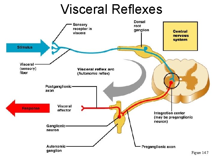
Visceral Reflexes • Visceral reflexes have the same elements as somatic reflexes • They are always polysynaptic pathways • Afferent fibers are found in spinal and autonomic nerves
Visceral Reflexes Figure 14. 7
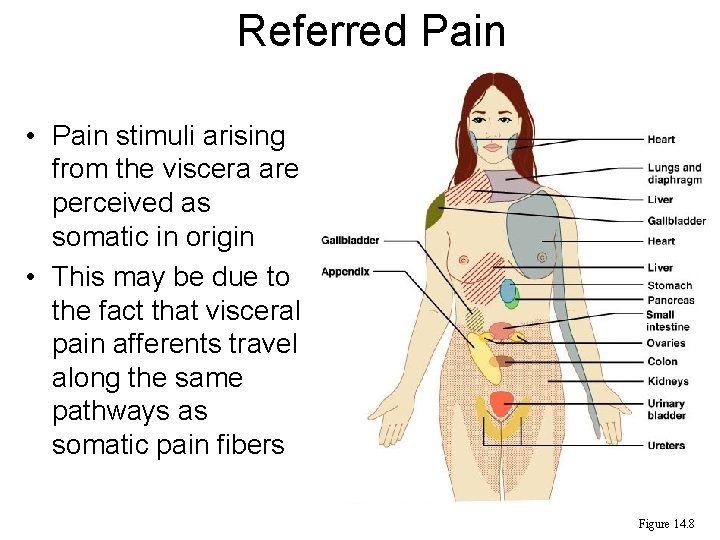
Referred Pain • Pain stimuli arising from the viscera are perceived as somatic in origin • This may be due to the fact that visceral pain afferents travel along the same pathways as somatic pain fibers Figure 14. 8
Neurotransmitters and Receptors • Acetylcholine (ACh) and norepinephrine (NE) are the two major neurotransmitters of the ANS • ACh is released by all preganglionic axons and all parasympathetic postganglionic axons • Cholinergic fibers – ACh-releasing fibers • Adrenergic fibers – sympathetic postganglionic axons that release NE • Neurotransmitter effects can be excitatory or inhibitory depending upon the receptor type
Cholinergic Receptors • The two types of receptors that bind ACh are nicotinic and muscarinic • These are named after drugs that bind to them and mimic ACh effects
Nicotinic Receptors • Nicotinic receptors are found on: – Motor end plates (somatic targets) – All ganglionic neurons of both sympathetic and parasympathetic divisions – The hormone-producing cells of the adrenal medulla • The effect of ACh binding to nicotinic receptors is always stimulatory
Muscarinic Receptors • Muscarinic receptors occur on all effector cells stimulated by postganglionic cholinergic fibers • The effect of ACh binding: – Can be either inhibitory or excitatory – Depends on the receptor type of the target organ
Adrenergic Receptors • The two types of adrenergic receptors are alpha and beta • Each type has two or three subclasses ( 1, 2, 1, 2 , 3) • Effects of NE binding to: – receptors is generally stimulatory – receptors is generally inhibitory • A notable exception – NE binding to receptors of the heart is stimulatory PLAY Inter. Active Physiology®: Nervous System II: Synaptic Transmission
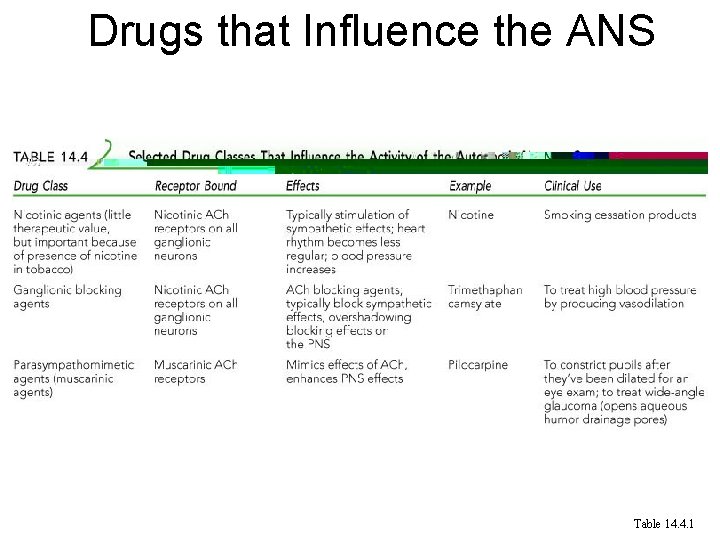
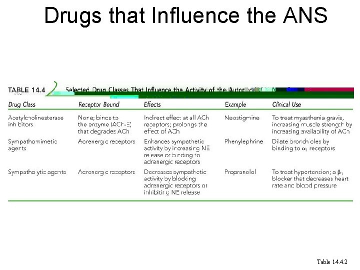
Effects of Drugs • Atropine – blocks parasympathetic effects • Neostigmine – inhibits acetylcholinesterase and is used to treat myasthenia gravis • Tricyclic antidepressants – prolong the activity of NE on postsynaptic membranes • Over-the-counter drugs for colds, allergies, and nasal congestion – stimulate -adrenergic receptors • Beta-blockers – attach mainly to 1 receptors and reduce heart rate and prevent arrhythmias
Drugs that Influence the ANS Table 14. 4. 1
Drugs that Influence the ANS Table 14. 4. 2
Interactions of the Autonomic Divisions • Most visceral organs are innervated by both sympathetic and parasympathetic fibers • This results in dynamic antagonisms that precisely control visceral activity • Sympathetic fibers increase heart and respiratory rates, and inhibit digestion and elimination • Parasympathetic fibers decrease heart and respiratory rates, and allow for digestion and the discarding of wastes
Sympathetic Tone • The sympathetic division controls blood pressure and keeps the blood vessels in a continual state of partial constriction • This sympathetic tone (vasomotor tone): – Constricts blood vessels and causes blood pressure to rise as needed – Prompts vessels to dilate if blood pressure is to be decreased • Alpha-blocker drugs interfere with vasomotor fibers and are used to treat hypertension
Parasympathetic Tone • Parasympathetic tone: – Slows the heart – Dictates normal activity levels of the digestive and urinary systems • The sympathetic division can override these effects during times of stress • Drugs that block parasympathetic responses increase heart rate and block fecal and urinary retention
Cooperative Effects • ANS cooperation is best seen in control of the external genitalia • Parasympathetic fibers cause vasodilation and are responsible for erection of the penis and clitoris • Sympathetic fibers cause ejaculation of semen in males and reflex peristalsis in females
Unique Roles of the Sympathetic Division • Regulates many functions not subject to parasympathetic influence • These include the activity of the adrenal medulla, sweat glands, arrector pili muscles, kidneys, and most blood vessels • The sympathetic division controls: – Thermoregulatory responses to heat – Release of renin from the kidneys – Metabolic effects
Thermoregulatory Responses to Heat • Applying heat to the skin causes reflex dilation of blood vessels • Systemic body temperature elevation results in widespread dilation of blood vessels • This dilation brings warm blood to the surface and activates sweat glands to cool the body • When temperature falls, blood vessels constrict and blood is retained in deeper
Release of Renin from the Kidneys • Sympathetic impulses activate the kidneys to release renin • Renin is an enzyme that promotes increased blood pressure
Metabolic Effects • The sympathetic division promotes metabolic effects that are not reversed by the parasympathetic division – Increases the metabolic rate of body cells – Raises blood glucose levels – Mobilizes fat as a food source – Stimulates the reticular activating system (RAS) of the brain, increasing mental alertness
Localized Versus Diffuse Effects • The parasympathetic division exerts shortlived, highly localized control • The sympathetic division exerts longlasting, diffuse effects
Effects of Sympathetic Activation • Sympathetic activation is long-lasting because NE: – Is inactivated more slowly than ACh – Is an indirectly acting neurotransmitter, using a second-messenger system – And epinephrine are released into the blood and remain there until destroyed by the liver
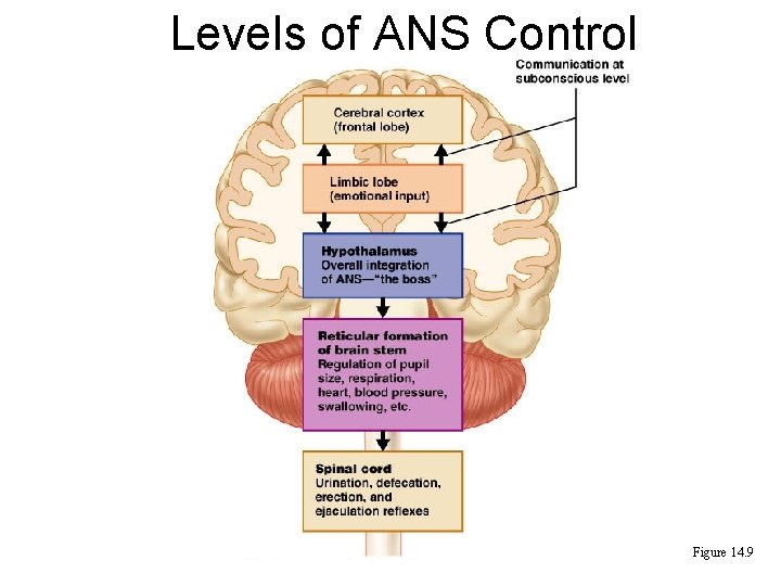
Levels of ANS Control • The hypothalamus is the main integration center of ANS activity • Subconscious cerebral input via limbic lobe connections influences hypothalamic function • Other controls come from the cerebral cortex, the reticular formation, and the spinal cord
Levels of ANS Control Figure 14. 9
Hypothalamic Control • Centers of the hypothalamus control: – Heart activity and blood pressure – Body temperature, water balance, and endocrine activity – Emotional stages (rage, pleasure) and biological drives (hunger, thirst, sex) – Reactions to fear and the “fight-or-flight” system
Embryonic Development of the ANS • Preganglionic neurons are derived from the embryonic neural tube • ANS structures in the PNS – ganglionic neurons, the adrenal medulla, and all autonomic ganglia – derive from the neural crest • Nerve growth factor (NGF) is a protein secreted by target cells that aids in the development of ANS pathways
Developmental Aspects of the ANS • During youth, ANS impairments are usually due to injury • In old age, ANS efficiency decreases, resulting in constipation, dry eyes, and orthostatic hypotension – Orthostatic hypotension is a form of low blood pressure that occurs when sympathetic vasoconstriction centers respond slowly to positional changes