NERVOUS SYSTEM Which direction is the dancer spinning


- the most inferior part of the")


")


")


 • • • The PNS includes all nervous tissue except")


- Muscle that alters the shape of the")

 • Cones")


")


- Slides: 76
NERVOUS SYSTEM
Which direction is the dancer spinning?
NERVOUS SYSTEM Functions: • Coordinates body systems • Communication system • Reacts to the environment (stimulus response)
Nervous System Organizational Chart Human Nervous System Central Nervous System Brain Peripheral Nervous System Motor Division Spinal Cord Somatic Sensory Division Autonomic Sympathetic Parasympathetic
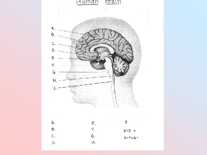
Brain 2 parts of the Central Nervous System: – Brain and Spinal Cord Brain • Made up of 100 billion neurons and 1 trillion neuroglia • Mass is approximately 3 lbs • 4 parts- cerebrum, cerebellum, upper brain stem and lower brain stem
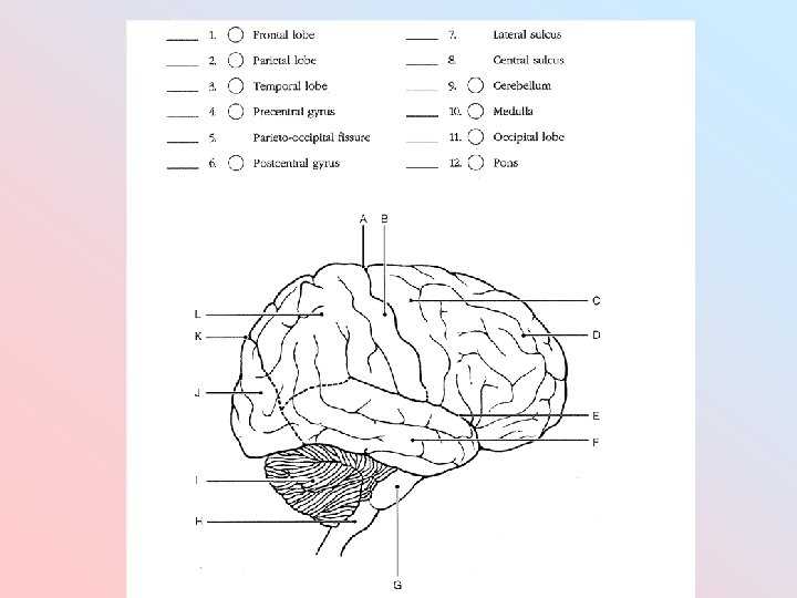
Cerebrum • Bulk of the brain • Outer folded layer- called the cerebral cortex- is made up of gray matter – Where messages are received in the brain – Peaks are called gyri – Deep grooves are called fissures – Shallow grooves are called sulci • Layer below the cortex is white matter • Divided into right and left sides called hemispheres • The deep connection between the two hemispheres is the corpus callosum
Lobes of the Cerebrum • Each hemisphere is divided into 4 lobes: – Frontal lobe- Speech, comprehension and muscle control – Occipital lobe- Vision – Parietal lobe- Touch, pain, taste and temperature – Temporal lobe- Hearing • Frontal and parietal lobes are separated by the central sulcus • The precentral gyrus is anterior to the central sulcus and the postcentral gyrus is posterior • The lateral sulcus outlines the temporal lobe • The parieto-occipital fissure separates the occipital and parietal lobes
Cerebellum • Second largest portion of the brain • Inferior to the occipital lobes • Surface is gray matter called cerebellar cortex • Below gray matter is white matter tractslook like tree branches. These tracts are called arbor vitae (type of tree). • Coordinates muscle contractions, regulates posture and balance and makes possible all skilled motor activities.
Upper Brain Stem • Called the diencephalon • Has 2 parts: – Thalamus- oval mass- Acts as a relay station for sensory impulses and also plays a role in cognition (acquiring knowledge) – Hypothalamus- below the thalamus- Has many functions: • Regulates hormones that control emotions, behaviors, eating and drinking, body temperature, circadian rhythms (sleep patterns), and heart rate
Lower Brain Stem • Medulla oblongata (or medulla)- the most inferior part of the lower brain stem and attaches to the spinal cord. It controls the rate and force of the heartbeat, diameter of the blood vessels and the medullary rhythmicity area which adjusts the rhythm of breathing. • Pons- In between the medulla and midbraincontains the pneumotaxic and apneustic areas for breathing • Midbrain- Most superior part of lower brain stem- Controls reflexes- swallowing
Reticular Formation • Neurons scattered throughout the upper and lower brain stem that alert the cerebral cortex to incoming sensory signals • Separates important from unimportant signals.
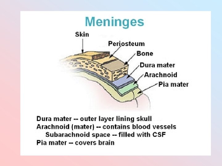
Protective Coverings of the Brain • Cranium • Meninges- 3 layers inside the cranium and superficial to the brain. From outside to inside: Dura mater Arachnoid layer Pia mater • Cerebrospinal Fluid (CSF)- circulates through cavities in the brain- called ventricles. This fluid “buoys” the brain so that it floats in the cranial cavity for protection. Typically, it is absorbed. If CSF accumulates, it builds up pressure. This is called hydrocephalus (water on the brain).
Hydrocephalussee Wendy’s story
Brain Protective Layers and Parts from the Outside to the Inside ( )
Sheep Brain Dissection 1. No food, drink or gum! 2. Wear gloves if handling the brain. 3. First, carefully remove the dura mater. Be especially careful around the optic chiasma and pituitary gland. 4. Observe the external anatomy. 5. Cut along the longitudinal fissure to view internal anatomy. 6. Use the packet as your guide. Complete the handout and turn it in.
External Sheep Brain
Brain-Blood Barrier • More than 100 years ago it was discovered that if blue dye was injected into the bloodstream of an animal, the tissues of the whole body EXCEPT the brain and spinal cord would turn blue. • The brain-blood barrier (BBB) exists because the brain capillaries are lined with endothelial cells that fit so tightly together that few substances can pass out of the blood stream. • It is permeable to water, oxygen, carbon dioxide, glucose, alcohol, and general anesthetics.
The functions of the BBB are to: • Protect the brain from "foreign substances" in the blood that may injure the brain. • Protect the brain from hormones and neurotransmitters in the rest of the body. • Maintain a constant environment for the brain. • The BBB can be broken down by: – – – High blood pressure Microwaves Radiation Infection Brain trauma
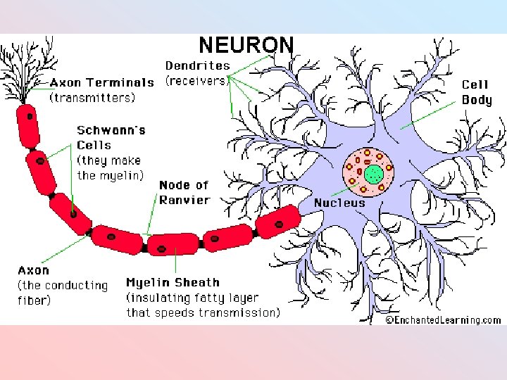
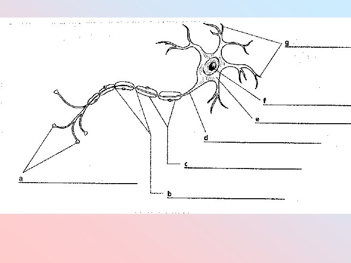
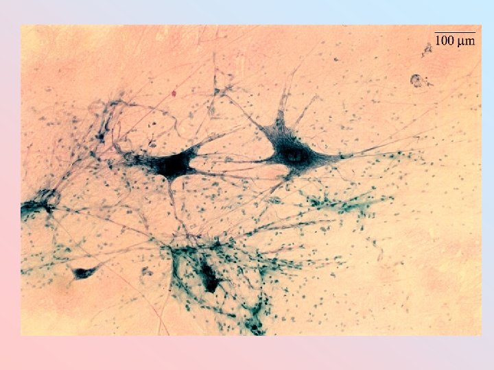
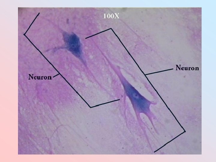
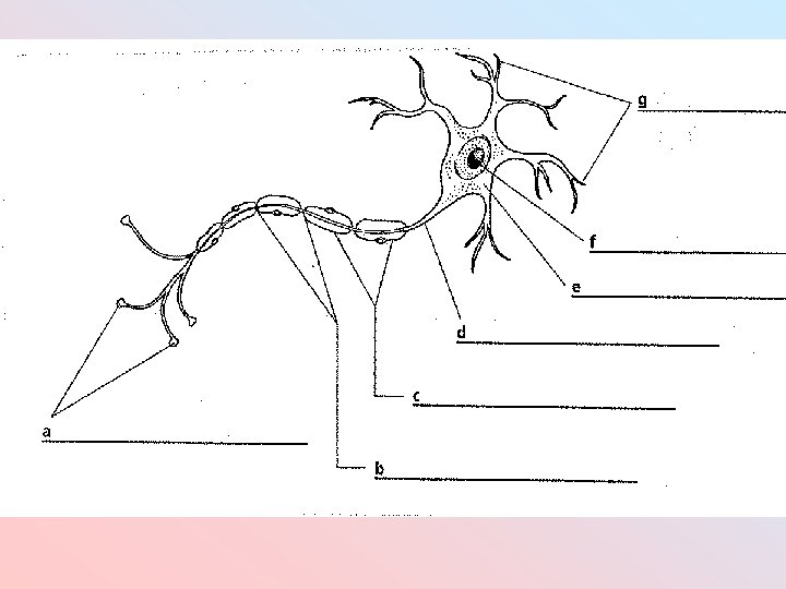
NERVE CELLS 1. Neuron = Information processing unit 2. Neuroglia= Support and protect neurons; Do not conduct messages; Smaller than neurons; 5 -50 times more numerous than neurons Draw a neuron in your notes……
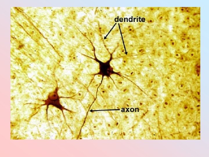
Parts of a neuron • Cell body- Contains the nucleus • Dendrites- Small extensions that receive information • Axon- Long extension that transmits information toward another neuron. • Axon terminal- End of an axon that stores neurotransmitters (chemicals that continue the message on to another neuron). • Synapse- Space between the axon of 1 neuron and the dendrites of another. Here is where neurotransmitters are dumped.
Parts of a neuron cont…. • Myelin sheath- Insulating fat around the axon- increases the speed of an impulse • Schwann cells or oligodendrocytes. Produce the myelin • Nodes of Ranvier- Gaps in the myelin sheath along an axon
AXON TERMINAL AND SYNAPTIC SPACE
Quick Review • 1. What are the 2 parts to the CNS? • 2. What is the bulk of the brain? • 3. What are the 4 lobes of the cerebrum? • 4. What connects the right and left halves of the cerebrum? • 5. What are the 2 parts to the diencephalon? • 6. What are the 3 parts of the lower brain stem? • 7. What are the 2 types of nerve cells? • 8. What are the 3 meninge layers? • 9. What part of a neuron receives messages? • 10. What part of a neuron sends messages on to another neuron (it is a long extension)?
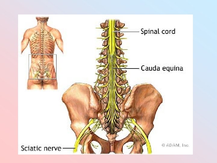
SPINAL CORD • Originates at the foramen magnum (opening at the base of the skull) • Protected by the vertebrae • Covered with the same 3 meninge layers as the brain- dura mater, arachnoid and pia mater • Also protected by fat tissue located in the epidural space- between the dura mater and the vertebral column • 16 -18 inches • Does not run the entire length of the vertebral column. Where it ends it is called the conus medullaris (between the L 1 and L 2 vertebrae)
Spinal Cord • 31 pairs of spinal nerves coming off of the spinal cord. Each pair originates at a vertebra (8 cervical, 12 thoracic, 5 lumbar, 5 sacral, 1 coccyx). Spinal nerves communicate between the spinal cord and the rest of the body. • Where the spinal cord ends there are nerves that arise that angle down like flowing hair. This is called cauda equina (horse’s tail).
Cross Section of the Spinal Cord • Deep anterior median fissure and the shallow posterior median sulcus divide the spinal cord into right and left halves. • Center of the spinal cord is the central canal which contains cerebrospinal fluid surrounded by gray matter and then surrounded by white matter. • Each pair of spinal nerves is connected to the cord at two points called a root.
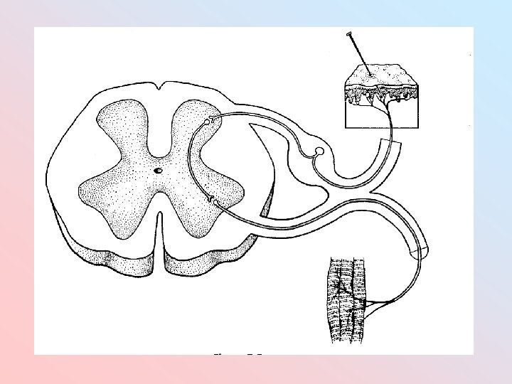
Roots of a spinal nerve • Posterior dorsal root- Contains sensory neurons (afferent neurons) that carry sensory information to the spinal cord. • Anterior ventral root- Contains motor neurons (efferent neurons) that carry motor information away from the spinal cord to the muscle.
Spinal Cord Video Clips • • • Levels of spinal cord injury Stem cell therapy research Stem cell therapy results Embryonic stem cell controversy
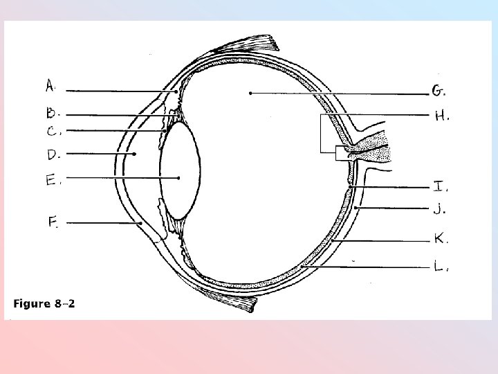
Quiz Picture Review
A. B. E. D. C.
Nervous System Organizational Chart Human Nervous System Central Nervous System Brain Peripheral Nervous System Motor Division Spinal Cord Somatic Sensory Division Autonomic Sympathetic Parasympathetic
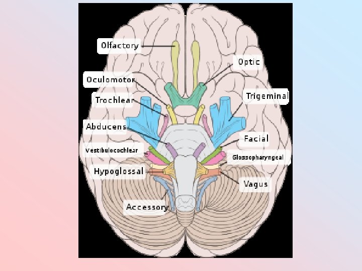
Cranial Nerves • There are 12 pairs of cranial nerves that emanate from brain tissue or sensory organs of the head and pass through openings in the cranium to send information to and from the brain. • Cranial nerves are numbered using Roman numerals.
Cranial Nerve Function I- Olfactory nerve Sends smell information to the olfactory bulb. II- Optic nerve Carries sight information from the retina of the eye and the optic chiasm is formed with the 2 optic nerves cross. III- Oculomotor nerve Controls muscle movement of the eyelid and the eyeball which allows the eye to turn, the pupil to constrict and the lens to change its shape. IV- Trochlear nerve Controls eye muscle movement. V- Trigeminal nerve Allows feeling in the head region- nose, eyes, tongue, teeth, skin, and sinuses. Also controls muscles for chewing (mastication). Has 3 branches coming off of it. VI- Abducent nerve Controls eye muscle movement.
Cranial Nerve Function VII- Facial nerve Allows feeling of the face, tongue and mouth. Also, controls muscles that cause facial expressions, chewing and signals for salivation. VIIIControls hearing and balance. Vestibulocochlear nerve VIIIICarries temperature, pressure and taste Glossopharyngeal information from the tongue, palate and pharynx nerve (throat). Signals for salivation X- Vagus nerve Constricts muscles of the pharynx, larynx (voice box), esophagus, trachea (air pipe) and even parts of the heart. XI- Spinal accesory nerve Controls muscles of the shoulders and neck. XII- Hypoglossal nerve Controls muscles of the tongue.
Peripheral Nervous System (PNS) • • • The PNS includes all nervous tissue except the brain and spinal cord. The 2 divisions of the PNS are the Sensory Division and the Motor Divison The Sensory Division involves sensory neurons (afferent) detecting a stimulus and sending the information to the brain or spinal cord (dorsal root of spinal nerves).
Motor Division • The Motor Division of the PNS involves responses from the spinal cord and brain to the muscles or glands to signal contraction or release of fluids. This division involves motor (efferent) neurons (ventral root of spinal nerves). • The 2 divisions of the Motor Division are the Somatic Division and the Autonomic Division.
Somatic Division • The Somatic Division signals for the contraction of skeletal muscles. – What are skeletal muscles? • This can be voluntary or involuntary. • Involuntary skeletal muscle action involves reflexes. – Patellar reflex – Withdrawal reflex
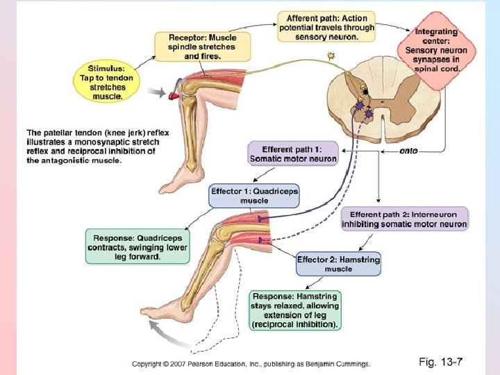
Patellar Relex • A type of stretch reflex- Protects the muscle against increases in length which may tear or damage muscle fibers. • This is also a spinal reflex where the spinal cord is the only part of the CNS involved in the reaction. • The main components of a spinal reflex are: 1. Sensory receptor- Distal end of a sensory neuron that responds to a stimulus. 2. Sensory neuron- Send messages via the dorsal root of a spinal nerve (axon terminals are in the gray matter).
3. Integrating center- Synapses between sensory and motor neurons 4. Motor neuron- Sends messages via the ventral root of a spinal nerve 5. Effector- Part of the body that responds to motor nerve impulses (muscles and glands) • The patellar tendon attaches the quadriceps muscle to the tibia bone of the lower leg. – Sensory receptor- Neuron in the quadriceps muscle that responds to the stretching of the muscle when the patellar tendon is stretched. – Effector #1 - Quadriceps muscle contracts, causing the lower leg to lift. – Effector #2 - Hamstring muscle relaxes allowing the lower leg to lift.
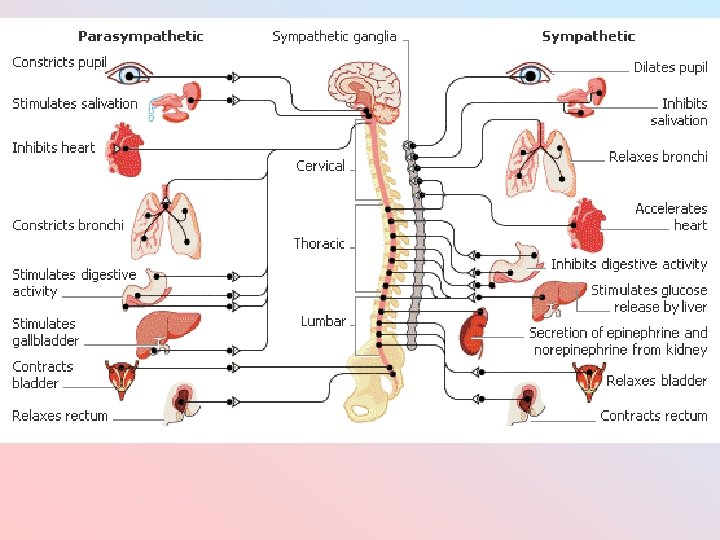
Autonomic Division • The Autonomic Division signals for the contraction of smooth and cardiac muscle. • This is involuntary. • The 2 divisions are the Sympathetic and Parasympathetic Divisions – Sympathetic- “Fight or Flight” activities • Due to the neurotransmitter Epinephrine/Norepinephrine (aka Adrenaline) • Originates from the T 1 -L 3 spinal nerves – Parasympathetic- “Rest and Digest” activities • Due to Acetylcholine • Originates from the cranial and sacral nerves
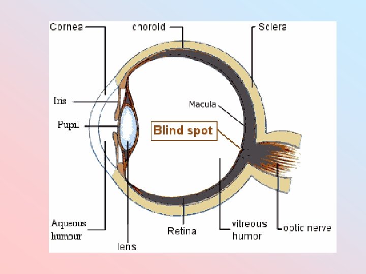
Layers of the Eyeball • The eye anatomy has 3 layers: Fibrous tunic, vascular tunic and retina
Fibrous tunic • This is the outer coat • The parts include: – Cornea- transparent covering that bends light rays to produce a clear image. If not curved properly, the image is blurred. – Conjunctiva- Epithelial layer that covers the outside edges of the cornea – Sclera- The white of the eye composed of dense connective tissue • Covers all of the eyeball except the cornea • Gives shape to the eyeball
Vascular tunic • Middle layer • The parts include: – Choroid- lines the internal surface of the sclera and provides nutrients to the eye – Pupil- The hole in the center of the eyeball – Iris- The colored circle around the pupil that is composed of muscle fibers. These fibers regulate the pupil opening- contract to reduce pupil size (parasympathetic) and relax to dilate (sympathetic) – Lens- Disk that focuses light rays on the retina
Vascular tunic continued… • Ciliary muscle (body)- Muscle that alters the shape of the lens to focus light rays onto the retina • Supensory Ligament (Ciliary zonule)- Attaches the lens to the ciliary body • Aqueous humor- Watery fluid that is found in a chamber anterior to the lens. This fluid provides nutrients to the lens and cornea and helps maintain appropriate pressure of the anterior eye. • Vitreous humor- Clear, jelly-like substance that prevents the eyeball from collapsing. This is posterior to the lens.
Retina • Lines the inner ¾ of the eyeball • Consists of a pigment epithelium and a neural portion • Pigment epithelium is a sheet of melanin that absorbs stray light rays and prevents scattering so that the image remains clear • Neural portion contains 2 types of photoreceptors that respond to light- rods and cones • Optic disk (blind spot)- Where the optic nerve meets the retina
Rods and Cones • Rods react to dim light (see at night) • Cones react to bright/colored light. – 3 types of cones that allow us to see all of the colors – Colorblindness is due to defective cones. This is more common in males because it is linked to the X sex chromosome and males only have one. – Fovea centralis (aka central fovea)- highest area of visual acuity (sharpest vision)- This is where cones are most densely concentrated.
Vision information transfer • After the light is absorbed by the rods and cones, the message is transferred to the optic nerve via electric impulse. Then it goes to the optic chiasm where the image from the right eye crosses to the left side of the brain and vice versa. The image then goes to the thalamus and then the appropriate occipital lobe (right or left).
EYE ANATOMY
Eye Activities • Path of Light Optic chiasma (Occipital lobes)
Eye Activities • Blind spot- When both eyes are open, you don’t experience a blind spot because the brain receives information from both eyes at the same time and blends it. When one eye is closed, a blind spot occurs in your open eye when the light falls on the retina where it connects with the optic nerve. Here, the retina lacks photoreceptors (rods and cones).
Eye Activities • A Hole in Your Hand- It looks like there is a hole through the left hand because of the brain’s function as a processor of information. Information from the right eye is superimposed with information from the left eye. The blended picture is a hole in your hand.
Eye Activities • After Images- Staring at an object allows the light to burn the image onto the retina. When you stop staring at the image, the cells in the eye remain stimulated for a short time, creating an after image. After images always occur in opposite colors because the stimulated rods and cones are fatigued and need a chance to get recalibrated.
PNS Review- Identify the lettered parts Human Nervous System Central Nervous System Brain A. B. Spinal Cord D. C. E. F. G.
PNS Review- Write the letter of the division of the PNS being described 2. Controls skeletal muscles 3. Causes dilation of eyes 4. Sends messages via the dorsal root of a spinal nerves 5. Controls smooth/cardiac muscle 6. Rest & Digest 7. Caused by adrenaline (epinephrine) 8. Can be involuntary or voluntary 9. Caused by acetylcholine 10. Relaxes the bladder
Eye Review- What part is being described? 1. 2. 3. 4. 5. 6. 7. 8. White of the eye Contains the highest # of rods/cones Controls pupil opening Provides nutrients to the lens/cornea Contains no rods/cones Controls the bending of the lens Provides nutrients to the eyeball Opening that allows light to hit the lens