Respiration Chapter 42 Respiration Gas exchange Movement of

 To improve gas absorption")


")

 Pharynx (back of throat) Larynx (voice box) Trachea")


 Bronchioles Alveoli Air sacs Gas exchange One cell layer thick Lung")


 Diaphragm contracts & flattens Intercostal muscles contract Raises the ribs Increases volume")
 Diaphragm relaxes & elevates Intercostal muscles relax Ribs lower Decreased volume Forces")


 & H+1 Buffer Blood")


- Slides: 42
Respiration Chapter 42
Respiration Gas exchange Movement of gas across membrane Diffusion (passive) To improve gas absorption Increase surface area for diffusion Decrease distance diffused gas travels Respiratory organs
Fish
Amphibians Lungs Cutaneous respiration
Amphibians
Birds Parabronchi Air sacs (lungs)
Fig. 42 -26 Anterior air sacs Posterior air sacs Air Trachea Lungs Air tubes (parabronchi) in lung INHALATION Air sacs fill EXHALATION Air sacs empty; lungs fill 1 mm
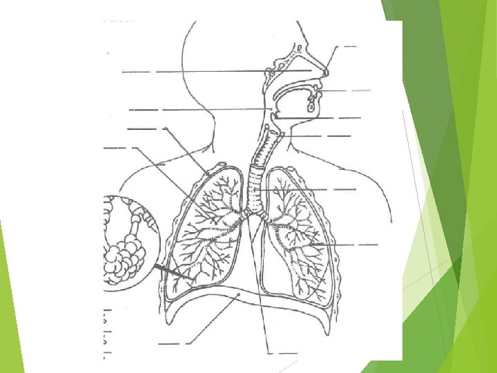
Anatomy Mouth (nose) Pharynx (back of throat) Larynx (voice box) Trachea
Anatomy
Anatomy
Anatomy Bronchi (Bronchus) Bronchioles Alveoli Air sacs Gas exchange One cell layer thick Lung tissue consists of millions
Anatomy
Anatomy
Anatomy Lungs 3 right lobes RUL, RML, RLL 2 left lobes LUL, LLL
Anatomy
Anatomy
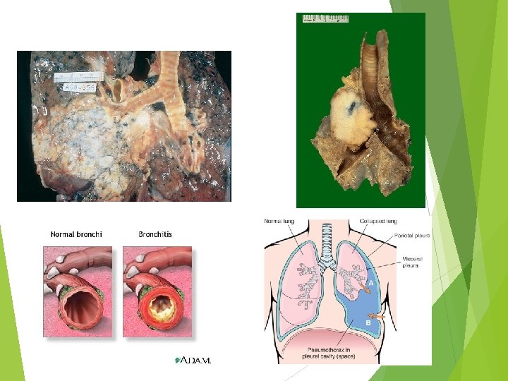
Anatomy Lung covered by a double folded membrane Visceral pleural membrane: Covers the lung Parietal pleural membrane: Lines inner wall of thoracic cavity
Anatomy Pleural cavity: Space between two membranes Filled with fluid Helps with movement of lungs
Breathing Diaphragm Muscle Separates thoracic cavity from abdominal cavity Intercostal muscles Muscles between the ribs
Inhalation (inspiration) Diaphragm contracts & flattens Intercostal muscles contract Raises the ribs Increases volume (decreases pressure) Air flows into the lungs
Exhalation (expiration) Diaphragm relaxes & elevates Intercostal muscles relax Ribs lower Decreased volume Forces air out
Breathing
Breathing measurements Tidal volume Amount of air moved into & out of lungs at rest Vital capacity Maximum amount of air that can be expired after forceful exhalation
Breathing measurements
Control of breathing Normal breathing Medulla oblongata Respiratory control center Neurons send impulse for muscles (diaphragm/intercostal) to contract Inspiration (inhalation) Stop sending impulse Expiration (exhalation)
Control of breathing Blood O 2 & CO 2 in normal range Neurons respond to a change in O 2 & CO 2 More response to CO 2 Increases carbonic acid (H 2 CO 3) CO 2 + H 2 O ⇆H 2 CO 3 ⇆ H + HCO 3 Lowers p. H
Control of breathing Stimulates peripheral chemoreceptors Aorta & carotids Send impulses to respiratory control center (medulla oblongata) Stimulates increased breathing
Control of breathing Central chemoreceptors Located in brain Respond to increased amount of CO 2 Peripheral receptors immediate response Central receptors maintained response until p. H is back to normal
Transport of gas Hemoglobin Contains four heme groups Center of each heme group is an iron Oxygen binds the iron (4 O 2 molecules) Oxygen in blood is mostly bound to Hgb Little is dissolved plasma
Transport of gas
Transport of gas Oxyhemoglobin Hemoglobin full of oxygen Lungs Deoxyhemoglobin Hemoglobin releases some oxygen Capillaries
Transport of gas Blood that leaves lungs 97% saturated Circulates oxygen diffuses into tissues 75% saturated Allows for reserves of oxygen Exercise Cardiac arrest
Transport of gas Decreased p. H Lower affinity of oxygen for hemoglobin Releases oxygen Increased temperature Lower affinity Exercise Increased CO 2, decreased p. H, increased temperature Increased release of O 2 to muscles
Transport of gas CO 2 In tissues Small amount bound to protein part of Hgb Remaining CO 2 in RBC Forms carbonic acid H 2 CO 3 Carbonic anhydrase
Transport of gas Carbonic acid separates Bicarbonate (HCO 3 -1) & H+1 Buffer Blood at alveoli Carbonic anhydrase reverses Forms water & CO 2 diffuses into lungs
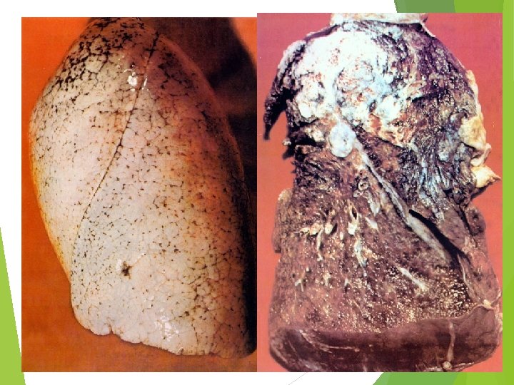
Abnormalities Emphysema Lung disease that destroys alveoli sacs Decreases vital capacity Traps air Hypoventilation Decreased air movement increased CO 2 Hyperventilation Increased air movement decreased CO 2
Emphysema