Osteoarthritis of the Hip Osteoarthritis of the Hip


")


- Slides: 24
Osteoarthritis of the Hip
Osteoarthritis of the Hip -The hip joint is one of the commonest sites of osteoarthritis In the past the traditional approach has been to consider hip OA as either primary or secondary Primary hip osteoarthritis was thought to occur when no underlying cause was apparent, and, when an obvious underlying cause was identified, the term secondary osteoarthritis was applied This system of classification is outdated, as is the use of the term ‘wear-and-tear’ arthritis. . .
A much more modern approach to the classification of hip OA is to consider the causes as mechanical or non-mechanical in origin
Causes.
Mechanical causes The hip is a ball-and-socket joint; as such, when there is ‘matched sphericity’ between the two articulating surfaces, a mechanical problem with the joint is unlikely. When there is loss of sphericity, then the articular surface is exposed to abnormal loads and contact forces that can initiate and then cause progressive hyaline cartilage damage
The common mechanical causes of hip OA include developmental dysplasia of the hip (DDH) where acetabular under-coverage leads to a loss of sphericity, and femoroacetabular impingement (FAI) where acetabular over-coverage or abnormal femoral shape leads to a loss of sphericit
Non-mechanical causes These are conditions or processes that can affect the hip joint in isolation or indeed be part of a more wide spread musculoskeletal disorder with the common end-stage effect being the destruction of hyaline cartilage
Clinical presentation The classic presentation of hip joint OA is groin pain associated with progressive stiffness and limp Initially this pain may be activity-related but later it is more persistent and can cause disturbed sleep antalgic gait is most common, Loss of internal rotation of the hip is one of the most consistent clinical Findings As loss of movement progresses, the patient may develop both a fixed external rotation contracture and fixed flexion deformity.
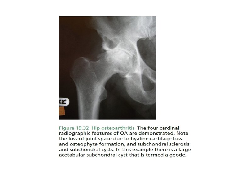
Radiological investigations The plain film radiograph represents the most common radiological investigation that is used to confirm the clinical suspicion of OA. The four important classical signs of OA equally apply to the hip joint: The earliest sign is 1)typically decreased joint space that represents loss of hyaline This is usually maximal in the superior weight bearing pole The later X-ray signs of OA are 2) Subarticular sclerosis, 3) subchondral cysts and 4)osteophyte formation
It is commonplace that the first presentation of a mechanical precipitating cause is end-stage OA. In younger women it is common to find the hallmarks of more subtle dysplasia that did not present earlier in life whereas in men in particular radiological hallmarks of femoroacetabular impingement (FAI) are often present.
TREATMENT OF HIP OA As the main presenting symptom is most often pain, the cornerstone of non-operative management of hip OA focuses on effective pain management Simple oral pharmacological agents such as (NSAIDs) Life-style modification TOTAL HIP ARTHROPLASTY: Total hip arthroplasty (THA) has established itself as one of the most successful operations performed across all surgical specialties and is the definitive treatment of end-stage OA of the hip. .
Hip fractures that occur between the articular margin of the femoral head to 5 cm below the lesser trochanter They are subdivided into intracapsular and extracapsular fractures The blood supply to the femoral head is typically damaged in intracapsular fractures and rarely in extracapsular Fractures. Extracapsular fractures are further subdivided into pertrochanteric and subtrochanteric fractures
Mechanism of injury: In the elderly patient the fracture usually results from a simple fall from standing height. In severe osteoporosis a fracture may occur from simple twisting moments and it is the hip fracture itself which causes the reported fall
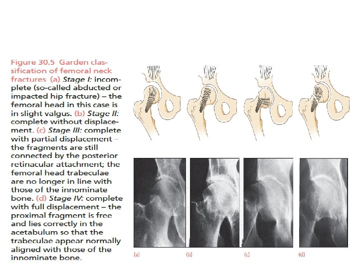
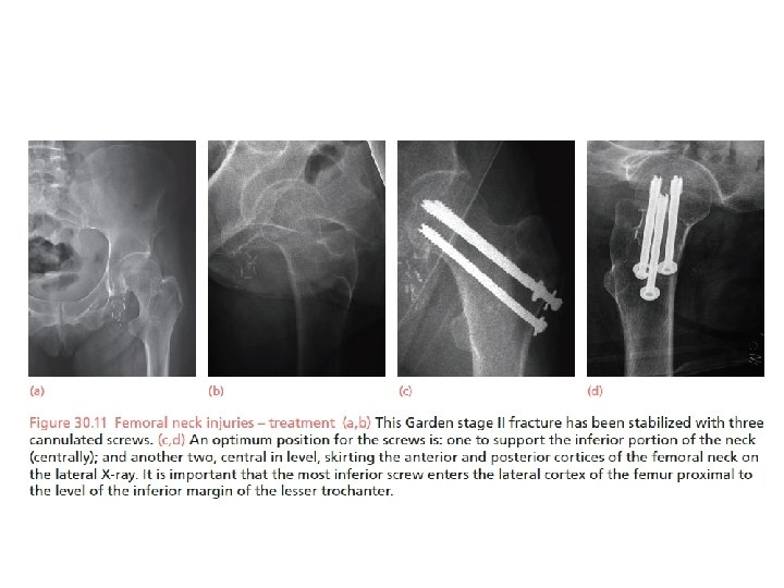
Pathological anatomy and classification INTRACAPSULAR HIP FRACTURES The most commonly used classification is the Garden classification, which is based on the amount of displacement apparent on X-rays of the hip
Stage I – an incomplete impacted fracture, including the so-called ‘abduction fracture’ in which the femoral head is tilted into valgus in relation to the neck • Stage II – a complete but undisplaced fracture • Stage III – a complete fracture with moderate displacement • Stage IV – a severely displaced fracture.
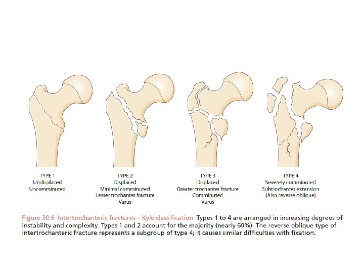
EXTRACAPSULAR HIP FRACTURES Intertrochanteric fractures These are divided into stable and unstable varieties. Reverse oblique intertrochanteric fractures These are a particularly unstable variant of this fracture pattern. As the plane of the fracture line is different from the usual pattern, the support offered by the medial column is lost
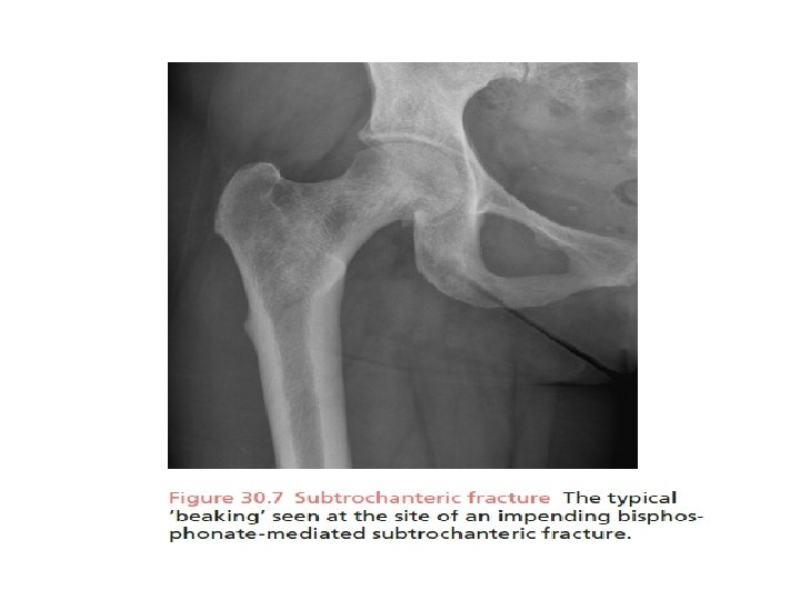
Subtrochanteric hip fractures : These fractures occur between the inferior margin of the lesser trochanter and 5 cm below this point. Fractures more distal than this are considered to be femoral shaft fractures
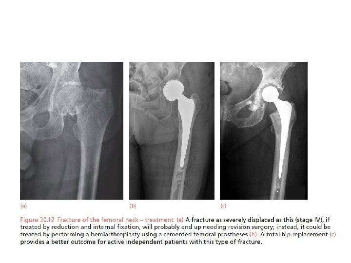
Treatment Initial treatment consists of pain-relieving measures including analgesia and a femoral nerve block. In the young patient every effort should be made to preserve the femoral head and fix the fracture; in contrast, reduction and fixation of fractures in the elderly patient is not recommended due to the associated high rates of subsequent re-operation (46%), nonunion (30%) and osteonecrosis of the femoral head(14%) Internal fixation An anatomical reduction is mandatory as a fracture fixed in a displaced position is at high risk of failure Prosthetic replacement Both displaced and undisplaced femoral neck fractures in the elderly patient should be treated by prosthetic replacement due to the high failure rates of open reduction and internal fixation in this group