Lecture 2 Introduction of nematode DrEman Sayed Mohammed


 through complex (life cycle o 1. 2.")


Development through extensive transomatic migration: nfective 3 rd stage penterating the")

 Superfamily: Oxyuroidea y: Oxyuridae (Pin worms) I- Genus: Oxyuris 1 - O. equi")


 * General characters: Family")
")


 (Rabbit pin-worm) *General characters: The")


")


- Slides: 37
Lecture 2 Introduction of nematode Dr/Eman Sayed Mohammed
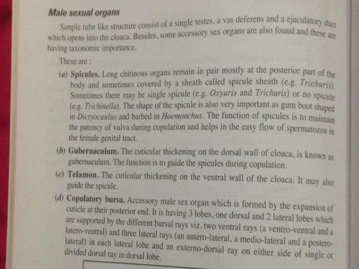
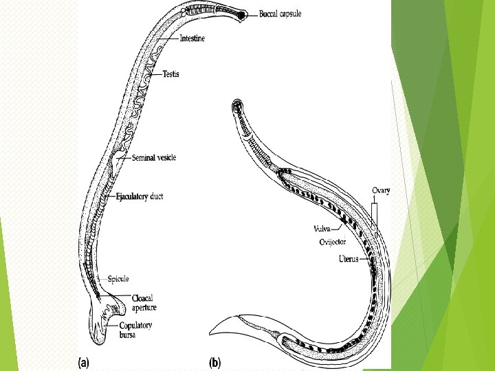
5 -Reproductive system q Male nematodes are usually smaller than their female. . q Basic male reproductive structures include: one testis, a seminal vesicle and a vas deferens into a cloaca q opening Male nematodes characteristic by curved q The genital system is tubular form andof male have posterior end , spicules and presence one tube bursa q In male intestine join with the genital duct to form the common cloaca which open exterior through colacal aperature q Syngamy, or cross fertilization, is common in most nematodes
Female Reproductive System tubular of form Consists 2 tube except for family trichinellidae and represented in a pair of ovaries attached to an 2 oviduct that has a swollen proximal end that forms a seminal receptacle. Each oviduct becomes a tubular uterus, and the two uteriaccording come together to Position of vulva varies to form a vagina that opens to the outside species : throughofa the genital pore(vulva)(vulvar flap) -middle body: Ascaridia and free living strongyloides. stercolaris -anterior third of the body: Ascaris and Oxyuris, filaria -posterior end of the body: Chabertia
Cross section of nematode
Development of nematodes o Parasitic nematode pass) through complex (life cycle o 1. 2. 3. life cycle either inside or outside the host Moulting (exsheathing, ecdysis): nematode undergo 4 moult during their development Formation of new cuticle Loosening of old one Rupture of old one and escape the larva Filariform infective stage
Development of nematodes Pass through 3 periods Nematode Post Embryonal developmen embryonal Development in developme definitive host t nt
evelopment of nematodes 1 - Embryonal development : its begin from egg fertilization untill formation of first stage larva and its occur either in uterus of female in their habitat in the definitive host or in the exterior after passage of developing egg with faeces So there is 3 possibilites of female nematode :
Female nematode oviparp us pass with faces to exterior then 1 st larval stage will develop and don’t hatch (Ascaridae, Tric huridae , Syngamidae) oviparou s pass with faces to exterior then 1 st larval stage will develop and hatch after certain period Stongyloididae, , strongylid ae. Ancylostomatidae, trichstr ongylidae oviparo useggs that stay lay in their habitat untill total embryonal development take place and egg pass contain 1 st larval stage Metastrongylidae , capillaria hepatica
oviviparp us oviviparo us pass with faces to exterior having 1 st larval exterior having stage and hatch in their 1 st larval stage habitat or in way to and don’t hatch exterior Dictyocaulus in their Some species of way to exterior metastrongylida e Larviparou s or viviparous lay 1 st larval stage pass to exterior with faces(some species of metastrongylidae pentrate) intestine and migtate in final host (trichinellidae) pentrate the blood vessels (filariidae)
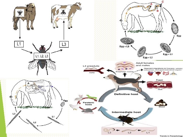
velopment of nematodes 2 - Post embryonal development : occur in the exterior and begin from formation of 1 st larval stage untill formation of infective stage larva Indirect Direct Without intermediate host geohelminth Inside eggs 1 st or 2 nd larval stage which don’t hatch untill taken by final host 3 rd larval stage hatch in exterior when reach to infective stage (Nematodirus and syngamus ) Outside eggs Begin after hatching 1 st larva from egg or after appearance of it in feces With intermediate host Biohelminth I. M. H will infected by ingestion of egg containing 1 st larva Metastrongylus, capillaria Ingestion, spirocerca of 1 st larval stage untill be infective stage then final host eat the I. M. H containing infective stage Protostrongylus, Habronema, Mullerius
velopment of nematodes 2 - Development in the final host : begin from infective stage of parasite untill formation of adult male and female with or without migration : A)Development through blood lung migration (pulmonary migration): Infective 3 rd stage burrow to blood or lymph …………right side heart ………. lung alveoli (3 rd strongyloides) or 4 th (ancylostoma)……. . bronchioles……. trachea……. . larynx……. . phary nx………oseophagus………. intestine …………adult B)Through blood liver lung migration (hepatopulmonary) : Infective 2 rd stage (ascaris) or 3 rd (syngamus) migrated from intestinal lumen penetrating mucous membrane ………………venous system……. portal vein……. liver……vena cava ………. heart………. lung……………. 4 th stage deveop……. . . . Trachea………. adult in (syngamus) or larynx………. pharynx…………. oseophagus…………………. . intestine ……………. adult in Ascaris.
velopment of nematodes D)Development through extensive transomatic migration: nfective 3 rd stage penterating the final host through the skin reach the hypodermis (filaria)While those entering by orally penterate m. m of intestinal tract. So by transomatic burrow inside different organ Untill develop to adult stage E)Through histotropic migration through m. m. of the gastrointestinal tract (histotropic migration): 1 rd stage(trichuris), 2 nd (Ascaridia) or 3 rd (trichostrongylidae
Mode of infection in nematode Ingestion: Mode of infection Penteration of skin 1 -Egg contain Active : direct 1 st larva(trichuris, penterate capillaria) 2 nd larva(Ascaris) Passive: by blood sucking arthropdes 3 rd larva( Inhalation of oxyuris) infected dust 2 - 3 rd stage 3 -intermediate host 4 - paratenic host 5 - milk from infected paratenic or definitive host Intrauterine (Toxocara) 2 nd stage larva
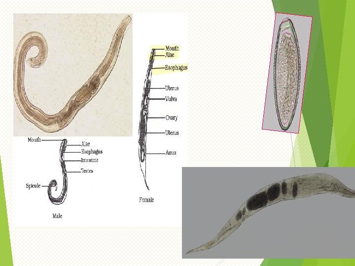
III) Superfamily: Oxyuroidea y: Oxyuridae (Pin worms) I- Genus: Oxyuris 1 - O. equi (Equine pin worm) (rectal worm) * General characters: Small to medium sized worm The male reaches about 1 cm. in length, while female reaches about 1. 5 cm in length. The oesophagus is double bulbed The male has one pin spicule. The young females are almost white in colour, slightly curved and has a relatively short, pointed tail. The mature females have a brownish colour. The vulva is situated near the anterior end of the body have a narrow long pointed tail The eggs are flattened on one side, curved in another side. ovoid and yellow. with mucoid a plug at one pole and usually contain larvae. Pin worm because of pointed tail in female worms * Habitat:
* Life cycle: After fertilization, the mature females wander down to the rectum and crawl out through the anal opening with the anterior parts of their bodies. The eggs are laid in clusters on the skin in the perineal region. Development of the egg is rapid, reaching the infective stage within 3 days. The infective stage may be reached on the perineal region or, more usually, the egg fall off to the ground. Infestation is by the ingestion of infective eggs or foods or bedding. Infective larvae are liberated in the small intestine and third stage larvae are found in the mucosal crypts of the ventral colon & cecum. . Histotropic migration
* Pathogenicity: Some ulceration can result from the mucosal feeding of the larvae. Most of clinical significance results from the intense itching caused by the sticky fluid which Oxyuris eggs are attached to the rump. In addition to loss of condition and poor appearance. biting and scratching may cause wounds open to infection. Severe cases can lead to nervousness and anorexia. Rubbing and scratching at the perineal region cause irritation, dull hair coat, and
II- Genus: Enterobius 1 - E. vermicularis (Human Pin worms) * General characters: Family worm Seat worm They are small white translucent worms. The males measure 2 -5 mm. while females measure 8 -13 mm. long. Double Bulbed oseophagus The males have a single spicule 70 u long and the caudal end of the body is curved ventrally. The tail of the female is long and pointed, equal to about one third of the total length of the body. The vulva opens just posterior to the middle of the body and the long vagina extends caudal. The eggs are translucent. The eggs measure 50 -60 u by 20 -30 u, and have a thick shell that is flattened on one side. The small size and colorlessness of the eggs make
D shape (planoconvex )
Retroinfection, or the migration of newly hatched larvae from the anal skin back into the rectum, may occur Some small number of eggs may become airborne and inhaled. These would be swallowed and follow the same development as
Transmission • • External Autoinfection Retroinfection Cross infection Inhalation Aerosol
* Habitat: The young and mature form occur in the small intestine, but the gravid female lives in the caecum , colon and rectum of man and higher primates such as Chimpanzee. * Pathogenicity: There is mild irritation at the points of attachment. In heavy infection, there intense itching of the preanal skin (perianal pruritis) due to external migration of the worms. Scratching of the area sometimes introduces secondary infections. Complications are much more common in women than in man for the fact that the female worm after depositing her eggs may losses its wax while trying to go back to the colon and enters the vagina. uterus and fallopian tubes and n both the pelvic and peritoneal regions.
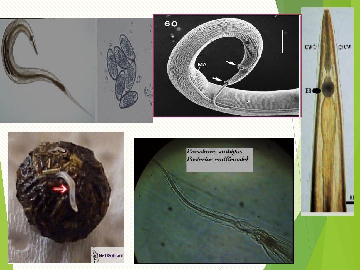
III- Genus Passalurus 1 - P. ambigus (Oxyuris ambigua) (Rabbit pin-worm) *General characters: The adult worm is white in colour. It is surrounded by transversaly striated cuticle The mouth opening is simple and surrounded by four papillae it opened into a double-bulbed oesophagus. Adult male measures 3. 2 -5. 2 mm long and with relatively long tail, while female measures 6. 3 -9. 2 mm long. The single spicule of the male is relatively short and no gubernaculum. The vulva at the anterior region just behind the oesophagus. The females, characterized by a long and narrow tail, are marked with about 40 circular, cuticular striations. It seems that the female worms deposit the eggs around the anus. The worms live about 106 days.
* Habitat: It occurs in the caecum and colon of rabbits. * Pathogenicity: ü In light infestation is relatively harmless. ü Desquamation of epithelial cells of small intestine. ü Eosinophilic exudate and some leukocytes and esinophils are observed.
Genus: Subulura S. brumpti * General characters: It is a small nematode with anterior end curved dorsally. The mouth part hexagonal, surrounded by 3 weakly developed lips, each with median papillae, two pairs of larger papillae located dorsally and venterally, anterior portions of oesphageal wall cuticularized forming 3 teeth like structures. Oesophagus is divided posteriorly and followed by a bulb. Cephalic alae extending to anterior portion of intestine in female and of middle of oesophagus in case of male. The male measures 7 -10 mm, long, tail curved ventrally, caudal papillae ( 10 pairs), 2 spicules are similar.
* Habitat: It occurs in the lumen of caecum of chickens, turkey, doves, duck, guinea fowl, pheasants and quail. * Life cycle: The embryonated eggs pass from the final host in caecal droppings. These eggs are eaten by beetles & cockroaches. Larvae hatch in 4 -5 hours, penetrate the intestinal wall and enter the body cavity where further development occurs. The infective stage (capsulated 3 rd larval stage) formed within 14 days post infestation. The definitive host eat the infected intermediate host (beetles & cockroaches), the larvae migrates to the caeca. the change to 4 th larval stage within 2 weeks. The final moult takes place on about the 18 th days postinfestation. Young adults continue to grow and develop and eggs appear in faeces m about 6 weeks post-infestation.
Family: Ascarididae Ascaridia galli Host Habitat Morphology Eggs Pathogenesis Species Fowl Small intestine Male up to 8 cm, female 12 cm There are 3 lips around the mouth The esophagus simple club shape There are 2 narrow lateral alae runs along the whole body of the worm The tail of male has weekly developed caudal alae and 10 pairs of caudal papillae The spicules are subequal A circular pericloacal sucker is present The female tail is straight and conical, with the vulva in the anterior portion of the body Oval, embryonated (one embryonal cell), yellowish, with smooth shell Hemorrhage and enteritis due to penetration of intestinal mucosa Bird become anemic, suffer from diarrhea Decrease egg production Intestinal obstruction or rupture due to mass accumulation of adult worms Sometimes the adult maybe found in the oviduct causing blockage and egg peritonitis Ascaridia galli � A. columbae � A. dissimilis Hetrakis gallinae (caecal worm) Fowl Caecum Male up to 1. 3 cm, female up to 16 mm, Whitish worm with long pointed tails There are 3 lips around the mouth The oesophagus has strong posterior bulb There are 2 narrow lateral alae runs along the whole body of the worm The tail of male has well developed caudal alae and 12 pairs of caudal papillae The spicules are unequal, the right one is slender thin and long and the other is short and thick A circular pericloacal sucker is prominent The female tail is straight and pointed, with the vulva opens directly behind the middle of the body Thickening of caecal mucosa, with petechial hemorrhage on the caecal surface typhilitis The principle economic importance of Heterakis species lies in its role as a carrier Histomonas meliagridis the causal agent of black head disease The organism passes from one bird to another in the gg of Heterakis spp.
Ascaridia galli Hetrakis gallinae
Ascaridia galli
Hetrakis gallinae (caecal worm)
*Pathogenicity of ascaridia galli: One of the most readily apparent effects of A. galli infestation is weight depression in the host. In severe infection, intestinal blockage can occur. In sometimes, the chickens infested with a large number of Ascarids suffer from loss of blood, reduced blood sugar content, increase urates. shrunken thymus glands, retarded growth, and greatly increased mortality. When large numbers of the young parasites penetrates into the intestinal mucosa. they cause hemorrhage, enteritis and the bird become anemic and suffer from diarrhea.
In heavy infestations, nodules form in the mucosa and sub mucosa. The large number produce some toxins that affect the metabolism of caecal digestion Black head disease due to Histomonas meliagridis Cyanosis at the head, comb and wattles (Give the head dark Typhilitis