Large intestine surgical anatomy 1 ascending and descending


- Slides: 59


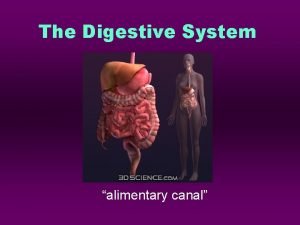
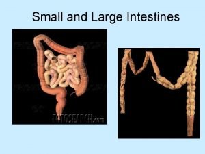
Large intestine surgical anatomy

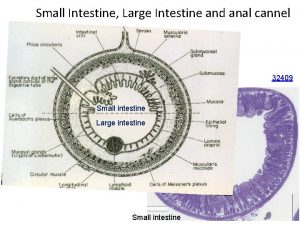
1 - ascending and descending colon >> intra and retro peritonium Don’t give abdominal symptoms until the inflammatory process go on , , , not mobile 2 - transverse colon >> intra peritonium >> there is a mesentery So its mobile and more prone to volvulus 3 - presence of tenia “ condense longitudinal fibers “ >> make the haustration 4 - presence of some fatty tissue “ can be very long in obese people >> on of the rare causes of acute abdominal pain ( non specific ) which is the most common cause of abdominal pain in the ER 5 - the caliber of the right side is wider than that of the left side >> so the tumors in the left side is presented earlier than in the right side

Clinical anatomy Haustration

Diet and large bowel diseases * Diet quality: - Fibre diet : “ help in decrease the contact time “ - Antioxident rich diet “ bind to the free radical decreasing its harmful effect “ - Dietary lectins - Carcinogens in diet * frequency of defecation &contact time

• Lectins are glycoproteins “ not digested easily , not cooked easily , it attaches to certain cells changing its nature >> causing antigen antibody reaction >> autoimmune disease It affect the enterokinase secretion , , also causing leaky bowel Part of lectins preferred certain blood group type. ******************************* The bile salt reacts with the bacteria in the large bowel liberating certain carcinogens

DIVERTICULOSIS 0 f THE COLON Is a sac like protrusion in the colonic wall, developes as a result of herniation of mucosa and sub mucosa through a point of weekness in the muscular wall.

DIVERTICULOSIS 0 f THE COLON 1 -false diverticulum 2 - between the mesenteric tenia And the lateral tenia 3 - acquired Colonic diverticulum 1 -true diverticulum 2 - anti-mesenteric 3 - congenital Meckle diverticulum

DIVERTICULAR DISEASE e congenital. 2. increase with age 5% befor age of 50 y 30%after age of 50 y 50 % over 70 y 66% over 85 y 3. common in left side sigmoid colon : 90% 4. M: F ratio equal 5. rare in the 3 rd world , is related to the fibre diet

ETIOLOGY 1. Precise etiology of this disease is unknown. - High intraluminal pressure and a weak colonic wall also may be caused by abnormal colonic motility 2. Genetic & enviromental factors may play a role - defective muscular structure, defects in collagen consistency. 3. Predisposing factors: a. obesity b. dec. physical activity c. alcohol, coffee , cigaret smoking , low fibre diet

pation is the main cause of increased n the colon, making the muscles strain to move stool that is too hard. >The excess pressure caused by the straining makes the weak spots in the colon bulge out, forming diverticula

PATHOLOGY: 1. It is a pseudo diverticulum tween mesenteric &anti mesenteric taenia ek sites in the circular m. , mesen. vesseles penetration. 4. Elevated I. L. pressure by tonic &rythmic contractions result in segmentation “ nonpropulsive contractions produce isolated segments”

5. Thickenning of long & circular muscles can lead to narrowing of colonic lumen So its considered as one of the causes of intestinal obstruction due to hypertrophic of the muscle Diverticulosis never occur in the rectum , because there is no tenia only circular and longitudinal muscles.

Pathology Weak points are the sites of entrance of the mesenteric arteries between the antimesenteric tenia and the mesenteric tenia. . So bleeding is sever


TERMINOLOGIES Diverticulosis: The presence of multiple diverticulae “generally implies to an absence of symptoms” Diverticular disease: Any clinical features caused by diverticulae including complications Diverticulitis: The presence of inflamatory process associated with diverticulae.

CLINICAL FEATURES DIVERTICULOSIS 1. Usually asymptomatic er abdominal colicky pain & flatulence which disapear after defecation

Symptoms & signs of acute diverticulitis: Migratory or shifting pain >> hypogastric to the left iliac fossa a. Acute lt. Lower quadrant pain : severe &deep b. Nausia & vomiting c. Fever, chills d. constipation, or alternating with bouts of diarrhoe inary symptoms: dys, freq. Urg. f. Tenderness, guardenning lt. lower quadrant ucocytosis h. Per rectal exam. : tender lt. side

DIAGNOSIS 1. 2. 3. 4. 5. Clinical features Radiology, Barium enema sigmoidscopy, colonoscopy CT U/S

Barium enema with double contrast

Barium enema with double contrast

Barium enema with double contrast

diverticulosis Normal

Sometimes fecal material is going to be impacted in the opening of the diverticulum causing pressure necrosis and sever bleeding


COMPLICATIONS 1. Abscess after acute inflammation Stage I - Pericolic abscess Stage II - Pelvic abscess Stage III - Purulent peritonitis Stage IV - Feculent peritonitis 2. Bleeding 3. fistula formation cutaneous , , vesicocolic , , recto-vaginal ) 4. Intestinal obstruction

TREATMENT DIVERTICULOSIS : 1. Hight residue diet : it lowers I. L. pressure, reduce symptoms, & prevents complications. 2. Encourage physical activity, i. e walking 3. antispasmodic for pain. 4. antibiotics some times needed

TREATMENT of DIVERTICULITIS: pon the severity of symptoms and clinical findings : a. Rest in bed uid diet or Administer intravenous fluid as ndicated. ert a nasogastric tube if patient is vomiting or colonic obstruction is suspected nfection &inflamation : antibiotics cover gram +ve, gram-ve, anaerobes &

Treatment of complications 1. perforation 2. abscess 3. Bleeding 4. Fistula formation 5. Large bowel obstruction

ULCERATIVE COLITIS a chronic disease characterized by diffuse mucosal inflammation of the colon and rectum

Epidemiology nic inflammatory disorder limited to the rectum and colon , relapses & remission is a character of the disease. cise etiology of ulcerative colitis is not well stood , abnormal activation of the immune system in the intestines is suggested. 3. sex ratio: nearly equal with a female preponderance. set of ulcerative colitis is most common between years of age, with a second peak in incidence between 50 and 80 years. 5. Cigarette smokers have a 40 percent lower risk of developing ulcerative colitis than do nonsmokers;

Ulcerative Colitis and Smoking smokers with ulcerative colitis tend to have fewer and less severe disease flare-ups. chers recently reported that smoking appears to alter the makeup of the various types of bacteria living in the intestinal tract ative colitis is an immune disease; it occurs when a person’s immune system mistakenly attacks and destroys the tissues of the colon. e relationships among intestinal microbes and the immune system are of particular relevance to inflammatory bowel disease.

PATHOLOGY 1. The disease starts in the rectum in 90% of the cases 2. Difuse inflamation of the mucosa , increase vascularity and congested mucosa with decrease ability to absorb water, lead to diarrhoe ple Minute Ulcer ”undermined ulcer” with numerous morrahgic spots the engorged vesseles can give rise to bleeding ulcerated areas are soon covered by granulation tissue , later end with scarring & shortenning. .

Cont. pathology 7. pseudopolyp formation found in about 15 -20% of cases ( not true polyps , no core vessels , , no mesincimal tissue , , only clusters of cells ) Due to hyperplasia of the mucosa at the edjes of the ulcers ic changes include: inflammation of the crypts of Lieberkuhn and crypts abscesses. (the most diagnostic feature in the pathology of ulcerative colitis) e patients due to an incompetent ileocecal valve , about 30 cm of the terminal ileum is affected.

pathology Pseudo polyops

EXTENT OF THE DISEASE The extent of colonic involvement can often, but not always, be predicted by the degree of symptomatology exhibited by the patient 1. Proctitis : Involvement limited to the rectum 2. Proctosigmoiditis: Involvement of the rectosigmoid colon -sided colitis: Involvement of the descending colon, which s along the patient's left side, up to the splenic flexure and the beginning of the transverse colon volvement of the entire colon, extending from the ectum to the cecum,


CLINICAL FEATURES 1. The hallmark symptoms of ulcerative colitis are : - intermittent bloody diarrhea - rectal urgency - tenesmus 2. Abdominal pain, cramping , subside after bowel movement 3. WT loss nal manefestation particularly arthritis, the mmonest Ankylosing spondylitis ( the most common ) skin lesions Primary sclerosing cholangitis While in crohns disease the most common is erythema nodosum

SIGNS Palor Dehydration ematiation Mild fever Tachycardia Abdominal tenderness Blood on digital rectal examination

EVALUATION OF SEVERITY Clinical scale: MILD : <4 motions /day , no systemic signs MODERATE : >4 motions /day , no systemic signs SEVER CASE : >4 motions /day +systemic signs : fever, tachycardia, WT loss, Hypoalbunaemia

Endoscopic scale a score of 0 is given for normal mucosa or inactive UC. a score of 1 is given for evidence of mild friability, reduced vascular pattern, and mucosal erythema. A score of 2 is indicative of moderate disease with friability, erosions, complete loss of vascular pattern, and significant erythema, a score of 3 indicates ulceration and spontaneous bleeding

Crohns dis. & ulcerative colitis Similarities : 1. Both are chronic inflamatory diseases. 2. Both are of unknown etiology 3. Both have no cure following medical treatment 4. Both have exrtra intestinal manefestations 5. Presence of diarrhoe in both cases. Non similarities: 1. Anatomical site in G. I. tract 2. Anatomical site in bowel wall. 3. Presence of skipped lesion. 4. Mucosal appearance 5. Surgical cure

Differential diagnosis of ulcerative colitis: includes any condition that produces chronic, intermittent diarrhea : 1. Crohn's disease, 2. ischemic colitis, 3. infectious colitis, 4. irritable bowel syndrome (IBS), 5. pseudomembranous colitis

CLINICAL DIAGNOSIS & DIAGNOSTIC TESTING linical history can be used to differentiate the various etiologies of chronic diarrhea 2. Lab. Tests and biopsy are the tests of choice to diagnose ulcerative colitis. 4. Barium enema

LAB. TESTS 1. stool examinations for ova and parasites and stool culture 2. CBC 3. Elevated sedimentation rate 4. elevated C-reactive protein (ie, >100 mcg/L): Both of these findings correlate with disease activity 5. Hypoalbuminemia (ie, albumin <3. 5 g/d. L 6. Hypokalemia (ie, potassium <3. 5 m. Eq/L eutrophil cytoplasmic antibody (p-ANCA), is found more mmonly in ulcerative colitis than in Crohn disease

Radiology Sewage like appearance early sign of uc. normal Loss of haustration Due to fibrosis Pipe like

Double contrast Pseudopolyps

typical vascular pattern, friability, exudates, ulcerations, and granularity in a continuous, circumferential pattern

*The choice of treatment depends on : a. location &severity of the disease. b. presence of complications. c. patient response to treatment

Medical treatment is always the first choice unless emergency surgery required. m of medical treatment is to control flare ups by reducing the mation that triger symptoms and reduce the chances of further flare ups & complications,

Medical treatment * First-line medical therapies : - 5 -aminosalicylic acid- (mesalamine) which acts topically from the colonic lumen to suppress the production of numerous proinflammatory mediators itis has been shown to respond better to suppositories than to oral 5 -ASA * response may take three to four weeks.

Cont. med. treatment tients with proctosigmoiditis require delivery of SA via an enema and may need four to six weeks of therapy to achieve remission. ts unable to tolerate the anal irritation of topical 5 -ASA may try oral preparations ents with pancolitis often require a combination d topical 5 -ASA compounds in addition to corticosteroids

Cont. med. treatment nts who fail to improve with the maximal ge of 5 -ASA compounds or who cannot tolerate the side effects, d therapy should be considered. nisone is given to these patients in dosag of 40 to 60 mg per day. -dose therapy is continued until symptoms are completely controlled (usually 10 to 14 days) sage is then tapered gradually by 5 mg per week

Cont. med. treatment When patients do not respond to orally administered steroids, they should be admitted to the hospital to receive intravenous corticosteroids, such as methylprednisolone sodium (Solu. Medrol) 40 mg daily *Hospitalized patients who fail to respond to intravenous corticosteroids after five to seven days are candidates for intravenous cyclosporine (Sandimmune)

COMPLICATIONS 1. Toxic colonic dilatation “Fulminating colitis”: a. The most common cause of death in ulcerative colitis b. characterized by a thin-walled, large, dilated colon that can eventually become perforated c. Symptoms & signs include abdominal pain and distension , fever and weakness , patient become disoriented d. Plain radiograph : colonic dilatation diameter> 6 cm 2. Perforation 3. Sever haemorrahge 4. Benign stricture may rarely cause intestinal obstruction 5. Colonic adenocarcinoma develops in 3 -5% of patients with ulcerative colitis. The risk increases with the duration of disease. The risk of colonic malignancy is higher in pancolitis and in cases in which disease occurs before the age of 15 years.

CANCER SCREENING *the risk of colon cancer is : 2 % in the first 10 years of ulcerative colitis, 8 % during the first 20 years, 18 % during the first 30 years *Patients who have only proctitis or proctosigmoiditis are considered to be at increased risk of developing colon cancer

INDICATION FOR SURGERY ses failing to respond to medical therapy or long-term steroid dependence 2. Chronic disease with frequent motions anaemia urgency tenesmu s or the disease being present for 7 -10 years 3. sever dysplasia, risk of neoplastic changes 4. Extra intestinal manefestations 5. Indications for urgent surgery include : Massive bleeding, perforation, toxic megacolon

SURGICAL PROCEDURES Surgery can often eliminate ulcerative colitis. EMERGENCY: Total colectomy + ileostomy. ELECTIVE : ocolectomy + ileo anal anastomosis with ouch

BAD PROGNOSTIC CRITERIA 1. age above 60 years 2. The whole colon is involved 3. Sever initial attack
 Haustrum of colon
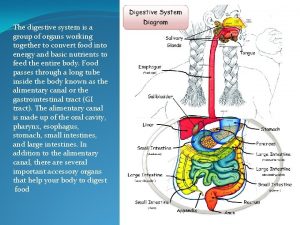
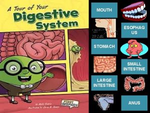
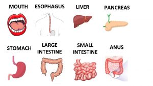
 Esophagus stomach small intestine large intestine
 What is chromatography
 Biochemistry chromatography
 Learning outcomes of ascending and descending order
 Similar fractions
 Corona radiata
 Descending bellows
 Intestine main function
 Large intestine relations
 Large intestine
 Rugae
Rugae Small intestine parts
 Lymphoid tissue
 Enteroendocrine cells
 Labelled diagram of a tooth
 Functions of the large intestine
 Left colic flexure
 Copyright
 Muscularis of large intestine
 Strongylus vulgaris
 Large intestine function in digestive system
 The main jobs of the large intestine are _____.
 Part of large intestine
 Large intestine function in digestive system
 Dr malak
 Large intestine function in digestive system
 Large intestine characteristics
 Large intestine histology
 Small intestine relations
 Major arteries of the systemic circulation
 Appendix (anatomy)
 The anatomy of a large scale hypertextual web search engine
 Oogoogle translate
 The anatomy of a large scale hypertextual web search engine
 The anatomy of a large-scale hypertextual web search engine
 Peristlasis
 Descending part of trapezius
 Bicep
Bicep Descending tracts
 Major arteries of the body
 Tongue deviation
 Nerve ganglia
 Descending clock auction
 Diatonic sequences
 Descending pathways
 Bridget riley descending
 What does raster mean
 Stretch reflex
 Bridget riley arrest 1
 Elsevier
 Les bêtes de la mer
 Weather how much do you know
 Overall shape of a melody
 George caleb bingham fur traders descending the missouri
 The metals having good weldability in descending order are
 Ventral spinocerebellar tract
 Ascending loop of henle
 Ascending reticular activating system
 Ascending levels of intellectual demand
