
Urinary System Chapter 18 Copyright 2016 by Elsevier
Urinary System Chapter 18 Copyright © 2016 by Elsevier Inc. All rights reserved. 1

Learning Objectives Lesson 18. 1: Urinary System 1. 2. 3. Identify the major organs of the urinary system and give the generalized function of each. Name the parts of a nephron and describe the role each component plays in the formation of urine. Explain how the kidneys act as vital organs in maintaining homeostasis. Copyright © 2016 by Elsevier Inc. All rights reserved. 2
Learning Objectives Lesson 18. 1: Urinary System (Cont. ) 4. 5. 6. Explain the importance of filtration, reabsorption, and secretion in urine formation. Discuss the mechanisms that control urine volume, including the normal amount and composition of urine. Explain the process of urine elimination. Copyright © 2016 by Elsevier Inc. All rights reserved. 3

Kidneys Location of the kidneys Ø Ø Under back muscles, behind parietal peritoneum, just above waistline Right kidney usually a little lower than left (Figure 18 -1) Gross structure: Internal anatomy (Figure 18 -2) Ø Renal cortex • Ø Renal medulla • Ø Outer layer of kidney substance Inner portion of kidney Renal pyramids • Triangular divisions of medulla Copyright © 2016 by Elsevier Inc. All rights reserved. 4

Kidneys (Cont. ) Gross structure: Internal anatomy Ø Renal papilla (pl. papillae) • Ø Renal pelvis • Ø Narrow, innermost end of pyramid Expansion of upper end of ureter; lies inside kidney Calyx (pl. calyces) • Division of renal pelvis Copyright © 2016 by Elsevier Inc. All rights reserved. 5
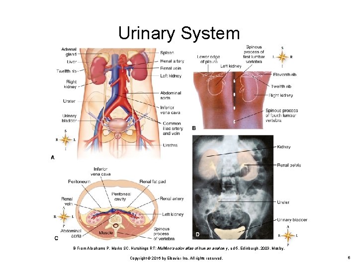
Urinary System B From Abrahams P, Marks SC, Hutchings RT: Mc. Minn’s color atlas of human anatomy, ed 5, Edinburgh, 2003, Mosby. Copyright © 2016 by Elsevier Inc. All rights reserved. 6
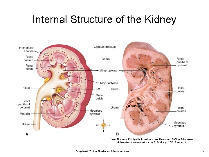
Internal Structure of the Kidney From Abrahams PH, Spratt JD, Loukas M, van Schoor AN: Mc. Minn & Abrahams’ clinical atlas of human anatomy, ed 7, Edinburgh, 2013, Elsevier Ltd. Copyright © 2016 by Elsevier Inc. All rights reserved. 7
Kidneys (Cont. ) Microscopic structure Ø Ø Ø Nephrons are microscopic units of kidneys Consist of renal corpuscle and renal tubule (Figure 18 -3) Renal corpuscle (Figure 18 -4) • Bowman capsule • Cup-shaped top of a nephron Glomerulus Network of blood capillaries tucked into the Bowman capsule Copyright © 2016 by Elsevier Inc. All rights reserved. 8
Kidneys (Cont. ) Microscopic structure Ø Renal tubule • Proximal convoluted tubule (PCT) • Nephron loop (Henle loop) • Extension of proximal tubule; consists of straight descending limb, hairpin turn, and straight ascending limb Distal convoluted tubule (DCT) • First segment of a renal tubule Part of tubal distal to ascending limb of nephron loop Collecting duct (CD) Straight part of a renal tubule Copyright © 2016 by Elsevier Inc. All rights reserved. 9
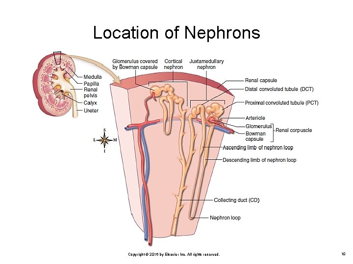
Location of Nephrons Copyright © 2016 by Elsevier Inc. All rights reserved. 10
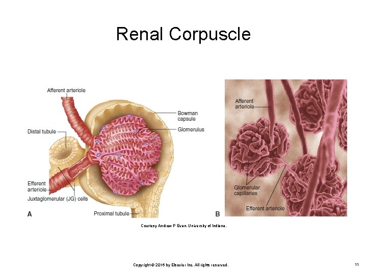
Renal Corpuscle Courtesy Andrew P Evan, University of Indiana. Copyright © 2016 by Elsevier Inc. All rights reserved. 11
Kidneys (Cont. ) Overview of kidney function Ø Ø Ø Excretes toxins and nitrogenous wastes Regulates levels of many chemicals in blood Maintains water balance Helps regulate blood pressure via secretion of renin Secretes erythropoietin (EPO) Copyright © 2016 by Elsevier Inc. All rights reserved. 12
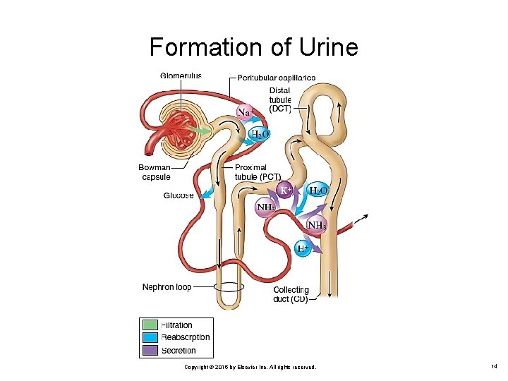
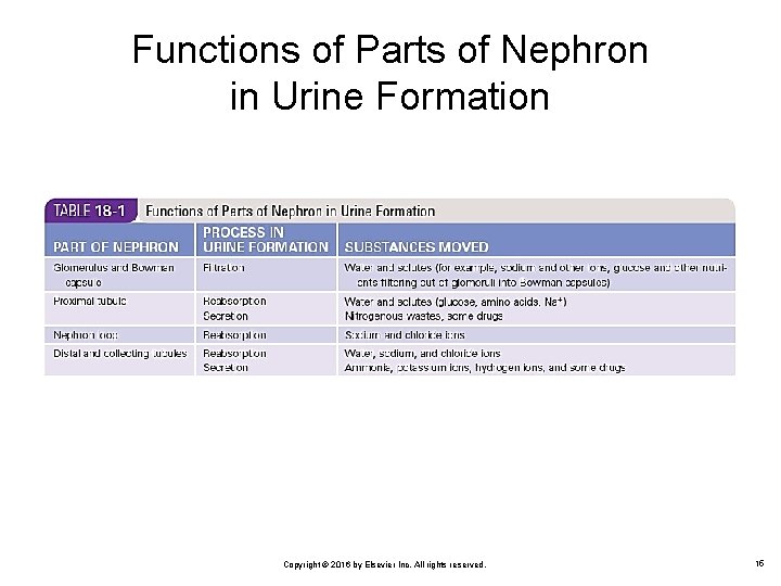
Formation of Urine Occurs by a series of three processes that take place in successive parts of nephron (Figure 18 -6 and Table 18 -1) Filtration Ø Ø Ø Goes on continually in renal corpuscles Glomerular blood pressure causes water and dissolved substances to filter out of glomeruli into Bowman capsule Normal glomerular filtration rate 125 m. L per minute Copyright © 2016 by Elsevier Inc. All rights reserved. 13
Formation of Urine Copyright © 2016 by Elsevier Inc. All rights reserved. 14
Functions of Parts of Nephron in Urine Formation Copyright © 2016 by Elsevier Inc. All rights reserved. 15
Formation of Urine (Cont. ) Reabsorption Ø Ø Ø Movement of substances out of renal tubules into blood in peritubular capillaries Water, nutrients, and ions are reabsorbed Water is reabsorbed by osmosis from proximal tubules Secretion Ø Ø Ø Movement of substances into urine in the distal and collecting tubules from blood in peritubular capillaries Hydrogen ions, potassium ions, and certain drugs are secreted by active transport Ammonia is secreted by diffusion Copyright © 2016 by Elsevier Inc. All rights reserved. 16
Control of Urine Volume Antidiuretic hormone (ADH) Ø Ø Aldosterone Ø Ø Secreted from the posterior pituitary gland Decreases the amount of urine by collecting ducts permeable to water Secreted by the adrenal cortex Stimulates the tubules to reabsorb sodium at a faster rate Atrial natriuretic hormone (ANH) Ø Ø Secreted from the heart’s atrial wall Stimulates kidney tubules to secrete more sodium and thus lose more water Copyright © 2016 by Elsevier Inc. All rights reserved. 17
Control of Urine Volume (Cont. ) Abnormalities of urine volume Ø Ø Ø Anuria: Absence of urine Oliguria: Scanty amount of urine Polyuria: Unusually large amount of urine Copyright © 2016 by Elsevier Inc. All rights reserved. 18
Elimination of Urine Ureters (Figure 18 -10) Ø Structure • Ø Narrow, long tubes with expanded upper end (renal pelvis) located inside kidney and lined with mucous membrane Function • Drain urine from renal pelvis to urinary bladder Copyright © 2016 by Elsevier Inc. All rights reserved. 19
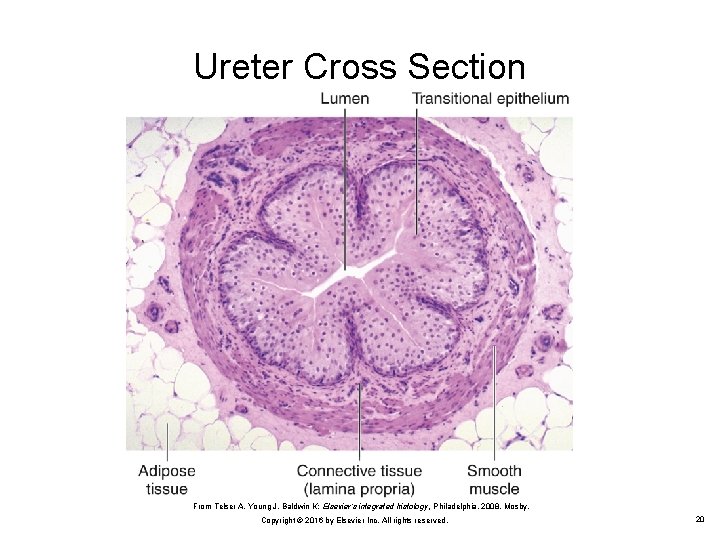
Ureter Cross Section From Telser A, Young J, Baldwin K: Elsevier’s integrated histology, Philadelphia, 2008, Mosby. Copyright © 2016 by Elsevier Inc. All rights reserved. 20
Elimination of Urine (Cont. ) Urinary bladder (Figure 18 -11) Ø Structure • • Ø Elastic muscular organ, capable of great expansion Lined with mucous membrane arranged in rugae, as is stomach mucosa Functions • • Storage of urine before voiding Voiding Copyright © 2016 by Elsevier Inc. All rights reserved. 21
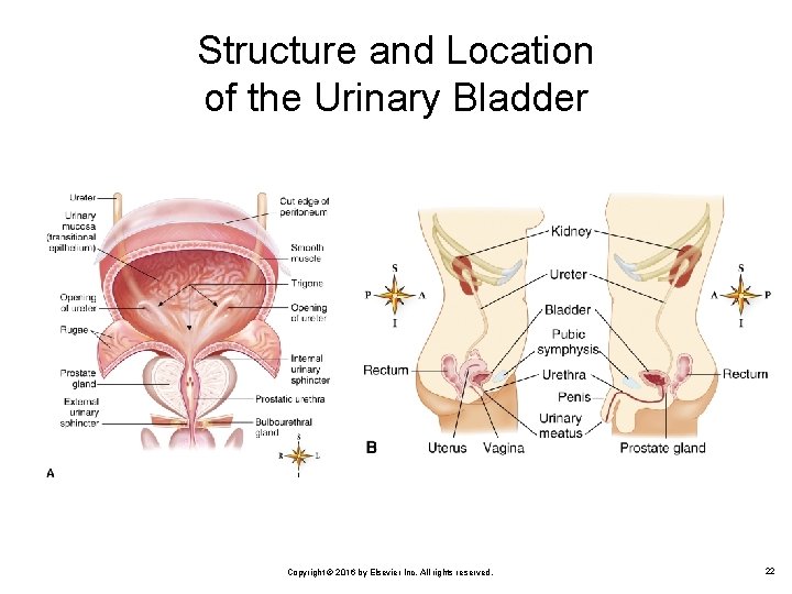
Structure and Location of the Urinary Bladder Copyright © 2016 by Elsevier Inc. All rights reserved. 22
Elimination of Urine (Cont. ) Urethra Ø Structure • • • Ø Functions • • Ø Narrow tube from urinary bladder to exterior Lined with mucous membrane Opening of urethra to the exterior called urinary meatus Passage of urine from bladder to exterior of the body Passage of male reproductive fluid (semen) from the body Cystitis • Bladder infection Copyright © 2016 by Elsevier Inc. All rights reserved. 23
Elimination of Urine (Cont. ) Micturition Ø Ø Passage of urine from body (also called urination or voiding) Regulatory sphincters • • Ø Internal urethral sphincter (involuntary) External urethral sphincter (voluntary) Bladder wall permits storage of urine with little increase in pressure Copyright © 2016 by Elsevier Inc. All rights reserved. 24
Elimination of Urine (Cont. ) Micturition Ø Emptying reflex • • Initiated by stretch reflex in bladder wall Bladder wall contracts Internal sphincter relaxes External sphincter relaxes, and urination occurs Copyright © 2016 by Elsevier Inc. All rights reserved. 25
Elimination of Urine (Cont. ) Micturition Ø Urinary retention • Ø Urinary suppression • Ø Urine produced but not voided No urine produced but bladder is normal Urinary incontinence • • • Urine is voided involuntarily; a common bladder control problem in elderly people May be caused by spinal injury or stroke Retention of urine may cause cystitis Copyright © 2016 by Elsevier Inc. All rights reserved. 26
Urinalysis Physical, chemical, and microscopic examination of urine Sign of a disease Ø Ø Changes in normal characteristics of urine Appearance of abnormal urine characteristics Copyright © 2016 by Elsevier Inc. All rights reserved. 27
Questions? Copyright © 2016 by Elsevier Inc. All rights reserved. 28
- Slides: 28