Digestive System Overview of Digestion n 2 main


. n The")


")


 n Helps the digestive process by: a. Mixes food with digestive enzymes")


 regulates flow into to large intestine. It also prevents")


. It is retroperitoneal. n The")


 n Bile salts")


- Slides: 77
Digestive System
Overview of Digestion n 2 main groups of organs in the digestive system. 1. Alimentary Canal (nutrition) a. Mouth b. Pharynx c. Esophagus d. Stomach e. Small bowel f. Large bowel
2. Accessory Digestive Organs a. Teeth b. Tongue c. Gall bladder d. Salivary glands e. Liver f. pancreas
Pharynx and Oral Cavity Superior to the ORAL CAVITY is the HARD PALATE composed of the MAXILLARY and PALATINE bones. n Superior and posterior to the oral cavity are the INTERNAL NARES. n From the internal nares, if we go anteriorly we will find the EXTERNAL NARES or NOSTRILS. n
n n n Posterior to the hard palate is the SOFT PALATE. This is muscular tissue that is moved during swallowing. Hanging from the soft palate is a conical structure called the UVULA. The two nasal cavities are separated by the NASAL SEPTUM which is formed by the union of the VOMER and PERPENDICULAR PLATE of the ETHMOID bones.
3 Areas of the Oral Cavity 1. OROPHARYNX -soft palate to epiglottis -two sets of TONSILS a. Palatine b. Lingual -the tonsils remove pathogens that enter the pharynx. They contain lymphocytes
n 2. NASOPHARYNX -located superior and posterior to the soft palate. -contains the PHARYNGEAL TONSILS and TUBAL TONSILS
n 3. LARYNGOPHARYNX -inferior to the epiglottis and posterior to the larynx. - this division opens into the esophagus and larynx.
Sagital section of cadaver head Notice the nasal conchae. They serve to expand the surface area to warm and moisten breathed air. Also, notice the position of the spinal cord within the vertebral canal.
How does “Digestion” occur? 6 step process: 1. Ingestion 2. Propulsion Peristalsis – alternate waves of muscular contraction and relaxation in the primary digestive organs. The end result is to squeeze food from one part of the system to the next.
3. 4. Mechanical Digestion - physical preparation of food for digestion. - Segmentation – mixing of food in the intestines with digestive juices. Chemical Digestion - Carbohydrates, Fat, and Proteins are broken down by enzymes.
5. Absorption - transfer of the digested portion of food into the blood from the digestive canal. 6. Defecation - removal/elimination of the waste products from the body.
Histology of the Digestive System n 1. 2. 3. 4. All alimentary canal organs have the same 4 layers. Mucosa (innermost layer) Submucosa (CT containing neurovascular bundles) Muscularis Externa (2 layers of smooth muscle) Serosa (outermost layer, visceral peritoneum)
Diagram of GI wall to show various kinds of glands -- some within the wall and some without (like the liver). These glands have ducts that empty into the lumen of the gut. In all cases, the epithelium lining the ducts and glands is continuous with the epithelium lining the lumen (cavity) of the gut.
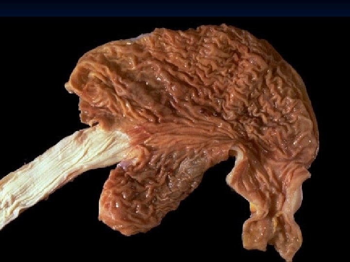
The image above shows a section of colon from a dog. Note the crypts extending from the lumen, and the numerous, foamy goblet cells that populate the epithelium of the crypts.
Secretion of mucus from goblet cells is elicited primarily by irritating stimuli rather than in response to hormones
Mouth and Associated Organs Food enters the GI tract at the mouth. It is chewed, manipulated by the tongue, and moistened with saliva. n Mouth has two parts: 1. vestibule – space between cheek and teeth. 2. oral cavity proper – space internal to the teeth. n
n Tongue - skeletal muscle - mixes food with saliva into a compact mass known as a BOLUS. - LINGUAL FRENULUM attaches the tongue to the floor of the mouth and prevents posterior movement of the tongue.
Salivary Glands When you dissect your cat, you will notice two muscles on the inside of the cheek. n The DIGASTRIC MUSCLE opens the jaw. n The MASSETER MUSCLE closes the jaw. n
The masseter inserts on the mandible. n Superficial to part of the masseter and anterior to the ear is the large PAROTID GLAND. This gland produces SALIVARY AMYLASE (ptyalin), a digestive enzyme. n The parotid gland is GRANULAR, it is attached by fascia. It is also the largest of the salivary glands. n
n n The parotid empties into the PAROTID DUCT which empties between the last two molars at the angle of the jaw. The parotid gland is an EXOCRINE GLAND. Exocrine glands empty via a duct to a specific location. The other type of gland is an ENDOCRINE GLAND that empties directly into the bloodstream.
Caudal and ventral to the parotid gland is the SUBMANDIBULAR GLAND (SUBMAXILLARY). n The SUBMAXILLARY DUCT empties this gland. It runs on the lateral aspect of the digastric muscle. n This gland carries saliva into the angle of the lower jaw. n
n n The SUBLINGUAL GLAND is on the submaxillary duct. It is wedge shaped and it is lateral to the digastric muscle. The DORSAL and VENTRAL FACIAL NERVES run around the outline of the masseter muscle. These nerves come out in front of the ear from the STYLOMASTOID FORAMEN and branch across the face.
1. Masseter Muscle 2. Parotid Gland 3. Parotid Duct 4. Submandibular Gland 5. Sublingual Gland 6. Lymph Nodes 7. Molar Gland
Masseter muscle Parotid Duct Parotid gland Submandibular gland Sublingual gland Submandibular Duct
5 Openings into the Pharynx 1. 2. 3. 4. 5. Mouth Left and right nasal passages Eustachian tubes (connect middle ear to the throat) Larynx Esophagus
Swallowing n n n Is a reflex. When the mouth closes, the soft palate is pushed superiorly and closes the nasal passages A sphincter valve closes off the eustachian tubes The glottis closes and respiration stops. The glottis also bends and closes the entrance into the larynx. The esophagus is opened by pressure of the food. This allows the epiglottis to open. Food then enters the esophagus.
Teeth Very similar to bone. n Three major components: 1. hydroxyapatite Ca (PO ) (OH) 2. bone collagen 3. cells n 5 4 3
The p. H of the mouth is usually 7. 2 n There acids in the mouth that come from three sources: 1. stomach acid during vomiting 2. foods 3. waste products of mouth bacteria n
Tooth Anatomy n n n n Enamel: hardest substance in the body Pulp Cavity: contains arteries, veins, and nerves. Alveolus: made of alveolar bone Root: made of dentin Gingiva: gum Periodontal membrane: periosteum found around the tooth Cementum: material that holds the tooth in the alveolus.
INCISORS – chisel shaped for nipping food. n CANINES – cone shaped for tearing n PREMOLARS – n MOLARS grinding food n 32 teeth in the Permanent Dentition n 20 teeth in the Deciduous Dentition n
Identify the Following: Incisors Molar Premolars Canines
The Digestive System n n Function: physically and chemically breakdown food products so that they can be absorbed and transported to cells. CARBOHYDRATES are the major source of biochemical energy. They include sugars and starches. These are eventually broken down into MONOSACCHARIDES (simple sugars)
n n PROTEINS are broken down to amino acids. AMINO ACIDS are the chemical building blocks of proteins. Proteins are necessary parts of cell membranes and nucleic acids (DNA and RNA). LIPIDS are broken down to fatty acids and glycerol. Lipids are very large molecules and cannot be directly absorbed. They are broken down by ENZYMES which are organic CATALYSTS. They are very specific for each chemical reaction and the function to speed up the reaction.
The name of an enzyme usually end in “-ase” and can give a clue as to its function. n For example, Lipase is an enzyme that catalyzes the breakdown of lipids (fats and oils) n
Chewing (Mastication) n Helps the digestive process by: a. Mixes food with digestive enzymes in saliva. b. Increases surfaces area of food c. Makes moving the food easier
Saliva Contains PTYALIN or SALIVARY AMYLASE. These are enzymes that break down starches. These enzymes are only active under certain p. H conditions. n The p. H of the mouth is about 7. 2 (slightly alkaline as 7 directly in the middle of the scale) n
When the swallowed food reaches the stomach, the p. H drops to 3 (very acidic). The ptyalin is no longer active at that p. H. n Once food is swallowed, smooth muscle in the esophagus carries the bolus by PERISTALSIS. n
Once food enters the esophagus, peristalsis is automatic. In fact, food can successfully reach the stomach while standing on your hands. n The bolus enters the stomach by passing through the GASTROESOPHAGEAL SPHINCTER. It is held shut by contraction of muscle. n
1. Diaphragm 7. Right Medial Lobe of Liver 2. Round Ligament 8. Right Lateral Lobe of Liver 3. Falciform Ligament 9. Gall Bladder 4. Left Lateral Lobe of Liver 10. Spleen 5. Left Medial Lobe of Liver 11. Greater Omentum 6. Quadrate Lobe of Liver
Gastroesophageal sphincter esophagus Fundus Pylorus Body Pyloric shpincter
esophagus GES stomach
The LESSER CURVATURE of the stomach is anchored to the liver with the LESSER OMENTUM. It cannot move. n The esophagus and duodenal ends are anchored. As food fills the stomach, it can sag on the left side. n
n n n The walls of the stomach have 3 muscle layers These muscles do not contract together-they contract out of sync. This enables the muscles to mix and churn the food in the body of the stomach. The mixing is with water, hydrochloric acid (produced in the stomach), and pepsin. This mixture is known as CHYME.
The release of the chyme is regulated by the pyloric sphincter. n The stomach also has longitudinal folds within the lumen. These folds, called RUGAE, increase the surface area of the stomach. n
Small Intestine n 3 parts: 1. Duodenum - 10 inches long 2. Jejunum - 8 feet long 3. Ileum - 11 feet long
mesentery
The small intestine produces 7 enzymes. There a total of 17 enzymes that are dumped into the duodenum for digestion. n The small intestine is the area where most digestion occurs. n It is also the place where 74% of the absorption of nutrients occur. n
The absorptive area is increased by: 1. circular folds called PLICAE CIRCULARIS. 2. Microscopic VILLI 3. MICROVILLI These structures increase the surface area of the small intestine by 600 x n
n n Within the plicae circularis are arteries, capillaries, and veins. The veins drain into the HEPATIC PORTAL SYSTEM which ultimately drain into the liver and INFERIOR VENA CAVA. There is also lymphatic drainage via LACTEALS which drain into the CISTERNA CHYLI. These drain into the THORACIC DUCT.
n SEGMENTAL PERISTLASIS occurs in the small intestine. This segmenting results in a sausage appearing structure.
n The ileocecal valve (sphincter) regulates flow into to large intestine. It also prevents backflow from the large intestine into the small intestine.
Sources of Intestinal Secretions Esophagus Stomach Cystic Duct Hepatic Ducts Spleen Common Bile Duct Gall Bladder pancreas Jejunum villi Duodenum microvilli
1. Cardiac Stomach 8. Ascending Colon 2. Fundic Stomach 9. Ileum 3. Stomach Body 10. Jejunum 4. Pyloric Stomach 11. Sigmoid Colon 5. Lesser Omentum 12. Spleen 6. Duodenum 13. Gastrospleenic Ligament 7. Pancreas (Ventral) 14. Bladder
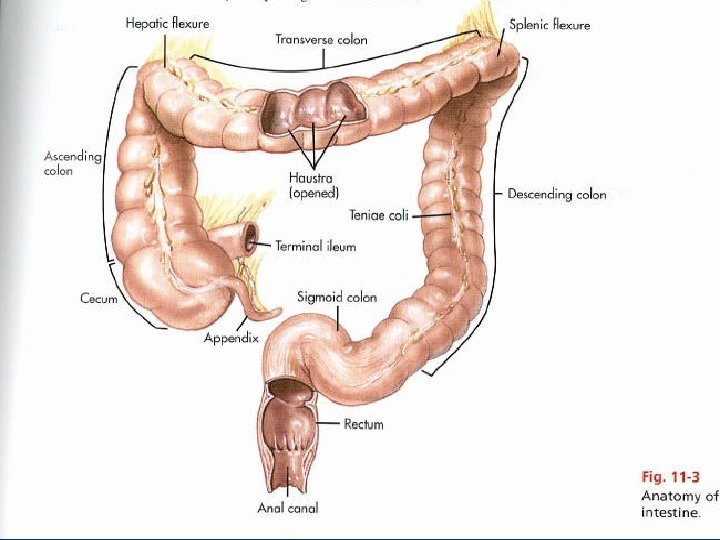
The Large Intestine 5 feet long n The CECUM extends as a 2. 5 inch blind sac caudally from the sphincter. n Off the cecum is the APPENDIX. There is currently no purpose for the appendix. Some research is pointing toward an immune function. n
From the cecum is the ASCENDING COLON (5 inches). It is retroperitoneal. n The RIGHT COLIC FLEXURE leads to the TRNASVERSE COLON (15 inches). n The LEFT COLIC FLEXURE leads to the DESCENDING COLON (10 inches) and it is also retroperitoneal. n
n n The descending colon leads to the SIGMOID COLON. The name change occurs at the SIGMOID FLEXURE. The RECTUM (5 inches) leads to the ANAL CANAL (7 inches). The final sphincter in the tract is the SPHINCTER ANI. The sigmoid and rectum are also retroperitoneal.
n n The transverse colon hands on a piece of the MESOCOLIC LIGAMENT. It hangs into the umbilical region. The longitudinal bands of muscle in the colon are three bands that do not completely surround the structure. The only part of the large intestine that have complete muscle coverage is the rectum.
Parotid Gland Liver esophagus Gastroesophageal sphincter Gall bladder cecum Fundus of Stomach pancreas Pylorus of stomach
n n The muscles act like a drawstring that contract the colon into little pouches. These pouches are called HAUSTRA. The material that reaches the colon is undigested and/or undigestable. Bacteria live in the colon. These are important for the synthesis of Vit. B 12 and K. Other bacteria are responsible for destroying the “bad” bacteria. E. coli is an example
n n The sphincter ani is an involuntary smooth muscle. The DEFECATION REFLEX which is kept in control by the sigmoid flexure and peristaltic activity. When peristalsis occurs the sphincter ani relaxes. An EXTERNAL SPHINCTER (skeletal muscle) can oppose the sphincter ani. This allows you to “hold it in” until you find a bathroom! The first part and part of the second third of the esophagus are also made of skeletal muscle. The rest of the GI tract is smooth muscle.
Transverse Colon c Ascending cecum appendix Haustra Tenia coli Descending Colon Terminal Ileum Si co gmo lon id rectum Anal Canal
1. Small Intestine 6. Transverse Colon 2. THE Mesentary 7. Descending Colon 3. Ileum 8. Sigmoid Colon 4. Cecum 9. Mesocolon 5. Ascending Colon 10. Greater Omentum
Types of Hernia
Some Definitions Secretion - discharge of materials synthesized by cells. Excretion - discharge of metabolic waste products from our cells. Occurs at skin, sweat glands, lungs, feces, and kidneys.
Liver n 5 functions: 1. Detoxification of blood Carbohydrate metabolism -glycogenesis – formation of glycogen from excess glucose in circulation. -glycogenolysis – breakdown of glycogen in times of fasting. -gluconeogenesis-formation of glucose in hepatocytes from raw materials. Lipid metabolism -synthesizes large quantities of cholesterol and phospholipids. -oxidizing triglycerides to produce energy. Protein synthesis Secretion of bile 2. 3. 4. 5.
Bile contains bile salts, water, pigments, cholesterol, and lecithin (a phospholipid) n Bile salts act like detergents and EMULSIFY fats. Makes fat form into small droplets that are more soluble. Greater surface area makes it more digestible. n
n n Bile is stored in the GALL BLADDER where it is concentrated. When fat is detected in the duodenum, the gall bladder contracts and bile is discharged into it. The COMMON BILE DUCT comes into the first inch of the duodenum. Its opening is called the AMPULLA OF VATER. This opening is controlled by the SPHINCTER OF ODDI. This sphincter relaxes when the gall bladder contracts.
Pancreas n n Produces approx. 10 enzymes which are responsible for digestion. The PANCREATIC DUCT carries these enzymes directly into the common bile duct. Sometimes it empties directly into the duodenum (anatomic variance). Also secretes BICARBONATE which neutralizes the duodenal contents. The ISLETS OF LANGERHANS produce INSULIN and GLUCAGON.
Spleen Stores blood n Produces WBC n Part of lymphatic system n Found midaxillary, deep to ribs 9 -11 and superior to the TPL. n