JOINTS DR Nazneen Qureshi DEPARTMENT OF ANATOMY MGM


 2. SECONDARY - SYMPHYSIS (Fibro Cartilagenous)")


 MGM Medical College, A’bad")


 Simple b)")

![NON AXIAL q. Plane synovial joints. q. Articular surfaces are flat [plane] q. Permits](https://slidetodoc.com/presentation_image_h2/a5b29a1c33ab40bf45078bb0d1b23466/image-49.jpg "NON AXIAL q. Plane synovial joints. q. Articular surfaces are flat [plane] q. Permits")
![UNI AXIAL q. One degre freedom of movement. 1. Hinge joint [ginglymus]. §Articular surfaces](https://slidetodoc.com/presentation_image_h2/a5b29a1c33ab40bf45078bb0d1b23466/image-50.jpg "UNI AXIAL q. One degre freedom of movement. 1. Hinge joint [ginglymus]. §Articular surfaces")
![2. Pivot[trochoid]. §Articular surfaces compromise central boney pivot surrounded by osseo -ligamentous ring. §Moves](https://slidetodoc.com/presentation_image_h2/a5b29a1c33ab40bf45078bb0d1b23466/image-51.jpg "2. Pivot[trochoid]. §Articular surfaces compromise central boney pivot surrounded by osseo -ligamentous ring. §Moves")


- Slides: 67
JOINTS DR. Nazneen Qureshi DEPARTMENT OF ANATOMY MGM MEDICAL COLLEGE
JOINTS INTRODUCTIONl Connection between two or more bones or cartilages. l Device that permit movements l Long bones articulates by their ends l Flat bones articulates by their margins l Irregular bones articulates by their surfaces l Joints are more in child.
Classification 1. Structural 2. Functional 3. Regional 4. According to no. of articulating bones
Structural classification Depends on type of connective tissue 1. Fibrous joints 2. Cartilaginous joints 3. Synovial joints
Functional classification - depends on degree of mobility 1. Synarthrosis -immovable 2. Amphiarthrosis – slightly movable 3. Diarthrosis –freely movable
Regional classification 1. Skull type 2. Vertebral type 3. Limb type
According to no. of articulating bones 1. Simple joint- interphalyngeal jt 2. Compound joint - elbow, wrist jt 3. Complex joint - AC jt, TM jt, SC jt
TYPES OF JOINTS Skull type Fibrous joint Synarthrosis Vertebral type Cartilagionous Amphiarthrosis Limb type Synovial joint Diarthrosis
COMPOUND JOINTS SIMPLE JOINTS COMPLEX JOINTS
JOINTS SYNARTHROSIS FIBROUS SUTURAL SYNDESMOSIS GOMPHOSIS DIARTHROSIS CARTELGENOUS SYNCHONDROSIS SYMPHISIS
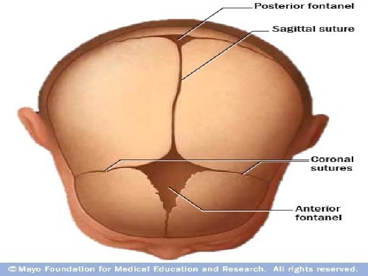
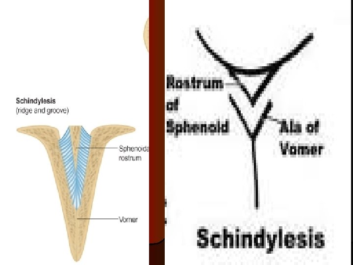
Fibrous Joints 1. SUTURAL –peculiar to skull appears in membranous type of bone a. Plane ---- interpalatine suture, internasal suture b. Squamous– parieto –temporal suture c. Serrate -- sagittal suture d. Denticulate – lambdoid suture e. Schindylesis [wedge and groove] ---rostrum of sphenoid and upper margin of vomer
Serrate suture
Denticulate suture
PLANE SUTURE INTERPALATINE INTERNASAL
SQUAMOUS SUTURE
2. SYNDESMOSIS -surfaces of bones united by interosseous ligaments -bones lie some distance apart. -slight amount of movement possible -ligaments persist throughout life Eg. – inferior tibiofibular joint -middle radio –ulnar joint
SYNDESMOSIS
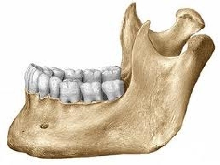
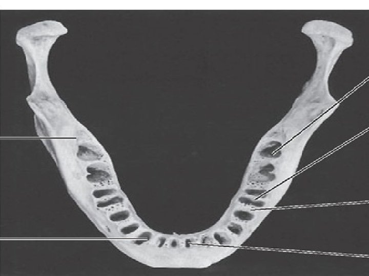
3. GOMPHOSIS – peg and socket joint eg. – root of teeth fit into socket of the jaw
CARTILAGENIOUS JOINTS 1. PRIMARY - SYNCHONDROSIS (Hyaline cartilagenous) 2. SECONDARY - SYMPHYSIS (Fibro Cartilagenous)
1. Primary cartilaginous joint- bones united by plate of hyaline cartilage which is temporary in nature. - Replaced completely by bone [synostosis] - no movement possible eg. 1. between basiocciput and basisphenoid 2. jt Between epiphysis and diaphysis 3. first chondrosternal joint
Chondro-sternal joint
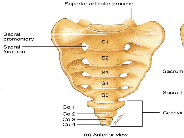
Secondary cartilaginous v v v Articular surface of bones are covered by hyaline cartilage and are united by a plate of fibrocartilage. Persist throughout life occupy median plane of body Limited movement possible eg. - intervertebral disc
Secondary CARTILAGENIOUS JOINTS
JOINT II Dr. Savita Kadam (Khiste) MGM Medical College, A’bad
DIARTHROSES INTRODUCTION q. Possess joint cavity filled with synovial fluid. q. Permit free movement. q. These are called synovial joints. q. Most of the joints of body are synovial.
SYNOVIAL JOINTS
CHARACTERISTICS OF SYNOVIAL JOINTS q. Presence of articular cartilage. q. Articular capsule. q. Joint cavity. q. Articulating bones have ligaments. q. Joint cavity divided by articular disc or meniscus.
ARTICULAR CARTILAGE q. Mostly hyaline. q. Avascular, non nervous and elastic. q. Porous in nature. q. Once damaged replaced by fibrous tissue. q. As age advances, degenrative and proliferative changes occur.
FUNCTIONS q. Provides smooth gliding surface. q. Reduces compression during weight bearing. NUTRITION q. From synovial fluid and qby diffusion from capillaries.
ARTICULAR CAPSULE q. Completely invest the joint. q. Formed by bundle of collagen fibres. q. Has outer fibrous capsule & inner synovial membrane. q. Pierced by blood vessels and nerves.
FUNCTIONS q. Binds articulating bones. q. Supports synovial membrane. q. Acts as watchdog.
SYNOVIAL MEMBRANE q. Vascular and cellular connective tissue membrane. q. Lines whole the joint cavity except for the articular surface covered by a hyaline cartilage.
FUNCTIONS q. Secrete synovial fluid. q. Liberates hyaluronic acid. q. Removes particulate matters.
SYNOVIAL FLUID q. Viscous and glairy fluid q. Contains Hyaluronic acid( Polymer of mucco polysaccharide q. Secreted by Synovial cells and mast cells. q. Contains monocytes, lymphocytes, macropha ges, etc.
FUNCTIONS q. Maintain nutrition of AC. q. Lubricate joint cavity. q. Prevent wear & tear. q. Particulate matters removed by macrophages.
LIGAMENTS q. True ligaments produced by thickening of fibrous capsule. q. Accessory ligaments may be intra/extra capsular. q. Sometimes produced by degeneration of muscle tendons. FUNCTIONS q. Permit desirable movements. q. Maintains stability of joints.
ARTICULAR DISC OR MINISCUS q. Divide joint cavity completely or incompletely. q. Present in joints where gliding movement is associated with angular movement. q. In foetal life covered by synovial membrane. FUNCTIONS q. Maintains interval between articular surfaces. q. Prevents wear and tear.
CLASSIFICATION OF SYNOVIAL JOINTS. 1. According to number of articulating bones. a) Simple b) Compound c) Complex 2. According to the axis of movement and shape of articular surfaces. a) Non axial (plane) b) Uniaxial c) Biaxial d) Polyaxial
COMPOUND JOINTS SIMPLE JOINTS COMPLEX JOINTS
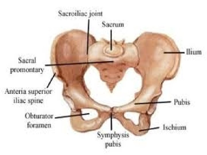
NON AXIAL q. Plane synovial joints. q. Articular surfaces are flat [plane] q. Permits gliding movements. Egs: ØIntercarpal joints. ØIntertarsal joints. ØCostovertebral joints. ØCostrotransverse joints. ØTarsometatarsal joints. ØIntermetatarsal joints. ØSacroiliac joints.
UNI AXIAL q. One degre freedom of movement. 1. Hinge joint [ginglymus]. §Articular surfaces are pulley shaped. §Bones united by strong collateral ligaments. §Moves around transverse axis. Egs: ØElbow ØAnkle ØInterphalangeal
2. Pivot[trochoid]. §Articular surfaces compromise central boney pivot surrounded by osseo -ligamentous ring. §Moves around vertical axis. Egs: ØSuperior and Inferior radio-ulnar joint. ØAtlanto-axial joint.
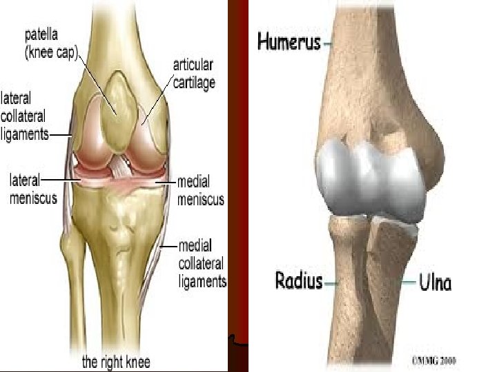
BI AXIAL q Two degree freedom of movements. 1. Condylar joint o Articular surfaces include two distinct condyles fiting into reciprocally concave surfaces. o Movement around transverse and vertical axis. Egs: Ø Knee joint. Ø TM joint.
2. Ellipsoid joint. o Articular surface oval and convex fitting into elliptical and concave surface. o Movements around transverse and antero posterior axis. Egs: Ø Wrist joint. Ø Radiocarpal joint. Ø Metacarpophalangeal joints. Ø Atlanto-Occipital joints.
Polyaxial joints. q. Three degree freedom of movement. 1. Saddle joint. • Articular surfaces reciprocally concavo-convex. • Movement occurs around transverse and AP axis with some rotation. Egs: Ø First carpometacarpal joint. Ø Sternoclavicular joint Ø Joint between femur and patella.
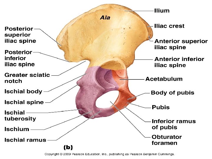
2. Ball and Socket joints. o. Articular surfaces include globular head fitting into cup shaped socket. o. Movement around transverse AP and vertical axis. Egs: ØShoulder joint ØHip joint.
JOINT POSITIONS. q. Close packed position of Joint surfaces completely congruent. q. Loose packed- All other positions of incongruency.
üBlood supply of joints. üNerve supply of joints
STABILITY OF SYNOVIAL JOINTS q. Muscles q. Ligaments q. Bones
FACTORS LIMITING THE RANGE OF MOVEMENT üShape of the articulating bones. üTension of ligaments. üTension of antagonistic muscles. üApproximation of soft parts.
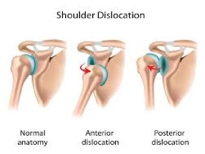
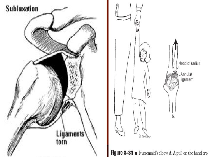
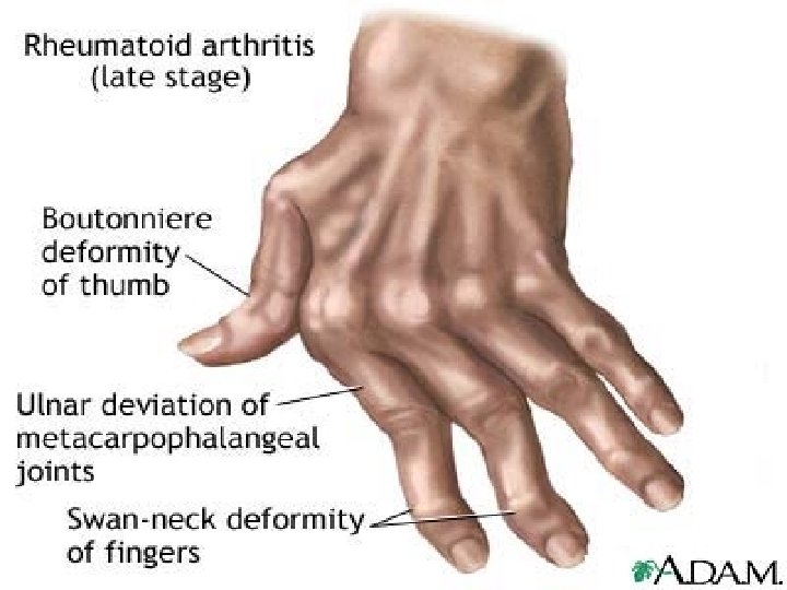
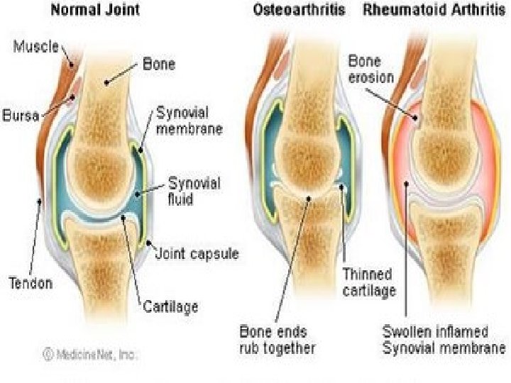
CLINICAL ANATOMY q q q q q PID Dislocation Subluxation Rheumatoid arthritis Osteoarthritis Spondylitis Stiffness of joint Neuropathic joints Joint replacement Sprains
PID
Cervical Spondylitis