Investigative Exam MD a The corneal topography shows


- Slides: 30
Investigative Exam MD
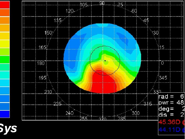
a. The corneal topography shows corneal steepening with downward displacement of the apex. b. Early keratoconus (the maximum power of cone is only 47. 50 D)
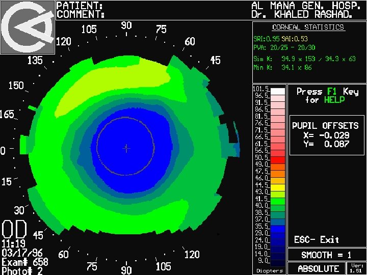
Post myopic ablation
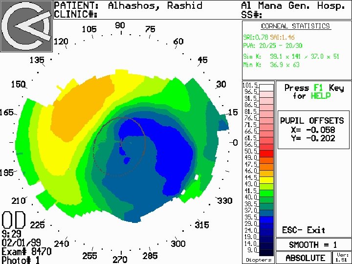
Decentered Abaltion
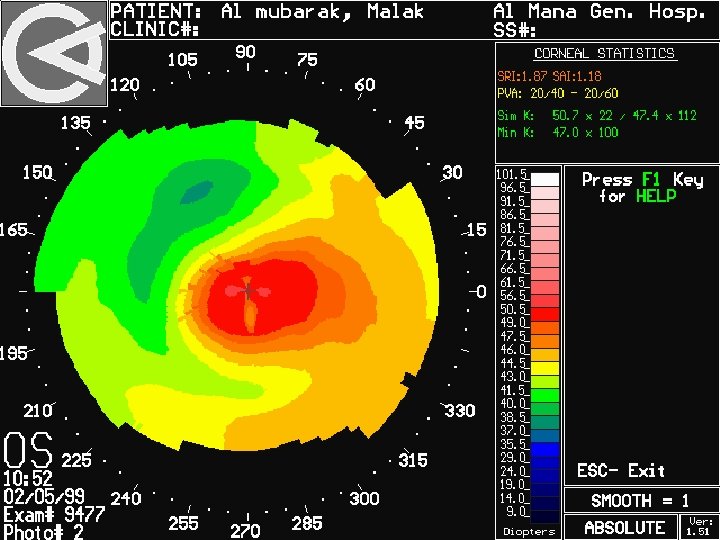
Post LASIK ectasia
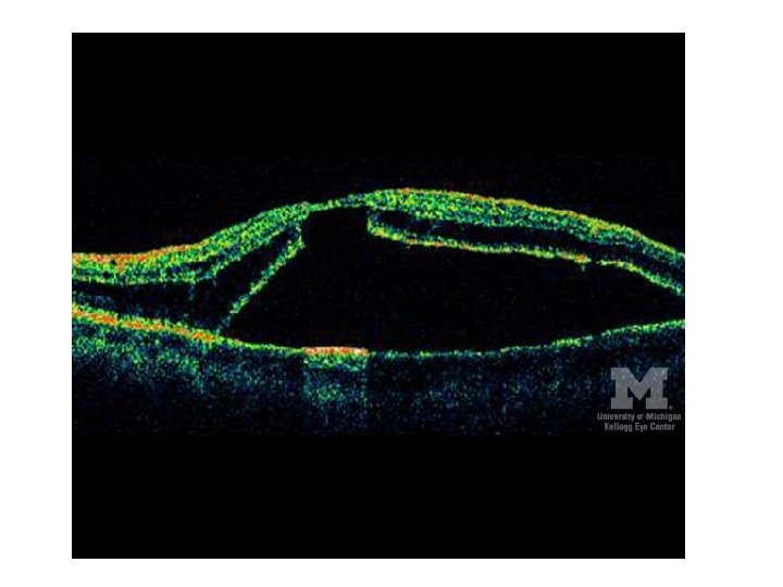
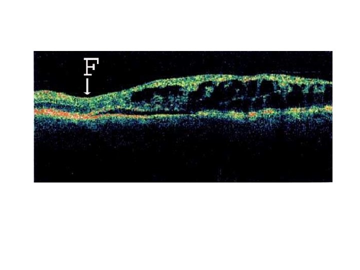
• Optical coherence tomography shows a neurosensory detachment involving the macula. • Note the schisis cavity overlying the neurosensory detachment as well as the outer layer hole.
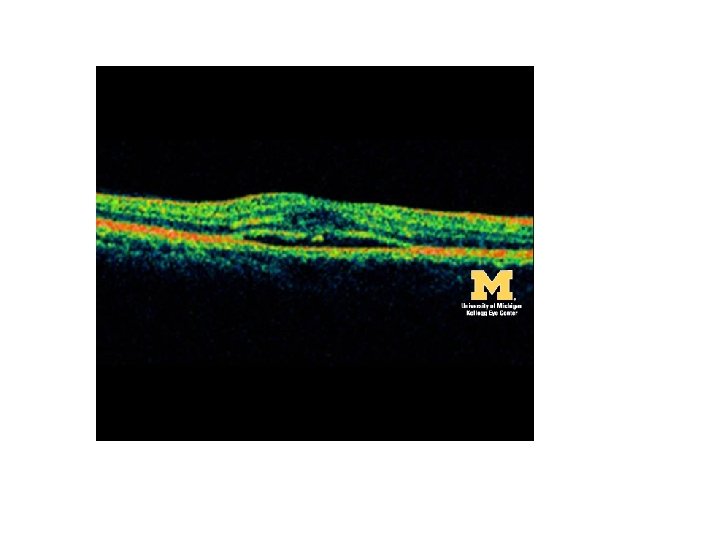
• Optical coherence tomography demonstrates a choroidal neovascular membrane and associated subretinal fluid
• Retinal pigment epithelium detachment
• Cystoid macular edema
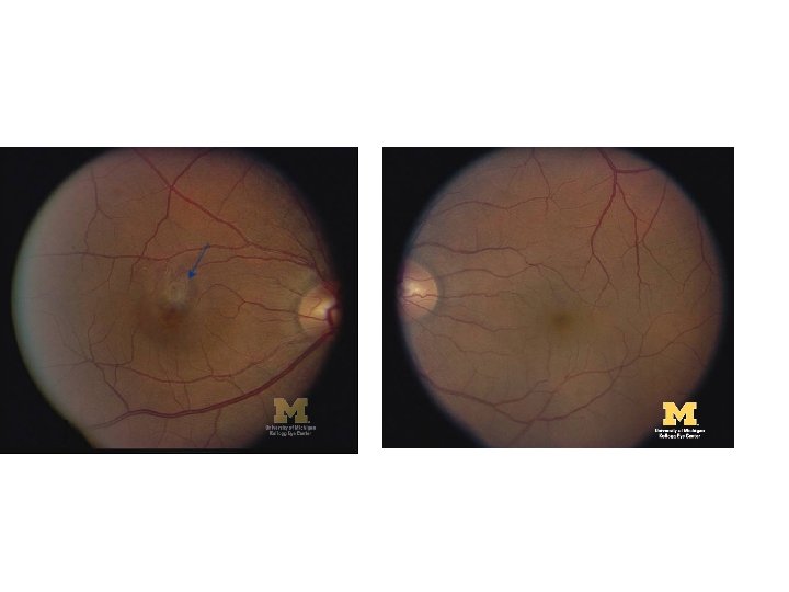
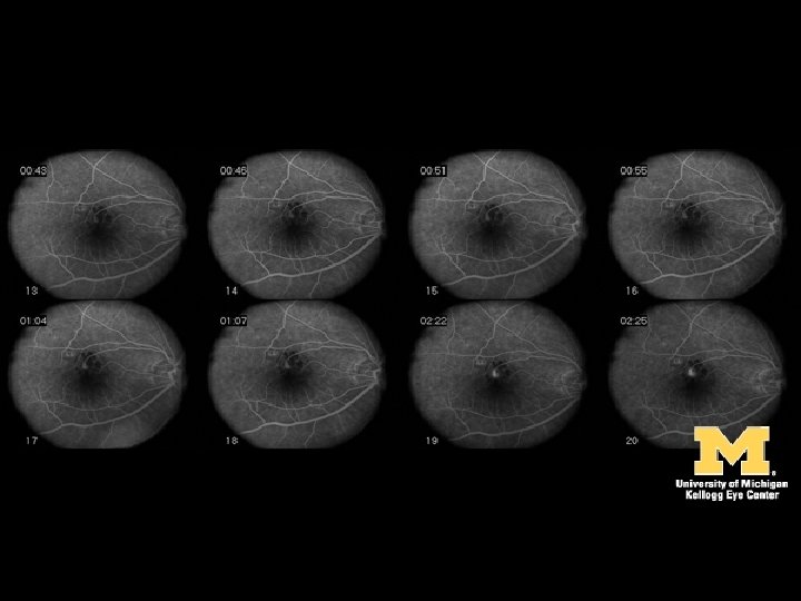
THE PICTURE AND FLUORESCEIN ANGIOGRAPHY ARE TAKEN FROM A PATIENT COMPLAINING OF DISTORTED RIGHT VISION. A. WHAT PHASE IS THE FLUORESCEIN ANGIOGRAPHY? B. WHAT IS THE DIAGNOSIS? C. WHAT UNDERLYING MEDICAL CONDITION
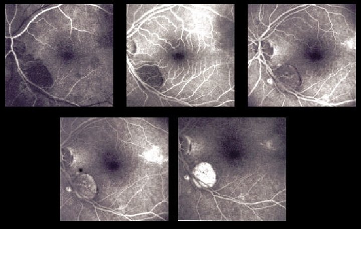
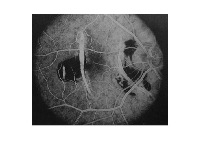
• Venous phase • Macroaneurysm • Hypertention
Choroidal rupture
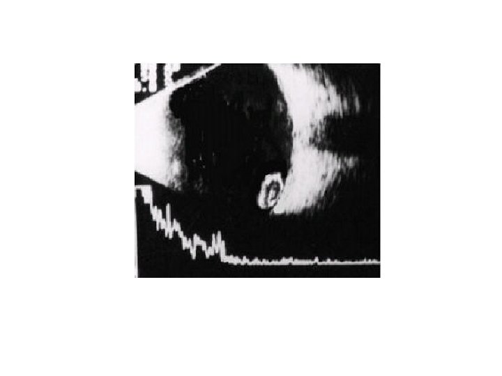
THE ABOVE B-SCAN WAS TAKEN FROM APATIENT WHO HAD A CATARACT SURGERY THE DAY BEFORE. A. WHAT IS THE DIAGNOSIS? B. SUGGEST TWO PREDISPOSING FACTORS.
• • • a. Expulsive suprachoroidal hemorrhage b. Multiple risk factors: Old age Hypertension Atherosclerosis Glaucoma or high preoperative IOP
THIS 70 YEAR-OLD MAN SUFFERED FROM PROGRESSIVE LEFT EXOPHTHALMOS. HIS CT SCAN IS SHOWN AS ABOVE. A. WHAT DOES THE CT SCAN SHOW? B. WHAT IS THE MOST LIKELY DIAGNOSIS?
a. Hyperostosis of the left lateral portion of the sphenoid with left proptosis. b. Sphenoid wing meningioma.
THE ABOVE MRI SCAN BELONGS TO A 3 YEAR-OLD GIRL. A. IN WHAT PLANE IS THIS SCAN TAKEN? B. IS THIS A T 1 OR T 2 WEIGHTED SCAN? C. WHAT IS THE MAIN ABNORMALITY SEEN? D. SUGGEST A POSSIBLE DIAGNOSIS?
Answer a. Axial scan of the orbit and brain b. T 1 (vitreous appears dark) C. Absence of the R. globe D. Enucleation of R. retinoblastoma