
4 2 2 3 01 2 1 R







- Slides: 22
4 2 2/ 3 01 2 / 1
R E D UN N L O I A T R A E IN EN D M M G A I X H E G E S J 4 2 2/ 3 01 2 / A H A N G A 2 0 1 3 - 1 3 9 1 D IR 2
ANESTHESIA IN CHILDHOOD PATIENT WITH GLAUCOMA. The mechanisms of glaucoma in childhood is often different from those seen in older patients. Clincian must be familiar with additional information for the evaluation and care of childhood glaucoma patients. 4 2 2/ 3 01 2 / 3
When enlargement of the cornea is associated with a deep anterior chamber glaucoma should be strongly suspected. The baby may be noticed to shy away from light or bury his head against his parent to prevent exposure to light. Clouding of the cornea is the most frequent physical abnormality. After age 4 years, glaucoma is most frequently detacted for decreased uncorrected visual acuity related to myopia. 4 2 2/ 3 01 2 / 4
4 2 2/ 3 01 2 / 5
THE OFFICE EVALUATION Before E. U. A the Office Evaluation of infants with suspected glaucoma is an important step in their evaluation. 4 2 2/ 3 01 2 / 6
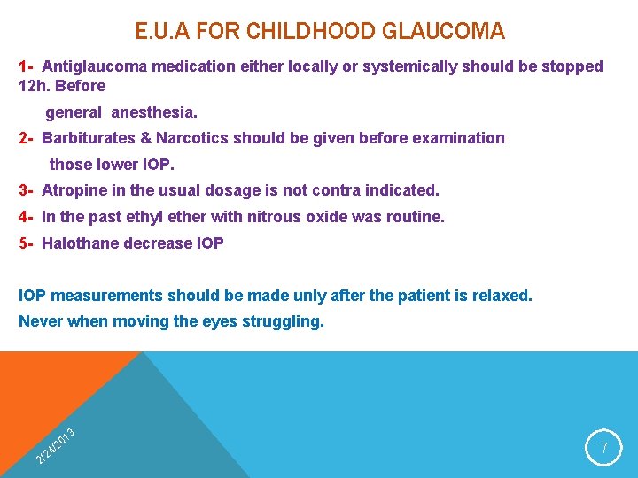
E. U. A FOR CHILDHOOD GLAUCOMA 1 - Antiglaucoma medication either locally or systemically should be stopped 12 h. Before general anesthesia. 2 - Barbiturates & Narcotics should be given before examination those lower IOP. 3 - Atropine in the usual dosage is not contra indicated. 4 - In the past ethyl ether with nitrous oxide was routine. 5 - Halothane decrease IOP measurements should be made unly after the patient is relaxed. Never when moving the eyes struggling. 4 2 2/ 3 01 2 / 7
5 STEPS IN E. U. A I. External examination II. Pressure measurements III. Corneal diameter measurements IV. Gonioscopy V. Ophthalmoscopy 4 2 2/ 3 01 2 / 8
4 2 2/ 3 01 2 / 9
EXTERNAL EXAMINATION 1. Evaluation of the corneal luster and clarity 2. Evaluation of the corneal epithelium 3. Evaluation of the Descemet’s m. not all leteaks in Descemet’s m. are caused by infantile glaucoma. (forceps injury at birth) 4 2 2/ 3 01 2 / 10
BREAKS DUE TO GLAUCOMA ARE: 1) Multiple 2) Curved 3) Horizontal 4 2 2/ 3 01 2 / 11
PRESSURE MEASUREMENTS 1) Tonometry is performed before Gonioscopy- IOP remain undisturbed 2) Methods of tonometry • Schiotz- indentation • Applanation – flatten the cornea • Goldman • Perkins applanation The eye should be still and in the primary position. 4 2 2/ 3 01 2 / 12
4 2 2/ 3 01 2 / 13
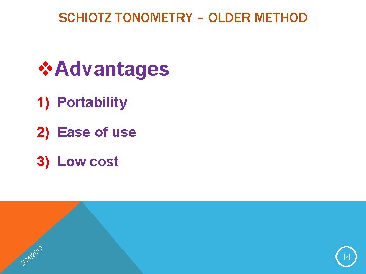
SCHIOTZ TONOMETRY – OLDER METHOD v. Advantages 1) Portability 2) Ease of use 3) Low cost 4 2 2/ 3 01 2 / 14
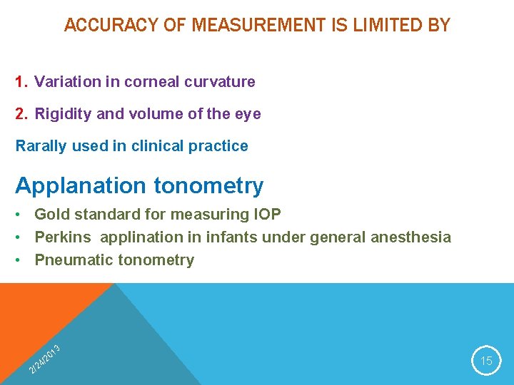
ACCURACY OF MEASUREMENT IS LIMITED BY 1. Variation in corneal curvature 2. Rigidity and volume of the eye Rarally used in clinical practice Applanation tonometry • Gold standard for measuring IOP • Perkins applination in infants under general anesthesia • Pneumatic tonometry 4 2 2/ 3 01 2 / 15
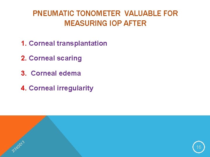
PNEUMATIC TONOMETER VALUABLE FOR MEASURING IOP AFTER 1. Corneal transplantation 2. Corneal scaring 3. Corneal edema 4. Corneal irregularity 4 2 2/ 3 01 2 / 16
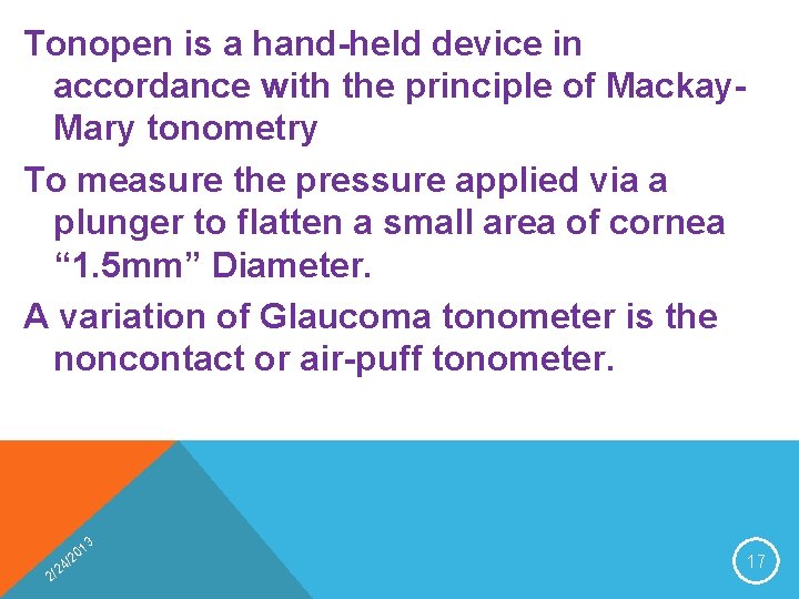
Tonopen is a hand-held device in accordance with the principle of Mackay. Mary tonometry To measure the pressure applied via a plunger to flatten a small area of cornea “ 1. 5 mm” Diameter. A variation of Glaucoma tonometer is the noncontact or air-puff tonometer. 4 2 2/ 3 01 2 / 17
4 2 2/ 3 01 2 / 18
CORNEAL DIAMETER MEASUREMENT v To measure the diametric distance from nasal limbus to temporal limbus. v Evaluating the severity and control of glaucoma in infants and children less than 2 years of age. v Normal range of corneal diameter in infants is between 9. 5 -10 mm increasing to 11. 5 -12 mm at one year of age and as much as 12. 50 in the first 3 years. v Under one year of age diameters of 12 -12. 50 mm are suggestive of abnormal enlargement. v 13 mm or more at any time in childhood is strongly suggestive of abnormality 3 2 2/ 1 20 / 4 19
GONIOSCOPY If the cornea is clear, Gonioscopy is performed by using koeppe Gonioscopy lenses of proper size and a hand-held binocular microscope Gonioscopy findings in infants and young children differ significantly from those of adults. 4 2 2/ 3 01 2 / 20
OPHTHALMOSCOPY Direct ophthalmoscopy is the best 1) For infants & children 2) Optic nerve head evaluation is crucial in ophthalmoscopy 3) Cupping of the nerve head from glaucoma in infants and children appears similar to that in adults. 4) Myopia is a common accompaniment of enlargement of the eye in infantile glaucoma. 4 2 2/ 3 01 2 / 21
THE END 4 2 2/ 3 01 2 / 22