Imaging in Hemangioma and Vascular Malformations Most superficial


 then, MRI with gadolinium")
 other method is")


 then,")


- Slides: 37
Imaging in Hemangioma and Vascular Malformations
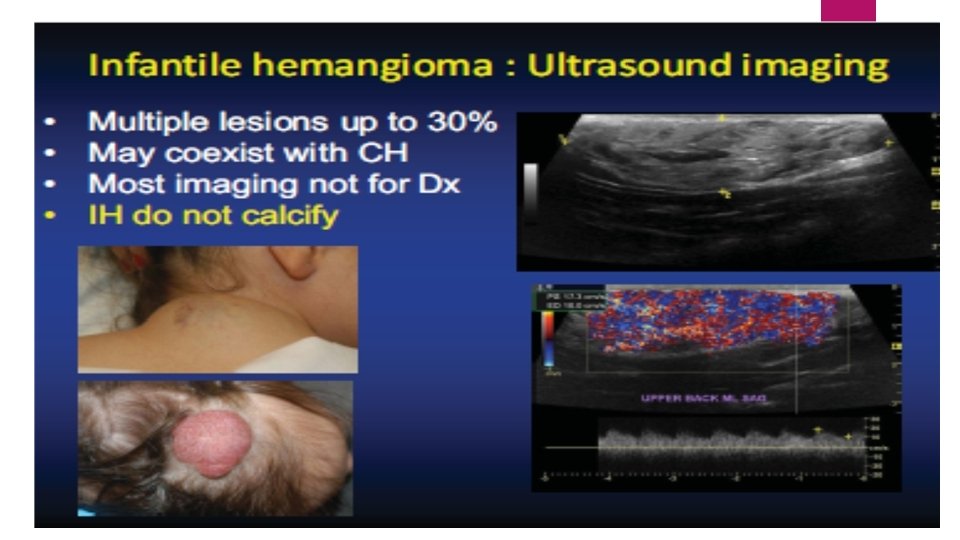
Most superficial and mixed infantile hemangiomas : diagnosed based upon their clinical features. imaging may be used for defining the extent and depth of the deep component of mixed lesions. Radiologic evaluation is particularly helpful for confirming the diagnosis of deep infantile hemangiomas that lack superficial changes.
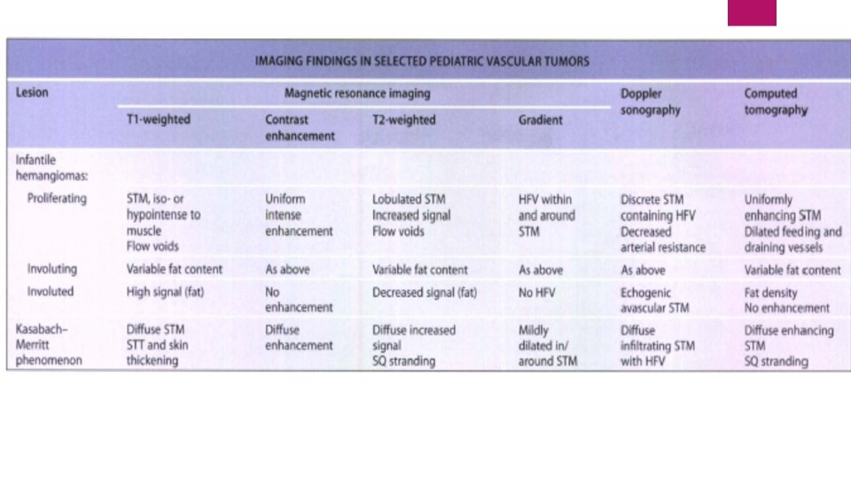
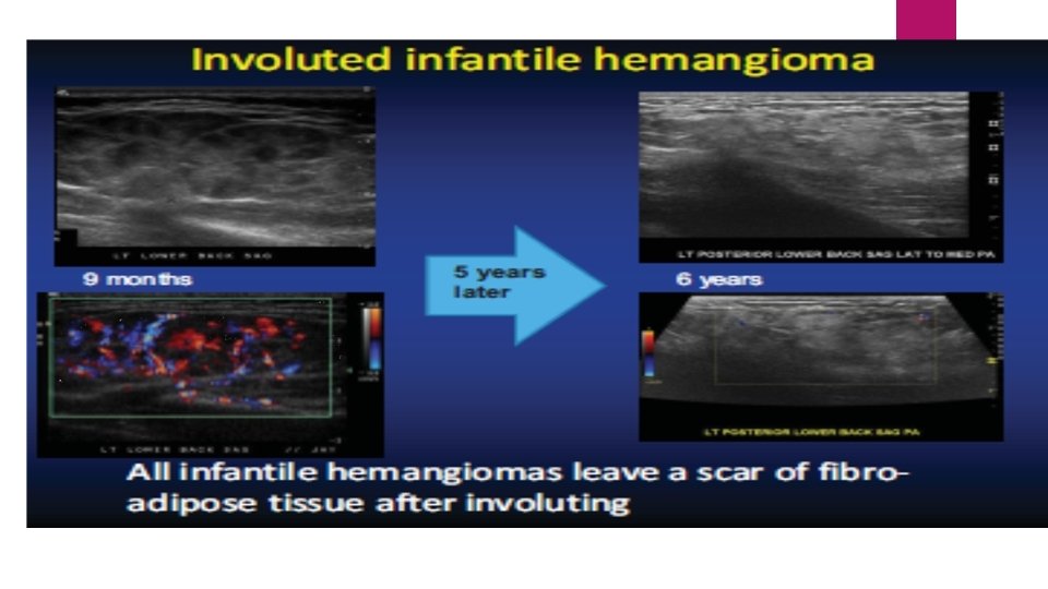
It is important to consider the phase of growth when assessing hemangiomas radiographically proliferating hemangiomas demonstrate different characteristics than their involuting and involuted counterparts.
Which technique is the most useful for helping to define the extent and tissue characteristics of vascular tumors? MRI
use of contrast can help to differentiate an infantile hemangioma from other tumors. If MRI does not confirm the diagnosis of infantile hemangioma or there are concerns regarding a possible malignancy histologic examination of the tumor is recommended.
Look for the presence of hemangiomas within internal organs and to detect other structural anomalies, including CNS malformations and arterial anomalies? MRI and/or MR angiography(MRA)
Imaging Characteristics of RICH: can be recognized by prenatal ultrasonography during the second and third trimester misdiagnosis as other vascular anomalies such as lymphatic or arteriovenous malformations is common Postnatal ultrasonographic evaluation of a RICH reveals uniformly hypoechoic lesions mostly confined to the subcutaneous fat. Diffuse vascularity is typical, with vessels showing venous or low-resistance arterial flow
NICH Doppler evaluation : fastflow vessels MRI: features similar to infantile hemangiomas
Kasabach. Merritt phenomenon MRI Findings: diffuse soft tissue masses that enhance after contrast administration Ill-defined margins involvement of multiple tissue planes overlying cutaneous thickening edema
Vascular Malformations
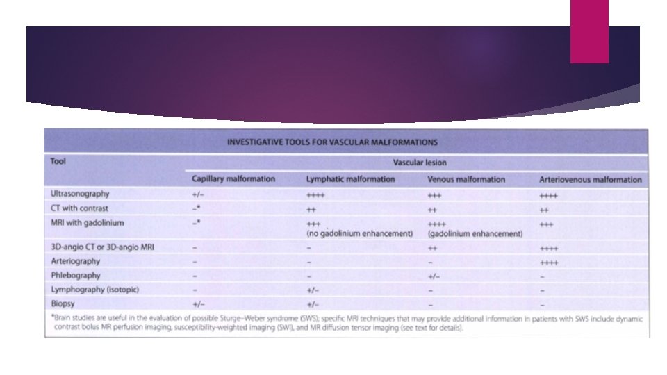
>90% of vascular malformations can be correctly categorized on the basis of their clinical features This helps in the selection of the best investigative tools avoiding redundant and unnecessary diagnostic imaging procedures.
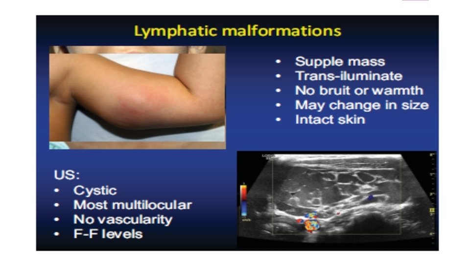
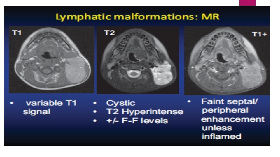
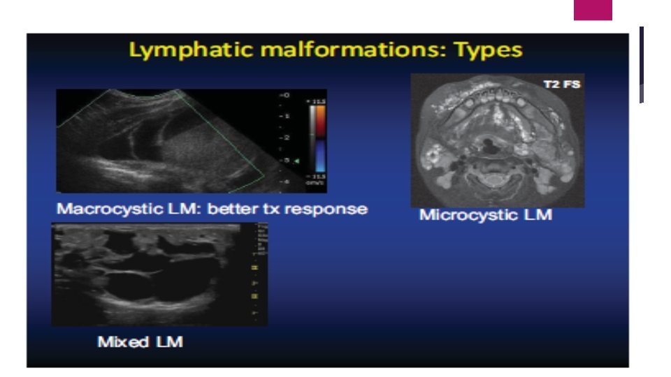
For Lymphatic Malformation: The Best option is: Sonography (4+) then, MRI with gadolinium (3+)
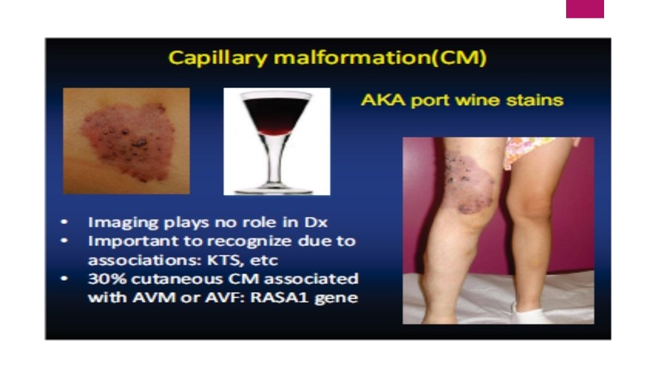
Capillary malformation Ultrasound No and biopsy are of little help (1+) other method is useful (0)
PWS in the V 1 or V 1 + other sites Examination: neurologic, ophthalmologic MRI with gadolinium favored over CT Consider SPECT or PET scan
Port-wine stain with any bluish hue, bluish mass or swelling in proclive position in the midforhead or cheek MRI + MRA + CT SCAN: DIAGNOSIS ASSESS EXTENT AND DEPTH OF INVOLVEMENT
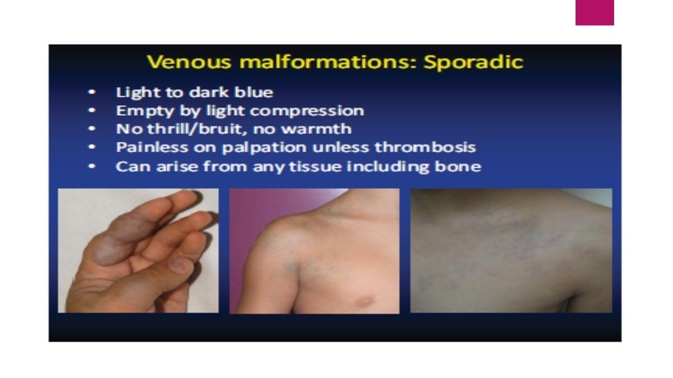
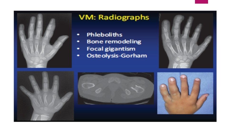
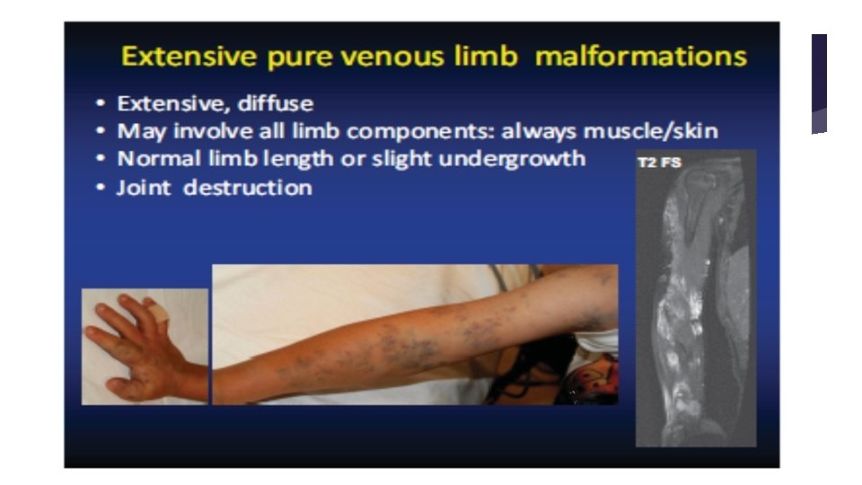
For Venous Malformation: The Best option is MRI with gadolinium : (4+) then, Sonography (3+) then CT Angio or MRI Angio or CT with contrast
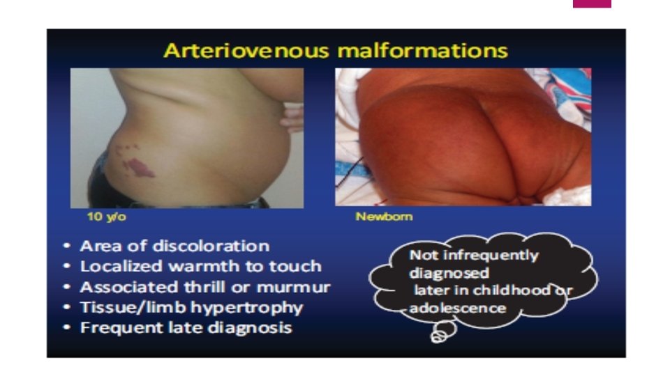
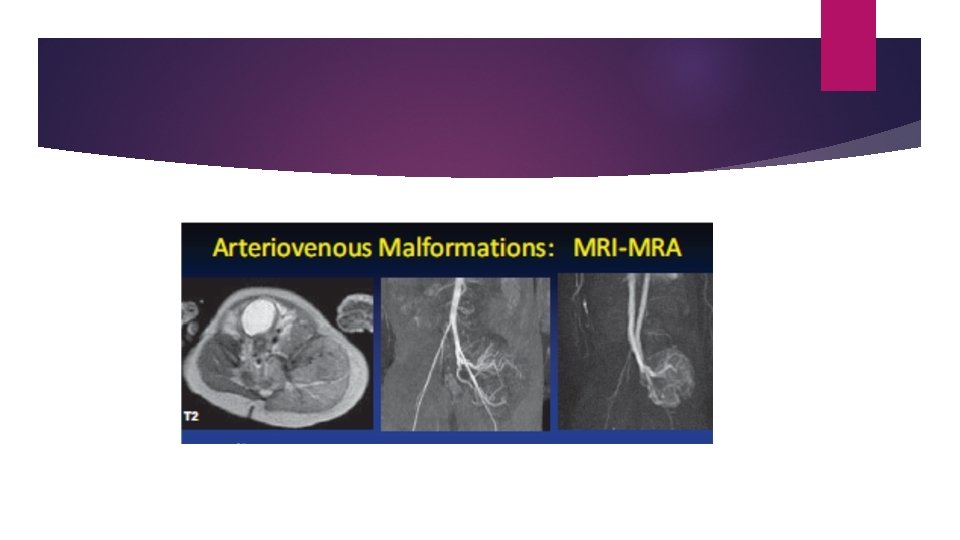
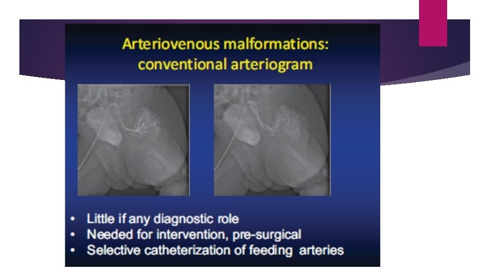
For Arterio-Venous Malformation: The Best options are Sonography, CT Angio or MRI Angio or arteriography (4+) then, MRI with gadolinium (3+)
Arteriovenous malformation
Final Points: