
Noninvasive detection of coronary inflammation by computed tomography
Non-invasive detection of coronary inflammation by computed tomography analysis of peri-coronary fat enhances cardiovascular risk prediction in 3912 individuals The CRISP-CT study Evangelos K. Oikonomou 1, Mohamed Marwan 2, Milind Y. Desai 3, Jennifer Mancio 1, Alaa Alashi 3, Erica Hutt Centeno 3, Cheerag Shirodaria 4, 5, John Deanfield 6, Stefan Neubauer 1, Keith M. Channon 1, Stephan Achenbach 2, Charalambos Antoniades 1 1 Division of Cardiovascular Medicine, Radcliffe Department of Medicine, University of Oxford, UK. of Cardiology, Friedrich-Alexander-Universität Erlangen-Nürnberg, Erlangen, Germany. 3 Cleveland Clinic Heart and Vascular Institute, Cleveland, USA. 4 Caristo Diagnostics Ltd. , Oxford, UK. 5 Oxford University Hospitals NHS Foundation Trust, Oxford, U. K. 6 UCL Institute of Cardiovascular Science, London, UK. 2 Department

Declaration of interest - Consulting/Royalties/Owner/ Stockholder of a healthcare company (Founder, shareholder and Chief Scientific Officer of Caristo Diagnostics) ESC Congress Munich 2018 •
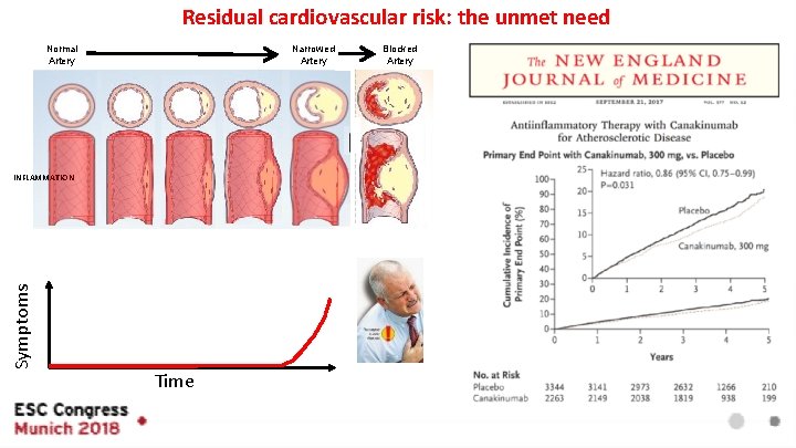
Residual cardiovascular risk: the unmet need Normal Artery Narrowed Artery Blocked Artery Symptoms INFLAMMATION Time Fishbein et al. Circulation. 1996; 94: 2662 -2666
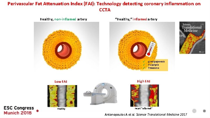
Perivascular Fat Attenuation Index (FAI): Technology detecting coronary inflammation on CCTA “Healthy, ” inflamed artery Healthy, non-inflamed artery ✓ ↓ Adipogenesis ✓ ↑Lipolysis ✓ ↑Oedema High FAI Low FAI Water Fat Healthy Water Fat Heart “inflamed” Antonopoulos A et al. Science Translational Medicine 2017
Can FAI predict cardiovascular risk?
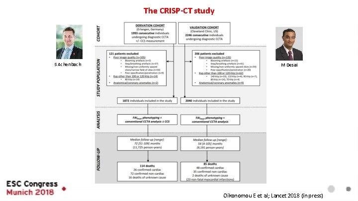
The CRISP-CT study S Achenbach M Desai Oikonomou E et al; Lancet 2018 (in press)
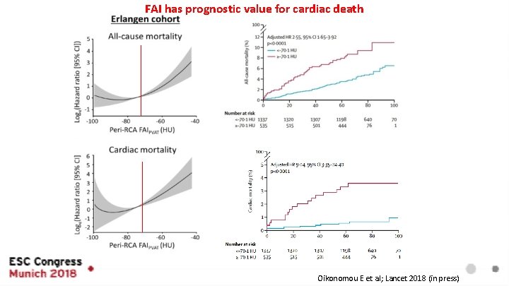
FAI has prognostic value for cardiac death Oikonomou E et al; Lancet 2018 (in press)
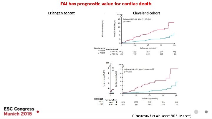
FAI has prognostic value for cardiac death Oikonomou E et al; Lancet 2018 (in press)
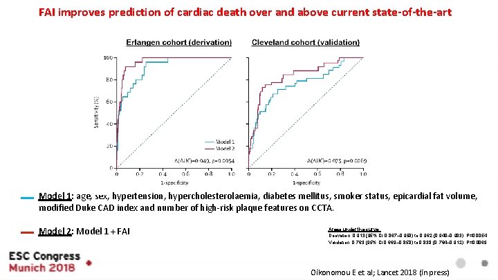
FAI improves prediction of cardiac death over and above current state-of-the-art Model 1: age, sex, hypertension, hypercholesterolaemia, diabetes mellitus, smoker status, epicardial fat volume, modified Duke CAD index and number of high-risk plaque features on CCTA. Model 2: Model 1 + FAI Areas under the curve: Derivation: 0· 913 (95% CI 0· 867– 0· 958) to 0· 962 (0· 940– 0· 983), P=0. 0054 Validation: 0· 763 (95% CI 0· 669– 0· 858) to 0· 838 (0· 764– 0· 912), P=0. 0069 Oikonomou E et al; Lancet 2018 (in press)
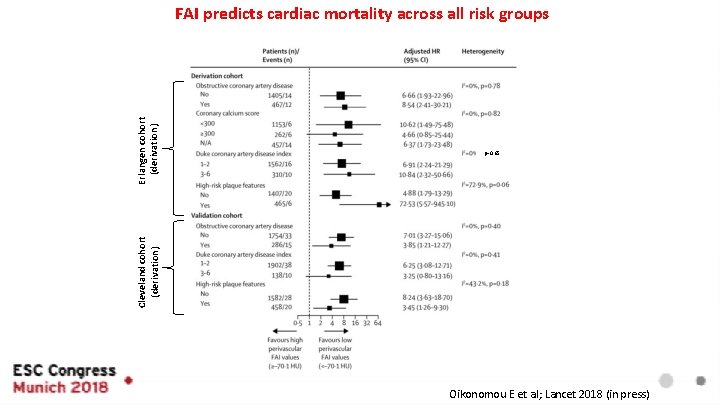
p=0. 65 Cleveland cohort (derivation) Erlangen cohort (derivation) FAI predicts cardiac mortality across all risk groups Oikonomou E et al; Lancet 2018 (in press)
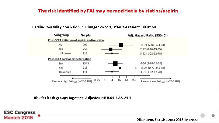
The risk identified by FAI may be modifiable by statins/aspirin Cardiac mortality prediction in Erlangen cohort, after treatment initiation Risk for both groups together: Adjusted HR 9. 04[3. 35 -24. 4] Oikonomou E et al; Lancet 2018 (in press)
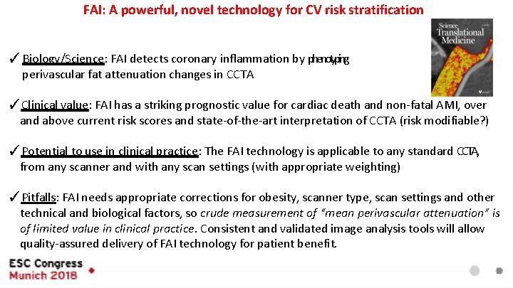
FAI: A powerful, novel technology for CV risk stratification ✓ Biology/Science: FAI detects coronary inflammation by phenotyping perivascular fat attenuation changes in CCTA ✓Clinical value: FAI has a striking prognostic value for cardiac death and non-fatal AMI, over and above current risk scores and state-of-the-art interpretation of CCTA (risk modifiable? ) ✓Potential to use in clinical practice: The FAI technology is applicable to any standard CCTA, from any scanner and with any scan settings (with appropriate weighting) ✓Pitfalls: FAI needs appropriate corrections for obesity, scanner type, scan settings and other technical and biological factors, so crude measurement of “mean perivascular attenuation” is of limited value in clinical practice. Consistent and validated image analysis tools will allow quality-assured delivery of FAI technology for patient benefit.
Acknowledgments E-mail: Charalambos. antoniades@cardiov. ox. ac. uk THE LANCET: Released 28/8/18, 14: 30 BST
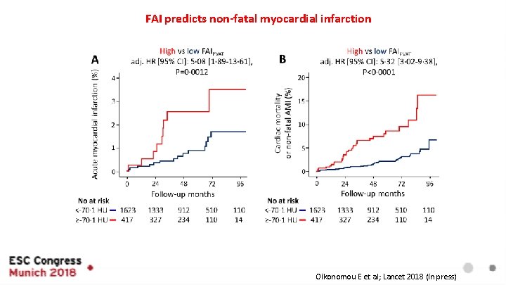
FAI predicts non-fatal myocardial infarction Oikonomou E et al; Lancet 2018 (in press)
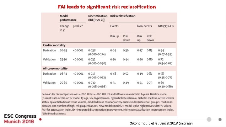
FAI leads to significant risk reclassification Oikonomou E et al; Lancet 2018 (in press)
- Slides: 15