Histology Lymphoid system Lymphoid System Collects excess interstitial


 are specialized post-capillary venous swellings characterized by plump endothelial cells")


) are structures found in the medulla of the")

 Stained with haematoxylin and eosin lymphoid follicle is circled with dotted line")

![Palatine tonsils occasionally called the faucial tonsils, [2] are the tonsils that can be](https://slidetodoc.com/presentation_image_h/d3f0aa4c9c97552c67b56b9d21420d5a/image-25.jpg "Palatine tonsils occasionally called the faucial tonsils, [2] are the tonsils that can be")

DEFINE THE FUNCTION OF T LYMPHOCYTES 2)DEFINE AND THE FUNCTION OF B")
- Slides: 28
Histology Lymphoid system
Lymphoid System *Collects excess interstitial. *Protects organism against invading pathogens or antigens by producing immune responses *Includes all cells, tissues and organs that contain lymphocytes *Major organs are lymph nodes , spleen, thymus, and tonsils
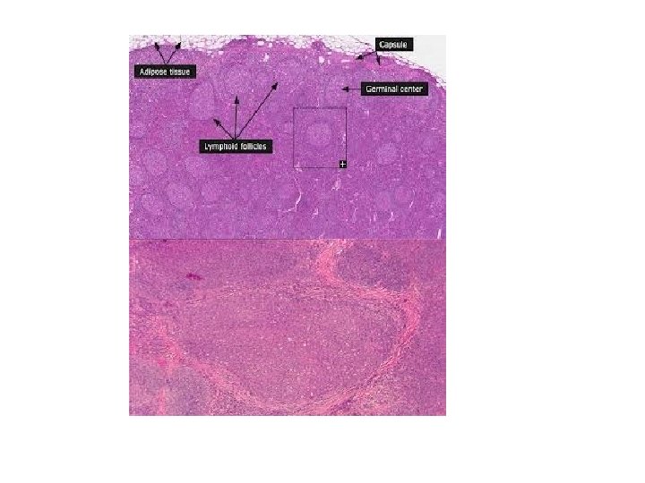
The lymphatic system is part of the circulatory system, comprising a network of conduits called lymphatic vessels that carry a clear fluid called lymph. directionally towards the heart. Lymph nodes are garrisons of B, T, and other immune cells. Lymph nodes act as filters or traps foreign particles and are important in the proper functioning of the immune system. They are packed tightly with the white blood cells called lymphocytes and macrophages. Structure The lymph node is surrounded by a fibrous capsule, and inside the lymph node the fibrous capsule extends to form trabeculae. The substance of the lymph node is divided into the outer cortex and the inner medulla surrounded by the cortex all around except for at the hilum, where the medulla comes in direct contact with the surface.
The liver LYMPH NODE Stained with haematoxylin and eosin 1 - cortex 2 - paracortical zone 3 - medulla 4 - medullary cords 5 - lymphoid follicle of the cortex 6 - capsule
The number and composition of follicles can change especially when challenged by an antigen, when they develop a germinal center. A lymph sinus is a channel within the lymph node lined by the endothelial cells along with fibroblastic reticular cells and allows for smooth flow of lymph through them. Thus, subcapsular sinus is a sinus immediately deep to the capsule, and its endothelium is continuous with that of the afferent lymph vessel. It is also continuous with similar sinuses flanking the trabeculae and within the cortex (cortical sinuses). The cortical sinuses and that flanking the trabeculae drain into the medullary sinuses, from where the lymph flows into the efferent lymph vessel.
Multiple afferent lymph vessels that branch and network extensively within the capsule bring lymph into the lymph node. This lymph enters the subcapsular sinus. The lymph gets slowly filtered through the substance of the lymph node and ultimately reaches the medulla. In its course it encounters the lymphocytes and may lead to their activation as a part of adaptive immune response. The concave side of the lymph node is called the hilum. The efferent attaches to the hilum by a relatively dense reticulum present there, and carries the lymph out of the lymph node.
Medulla There are two named structures in the medulla: The medullary cords are cords of lymphatic tissue, and include plasma cells, macrophages, and B cells The medullary sinuses (or sinusoids) are vessel-like spaces separating the medullary cords. Lymph flows into the medullary sinuses from cortical sinuses, and into efferent lymphatic vessels. Medullary sinuses contain histiocytes (immobile macrophages) and reticular cells. The medulla contains large blood vessels, sinuses and medullary cords that contain plasma cells secreting antibody
1. Dense connective tissue capsule 2. Dense connective tissue trabeculae 3. Cortex Je 3 a. Cortical nodules with germinal centers Je 3 b. Paracortex 4. Medulla j. Je 4 a. Medullary cords j. Je 4 b. Medullary sinus 5. Subcapsular sinus
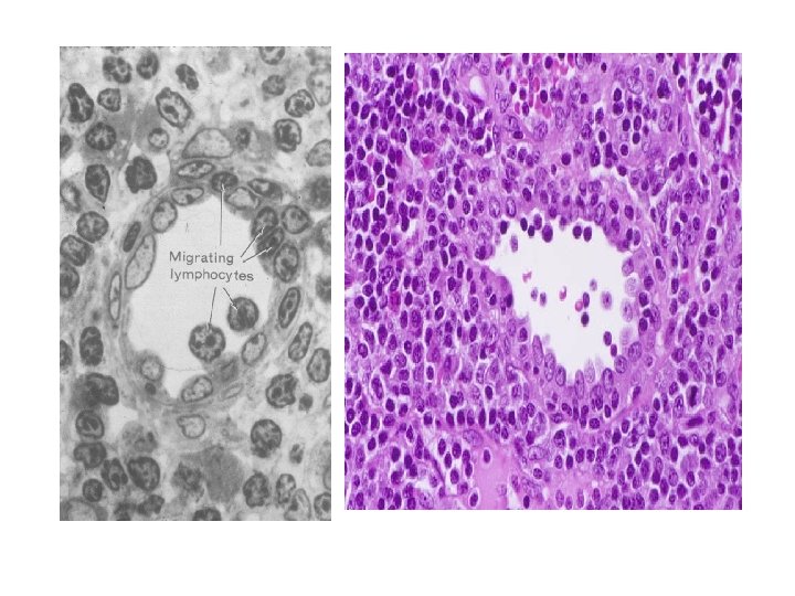
High endothelial venules (HEV) are specialized post-capillary venous swellings characterized by plump endothelial cells as opposed to the usual thinner endothelial cells found in regular venules. HEVs enable lymphocytes circulating in the blood to directly enter a lymph node (by crossing through the HEV.
Thymus gland anterior view of chest showing location and size of adult thymus
The thymus is a specialized organ of the immune system. The thymus "educates" T-lymphocytes (T cells), which are critical cells of the adaptive immune system. Each T cell attacks a foreign substance which it identifies with its receptor. Each T cell attacks a different antigen. T cells that attack the body's own proteins are eliminated in the thymus. Thymic epithelial cells express major proteins from elsewhere in the body, and T cells that respond to those proteins are eliminated through programmed cell death (apoptosis).
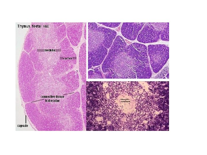
The thymus is composed of two identical lobes and is located anatomically in the anterior superior mediastinum, in front of the heart and behind the sternum. Histologically, the thymus can be divided into a central medulla and a peripheral cortex which is surrounded by an outer capsule. The cortex and medulla play different roles in the development of T-cells. Cells in the thymus can be divided into thymic stromal cells and cells of hematopoietic origin (derived from bone marrow resident hematopoietic stem cells). Developing T-cells are referred to as thymocytes and are of hematopoietic origin. Stromal cells include thymic cortical epithelial cells, thymic medullary epithelial cells, and dendritic cells.
Structure Each lateral lobe is composed of numerous lobules held together by delicate areolar tissue; the entire organ being enclosed in an investing capsule[12] of a similar but denser structure. The primary lobules vary in size from that of a pin's head to that of a small pea, and are made up of a number of small nodules or follicles. Cortex The cortical portion is mainly composed of lymphoid cells, supported by a network of finely-branched epithelial reticular cells, which is continuous with a similar network in the medullary portion. This network forms an adventitia to the blood vessels. The cortex is the location of the earliest events in thymocyte development, where T cell receptor gene rearrangement and positive selection takes place
Medulla In the medullary portion, the reticulum is coarser than in the cortex, the lymphoid cells are relatively fewer in number, and there are found peculiar nest-like bodies, the concentric corpuscles of Hassall. These concentric corpuscles are composed of a central mass, consisting of one or more granular cells, and of a capsule formed of epithelioid cells. They are the remains of the epithelial tubes, which grow out from the third branchial pouches of the embryo to form the thymus. Each follicle is surrounded by a vascular plexus, from which vessels pass into the interior, and radiate from the periphery toward the center, forming a second zone just within the margin of the medullary portion. In the center of the medullary portion there are very few vessels, and they are of minute size.
Hassall's corpuscles (or thymic corpuscles (bodies)) are structures found in the medulla of the human thymus, formed from eosinophilic type VI epithelial reticular cells arranged concentrically.
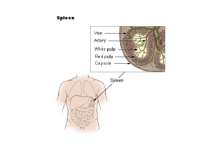
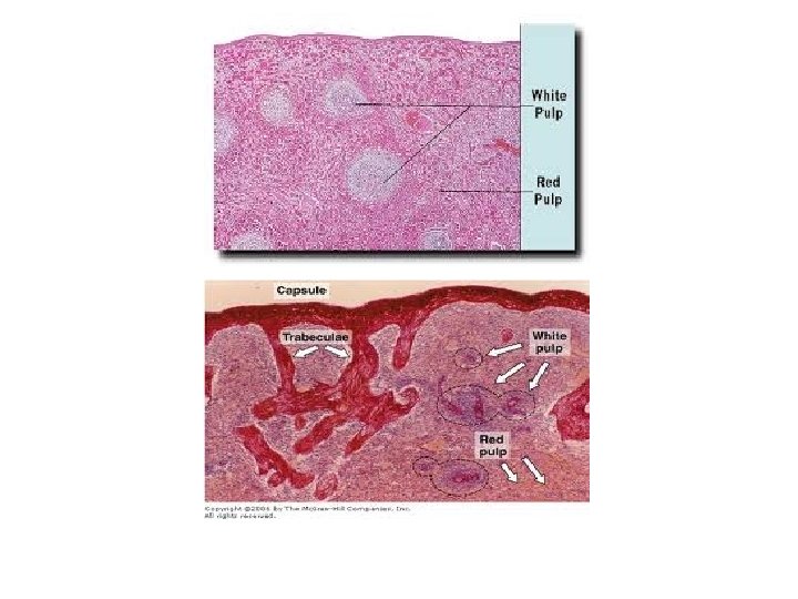
Spleen is an organ found in virtually all vertebrate animals. Similar in structure to a large lymph node, the spleen acts primarily as a blood filter. As such, it is a non-vital organ, with life possible after removal. The spleen plays important roles in regard to red blood cells (also referred to as erythrocytes) and the immune system. In humans, it is located in the left upper quadrant of the abdomen. It removes old red blood cells and holds a reserve of blood in case of hemorrhagic shock while also recycling iron. As a part of the mononuclear phagocyte system, it metabolizes hemoglobin removed from senescent erythrocytes. The globin portion of hemoglobin is degraded to its constitutive amino acids, and the heme portion is metabolized to bilirubin, which is subsequently shuttled to the liver for removal. It synthesizes antibodies in its white pulp and removes antibody-coated bacteria along with antibody-coated blood cells by way of blood and lymph node circulation. The spleen is brownish in color.
SPLEEN (follicle) Stained with haematoxylin and eosin lymphoid follicle is circled with dotted line 1 - germinal center of the follicle 2 - mantie zone of the follicle 3 - marginal zone of the follicle 4 - periarterial area of the follicle 5 - central arteriole 6 - red pulp 7 - trabeculae
The red pulp of the spleen is composed of connective tissue known as the cords of Billroth and many splenic sinuses that are engorged with blood, giving it a red color. Its primary function is to filter the blood of antigens, microorganisms, and defective or worn-out red blood cells. The spleen is made of red pulp and white pulp, separated by the marginal zone; 76 -79% of a normal spleen is red pulp. Unlike white pulp, which mainly contains lymphocytes such as T cells, red pulp is made up of several different types of blood cells, including platelets, granulocytes, red blood cells, and plasma.
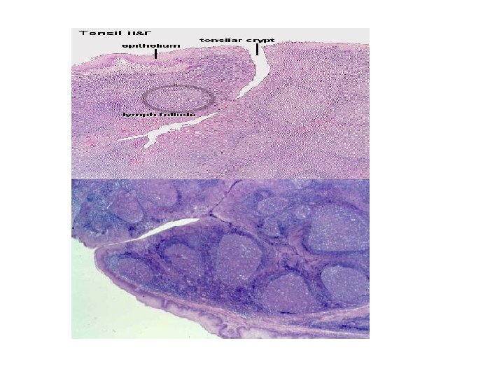
Palatine tonsils occasionally called the faucial tonsils, [2] are the tonsils that can be seen on the left and right sides at the back of the throat. Tonsillitis is an inflammation of the tonsils and will often, but not necessarily, cause a sore throat and fever. In chronic cases tonsillectomy may be indicated. Palatine tonsils consist of approximately 15 crypts, which result in a large internal surface. The tonsils contain four lymphoid compartments that influence immune functions, namely the reticular crypt epithelium, the extrafollicular area, the mantle zones of lymphoid follicles, and the follicular germinal centers. In human palatine tonsils, the very first part exposed to the outside environment is tonsillar epithelium.
PALATINE TONSIL Stained with haematoxylin and eosin 1 - lymphoid follicle 2 - diffuse lymphoid tissue 3 - crypt 4 - epithelium of the oral cavity musosa 6 - submucosa of the inner cover of oral cavity forms hemi-capsule of the tonsil
Home work 1)DEFINE THE FUNCTION OF T LYMPHOCYTES 2)DEFINE AND THE FUNCTION OF B LYPHOCYTES 3)TYPES OF IMMUNE RESPONCES