Histology of Lymphoid Organs Immune system The immune


 lymphoid organ: Where lymphatic cells are formed and matured. –")


- Slides: 43
Histology of Lymphoid Organs
Immune system • The immune system is the body's defense against infectious organisms, organs include thymus, bone marrow, lymph nodes, spleen and palatine tonsils. • The first line of defence consists of skin and mucosal linings of alimentary, respiratory and urogenital system. • Types of immune system : Innate immune system Adaptive immune system
Innate Immune System • • • Natural or nonspecific immune system Second line defence against harmful agents Responds is rapid within few hours Has no immunological memory Consists of proteins, peptides, cytokines, phagocytic cells and natural killer cells.
Adaptive Immune System • • • Specific immune system Third line of defence Responds slower than innate immune system Has immunological memory Consists of B lymphocytes , T lymphocytes and antigen presenting cells.
Antigen is a harmful substance which enters the body which causes the body to make antibodies as a response to fight off disease. OR A substance that is capable of stimulating an immune response, specifically activating lymphocytes. Antibody also known as an immunoglobulin (Ig), is a large, Yshaped protein produced mainly by plasma cells that is used by the immune system to neutralize pathogens such as bacteria , viruses and other toxic substance.
Lymphoid Organs Central (primary) lymphoid organ: Where lymphatic cells are formed and matured. – T cells in thymus – B cells in bone marrow Peripheral (secondary) lymphoid organ: Whenever there is an antigenic stimulation, their lymphocytes increase in number and take part in the immune response against the antigen. Including lymph nodes, spleen and tonsils.
Lymph Nodes
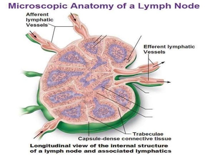
• Entire node is bean shaped • The concavity constituting a hilum through which blood vessels enter and leave the node • Several lymph vessels enter the node on its convex aspect • Usually a single lymph vessel leaves the node through its hilum.
• Each lymph node consists of – Capsule connective tissue framework densely packed collagen fibers – Parenchyma Outer darkly stained Cortex Inner lightly stained Medulla
The connective tissue framework The lymph node is surrounded by a pericapsular adipose tissue that contains arterioles and venules. The lymph node is covered by a capsule consisting mainly of collagen fibres. Some elastic fibres and some smooth muscles may be present.
A number of septa/trabeculae extend into the node from the capsule and divide the node into the lobules The hilum is occupied by a mass of dense fibrous tissue
Lymph node
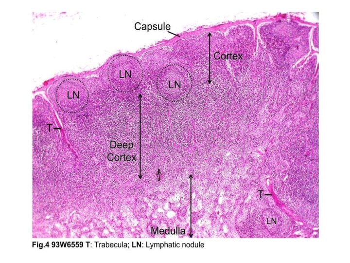
• On section, – The node has an outer zone containing densely packed lymphocytes, stains darkly • Cortex does not extend into the hilum
• Surrounded by the cortex, there is a lighter staining zone in which lymphocytes are fewer • Medulla
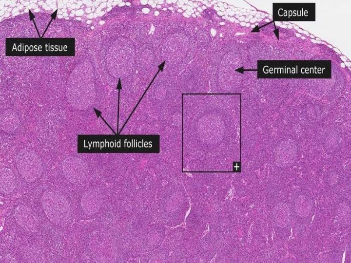
• Within the cortex The cortex can further divided into two regions Outer region contains lymphatic nodules known as nodular cortex Inner region is free of lymphatic nodules called paracortex or deep cortex The cortex contains numerous lymphocyte aggregations called lymphatic follicles or lymphatic nodules. Each nodule has a paler staining germinal centre surrounded by a zone of densely packed lymphocytes.
Lymph node
• Within the medulla The lymphocytes are arranged in the form of branching and anastomosing cords known as medullary cords containing B lymphocytes, plasma cells and macrophages. The medullary cords interspersed with medullary sinuses that drain lymph from the node into the efferent vessels at the hilus.
Lymph node
Medullary cord and sinus
• Central lymphoid organ • Present in the superior and anterior mediastinum of thorax
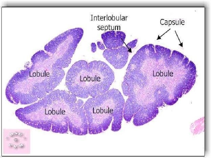
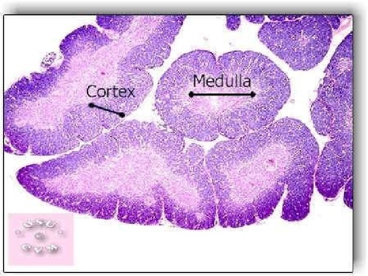
It is bilobed organ Each lobe has a connective tissue capsule Connective tissue septa passes inward Subdivide the lobe Lobule has an outer cortex & inner medulla
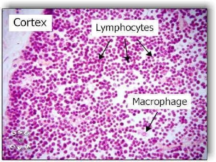
Thymic Cortex In the cortex of each lobule the epithelioreticulur cells is densely packed with T lymphocytes also known as thymocytes. Lymphatic nodules are not present in the normal thymus. There are three types of epithelioreticulur cells present in cortex. Type I epithelioreticulur cell (located in outer cortex) Type II epithelioreticulur cell (located in midcortex) Type III epithelioreticulur cell (located at the juction of cortex and medulla)
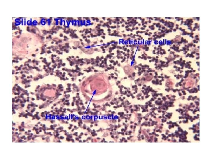
Thymic medulla • The medulla , inner portion of thymic lobule contain fewer T lymphocytes and a large number of epithelioreticular cells. • Three types of epithelioreticular cells are present. Type IV epithelioreticular cells Type VI epithelioreticular cells
Thymus
Corpuscles of Hassall • Small rounded structures present in the medulla of the thymus • Has a central core formed by epithelial cells • Around this mass there is a wall formed by concentrically arranged epithelial cells
Thymus
• Stem cells formed in bone marrow travel to the thymus • Here they come to lie in the superficial part of the cortex, & divide repeatedly to form small lymphocytes known as thymocyte. • About 95% of thymocytes produced in the thymic cortex fail to become properly immunocompetent. • Only 5% have received proper thymic education and become properly immunocompetent T lymphoctes.
• As lymphocytes divide they pass into the cortex, & into the medulla • They leave thymus by passing into blood vessel and lymphatics
• The central mass also contains degenerating macrophages
MCQS Q. 1 When looking at a lymph node, where are lymphatic nodules? a. Deep cortex b. Tertiary cortex c. Medulla d. Paracortical zone e. Outer cortex
Q 2. What is another term for lymphatic nodules? a. Lymph follicles b. White pulp c. Peyer's patches d. Lymph node e. Diffuse lymphatic tissue
Q 3. Which of the following is composed of epithelioreticular cells? a. Spleen b. Thymus c. Bone marrow d. Lymph node e. None of the above
Q 4. What are the spherical structures seen in the medulla of the thymus called? a. Psammoma bodies b. Corpora arenacea c. Hassall's corpuscles d. Prostatic concretions e. Pacinian corpuscles