IMMUNITY DEFINITION AND TYPES OF IMMUNITY Immunity is


. HLA is")


 is present")


 is a laboratory test done to determine")


- Slides: 72
IMMUNITY
DEFINITION AND TYPES OF IMMUNITY Immunity is defined as the capacity of the body to resist the pathogenic agents. It is the ability of the body to resist the entry of different types of foreign bodies like bacteria, virus, toxic substances, etc. Immunity is of two types: I. Innate immunity II. Acquired immunity.
INNATE IMMUNITY OR NONSPECIFIC IMMUNITY Innate immunity is the inborn capacity of the body to resists the pathogens. By chance, if the organisms enter the body, innate immunity eliminates them before the development of any disease. This type of immunity represents the first line of defense against any type of pathogens. Therefore, it is also called nonspecific immunity.
Examples of innate immunity are: 1. Destruction of toxic substances or organisms entering digestive tract through food by enzymes in digestive juices. 2. Destruction of bacteria by salivary lysozyme 3. Destruction of bacteria by acidity in urine and vaginal fluid.
ACQUIRED IMMUNITY OR SPECIFIC IMMUNITY Acquired immunity is the resistance developed in the body against any specific foreign body like bacteria, viruses, toxins, vaccines or transplanted tissues. So, this type of immunity is also known as specific immunity. It is the most powerful immune mechanism that protects the body from invading organisms or toxic substances. Lymphocytes are responsible for acquired immunity (Fig. 2) Types of Acquired Immunity Two types of acquired immunity develop in the body: 1. Cell mediated immunity or cellular immunity. 2. Humoral immunity.
DEVELOPMENT AND PROCESSING OF LYMPHOCYTES In fetus, lymphocytes develop from bone marrow. All the lymphocytes are released in the circulation and are differentiated into two categories: 1. T lymphocytes 2. B lymphocytes.
T. LYMPHOCYTES T lymphocytes are processed in thymus. The processing occurs mostly during the period between just before birth and few months after birth. Thymus secretes thymosin which accelerates the proliferation and activation of lymphocytes in thymus. It also increases the activity of lymphocytes in lymphoid tissues. Types of T Lymphocytes During the processing, T lymphocytes are transformed into four types: 1. Helper T cells or inducer T cells 2. Cytotoxic T cells or killer T cells 3. Suppressor T cells 4. Memory T cells.
Storage of T Lymphocytes After the transformation, all the types of T. lymphocytes leave thymus and are stored in lymphoid tissues of lymph nodes, spleen, bone marrow and the GI tract.
B LYMPHOCYTES B lymphocytes were first discovered in the bursa of Fabricius in birds hence the name B lymphocytes. The bursa of Fabricius is a lymphoid organ situated near the cloaca of birds. The bursa is absent in mammals, and the processing of B lymphocytes takes place in bone marrow and liver.
Types of B Lymphocytes After processing, the B lymphocytes are transformed into two types: 1. Plasma cells 2. Memory cells. Storage of B Lymphocytes After the transformation, B lymphocytes are stored in the lymphoid tissues of lymph nodes, spleen, bone marrow and the GI tract.
development of immunity
ANTIGENS DEFINITION AND TYPES Antigens are the substances, which induce specific immune reactions in the body. The antigens are mostly the conjugated proteins like lipoproteins, glycoproteins and nucleoproteins. Antigens are of two types: 1. Autoantigens or self antigens which are present on the body's own cells like 'A' antigen and 'B' antigen on the RBCs. 2. Foreign antigens or nonself antigens which enter the body from outside.
DEVELOPMENT OF CELL MEDIATED IMMUNITY The cell mediated immunity is offered by T lymphocytes. It involves several types of cells such as macrophages, T lymphocytes and natural killer cells and hence the name cell mediated immunity. It is also called cellular immunity or T cell immunity. It does not involve antibodies. Cellular immunity is the major defense mechanism against infections by viruses, fungi and few bacteria. It is also responsible for delayed allergic reactions and rejection of transplanted tissues.
Cell mediated immunity starts developing when T cells come in contact with the antigens. Usually, the invading microbial or nonmicrobial organisms carry the antigenic materials. These antigenic materials are released from invading organisms and are presented to the helper T cells by antigen presenting cells.
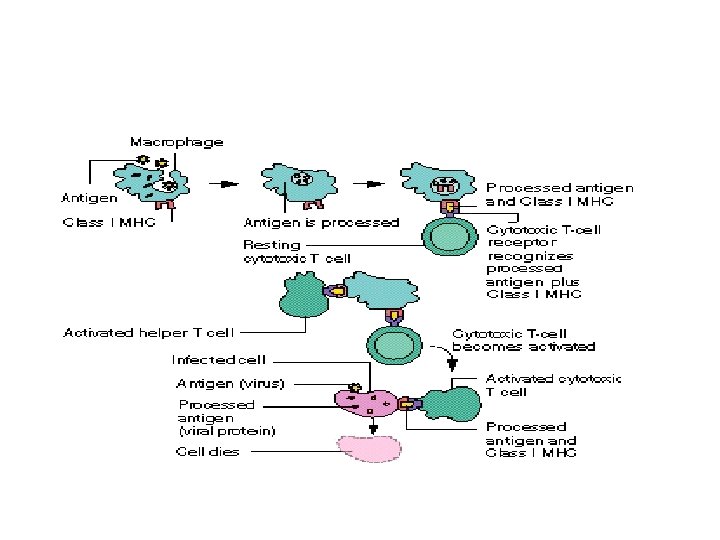
ANTIGEN PRESENTING CELLS Antigen presenting cells are the special type of cells in the body which induce the release of antigenic materials from invading organisms and later present these materials to the helper T cells. Major antigen presenting cells are macrophages. nodes and skin Dendritic cells in spleen, lymph also function like antigen presenting cells.
Role of Antigen Presenting Cells Invading foreign organisms are either engulfed by macrophages through phagocytosis or trapped by dendritic cells. Later, the antigen from these organisms is digested into small peptides. The antigenic peptide products are moved towards the surface of the antigen presenting cells and loaded on
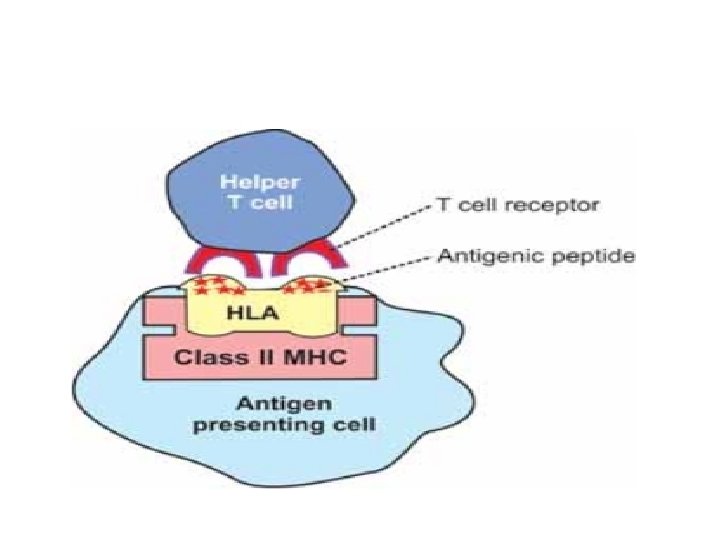
genetic matter of the antigen presenting cells called human leukocyte antigen (HLA). HLA is present in the molecule of class II major histocompatiblility complex (MHC) which is situated on the surface of the antigen presenting cells.
Presentation of Antigen The antigen presenting cells present their class II MHC molecules together with antigen bound HLA to the helper T cells. This activates the helper T cells through series of events Sequence of Events during Activation of Helper T Cells 1. Helper T cell recognizes the antigen bound to class II MHC molecule which is displayed on the surface of the antigen presenting cell. It recognizes the antigen with the help of its own surface receptor protein called T cell receptor.
2. The recognition of the antigen by the helper T cell initiates a complex interaction between the helper T cell receptor and the antigen. This reaction activates helper T cells. 3. At the same time macrophages (the antigen presenting cells) release interleukin-1 which facilitates the activation and proliferation of helper T cells. 4. The activated helper T cells proliferate and the proliferated helper T cells enter the circulation for further actions. 5. Simultaneously, the antigen bound to class II MHC molecules activates the B cells also resulting in development of humoral immunity.
ROLE OF HELPER T CELLS The helper T cells which enter the circulation activate all the other T cells and B cells. The helper T cells are of two types: 1. Helper-1 (TH 1) cells 2. Helper-2 (TH 2) cells. Role of TH 1 Cells TH 1 cells are concerned with cellular immunity and secrete two substances: i. Interleukin-2 which activates the other T cells ii. Gamma interferon which stimulates the phagocytic activity of cytotoxic cells, macrophages and natural killer (NK) cells.
Role of TH 2 Cells TH 2 cells are concerned with humoral immunity and secrete interleukin-4 and interleukin-5 which are concerned with: i. Activation of B cells ii. Proliferation of plasma cells iii. Production of antibodies by plasma cell HLA = Human leukocyte antigen.
ROLE OF CYTOTOXIC T CELLS The cytotoxic T cells that are activated by helper T cells circulate through blood, lymph and lymphatic tissues and destroy the invading organisms by attacking them directly. Mechanism of Action of Cytotoxic T Cells 1. The receptors situated on the outer membrane of cytotoxic T cells bind the antigens or organisms tightly with cytotoxic T cells. 2 -Then, the cytotoxic T cells enlarge and release cytotoxic substances like the lysosomal enzymes which destroy the invading organisms. 3. Like this, each cytotoxic T cell can destroy a large number of microorganisms one after another.
Other Actions of Cytotoxic T Cells. 1. The cytotoxic T cells also destroy cancer cells, transplanted cells such as those of transplanted heart or kidney or any other cells, which are foreign bodies. 2. Cytotoxic T cells destroy even body's own tissues which are affected by the foreign bodies, particularly the viruses. Many viruses are entrapped in the membrane of affected cells. The antigen of the viruses attracts the T. cells. And the cytotoxic T cells kill the affected cells also along with viruses. Because of this cytotoxic T cell is called killer cell.
ROLE OF SUPPRESSOR T CELLS The suppressor T cells are also called regulatory T cells. These T cells suppress the activities of the killer T cells. Thus, the suppressor T cells play an important role in preventing the killer T cells from destroying the body's own tissues along with invaded organisms. The suppressor cells suppress the activities of helper T cells also.
ROLE OF MEMORY T CELLS Some of the T cells activated by an antigen do not enter the circulation but remain in lymphoid tissue. These T cells are called memory T cells. In later periods, the memory cells migrate to various lymphoid tissues throughout the body. When the body is exposed to the same organism for the second time, the memory cells identify the organism and immediately activate the other T cells. So, the invading organism is destroyed very quickly. The response of the T cells is also more powerful this time.
SPECIFICITY OF T CELLS Each T cell is designed to be activated only by one type of antigen. It is capable of developing immunity against that antigen only. This cells. property is called the specificity of T
DEVELOPMENT OF HUMORAL IMMUNITY Humoral immunity is the immunity mediated by antibodies. Antibodies are secreted by B-lymphocytes and released into the blood and lymph. The blood and lymph are the body fluids (humours or humors in Latin). Since the B-lymphocytes provide immunity through humors, this type of immunity is called humoral immunity or B cell immunity. The antibodies are the gamma globulins produced by B lymphocytes. These antibodies fight against the invading organisms. The humoral immunity is the major defense mechanism against the bacterial infection. As in the case of cell mediated immunity, the macrophages and other antigen presenting cells play an important role in the development of humoral immunity also.
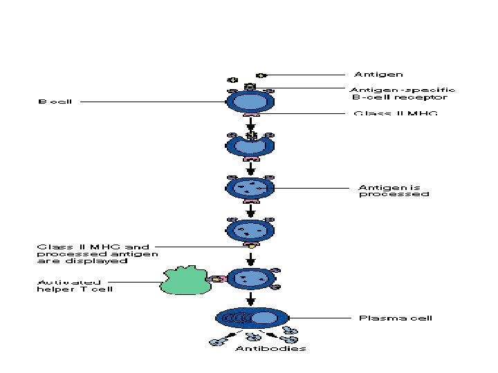
ROLE OF ANTIGEN PRESENTING CELLS The ingestion of foreign organisms and digestion of their antigen by the antigen presenting cells are already explained. Presentation of Antigen The antigen presenting cells present their class II MHC molecules together with antigen bound HLA to B cells. This activates the B cells through series of events.
Sequence of Events during Activation of B Cells on the surface of 1. The B cell recognizes the antigen bound to class II MHC molecule which is displayed the antigen presenting cell. It recognizes the antigen with the help of its own surface receptor protein called B cell receptor. 2. The recognition of the antigen by the B cell initiates a complex interaction between the B cell receptor and the antigen. This reaction activates B cells. 3. At the same time macrophages (the antigen presenting cells) release interleukin-1 which facilitates the activation and proliferation of B cells. 4. The activated B cells proliferate and the proliferated B cells carry out the further actions. 5. Simultaneously the antigen bound to class II. MHC molecules activates the helper T cells also resulting in development of cell mediated immunity (already explained). Transformation of B Cells the proliferated B cells are transformed into two types of cells: 1. Plasma cells 2. Memory cells.
ROLE OF PLASMA CELLS The plasma cells destroy the foreign organisms by producing the antibodies. Antibodies are globulin in nature. The rate of the antibody production is very high, i. e. each plasma cell produces about 2000 molecules of antibodies per second. The antibodies are also called immunoglobulins. The antibodies are released into lymph and then transported into the circulation. The antibodies are produced until the end of lifespan of each plasma cell which may be from several days to several weeks.
ROLE OF MEMORY B CELLS Memory B cells occupy the lymphoid tissues throughout the body. The memory cells are in inactive condition until the body is exposed to the same organism for the second time. During the second exposure, the memory cells are stimulated by the antigen and produce more quantity of antibodies at a faster rate, than in the first exposure. The antibodies produced during the second exposure to the foreign antigen are also more potent than those produced during first exposure. This phenomenon forms the basic principle of vaccination against the infections.
ROLE OF HELPER T CELLS Helper T cells are simultaneously activated by antigen. The activated helper T cells secrete two substances called interleukin 2 and B cell growth factor, which promote: 1. Activation of more number of B lymphocytes 2. Proliferation of plasma cells 3. Production of antibodies.
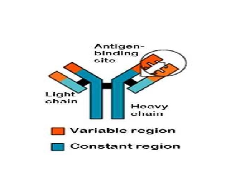
ANTIBODIES An antibody is defined as a protein that is produced by B lymphocytes in response to the presence of an antigen. Antibody is globulin in nature and it is also called immunoglobulin (Ig). The immunoglobulins form 20 percent of the total plasma proteins. The antibodies enter almost all the tissues of the body. Types of Antibodies Five types of antibodies are identified: 1. Ig. A (Ig alpha) 2. Ig. D (Ig delta) 3. Ig. E (Ig epsilon) 4. Ig. G (Ig gamma) 5. Ig. M (Ig mu).
Immunoglobulin
Among these antibodies, Ig. G forms 75 percent of the antibodies in the body. Structure of Antibodies are gamma globulins and are formed by two pairs of chains namely, one pair of heavy or long chains and one pair of light or short chains Mechanism of Actions of Antibodies The antibodies protect the body from the invading organisms in two ways: 1. By direct actions 2. Through complement system.
1. Direct Actions of Antibodies directly inactivate the invading organism by any one of the following methods: i. Agglutination: In this, the foreign bodies like RBCs or bacteria with antigens on their surfaces are held together in a clump by the antibodies. ii. Precipitation: In this, the soluble antigens toxin are converted into insoluble forms and then precipitated. iii. Neutralization: During this, the antibodies cover the toxic sites of antigenic products. iv. Lysis: In this, the antibodies rupture the cell membrane of organisms and then destroy them.
2. Actions of Antibodies through Complement System The complement system is the one that enhances or accelerates various activities during the fight against the invading organisms. It contains plasma enzymes, which are identified by numbers from C 1 to C 9. . Functions of Different Antibodies 1. Ig. A plays a role in localized defense mechanism in external secretions like tear. 2. Ig. D is involved in recognition of the antigen by B lymphocytes. 3. Ig. E is involved in allergic reactions. 4. Ig. G is responsible for complement fixation. 5. Ig. M is also responsible for complement Fixation.
Specificity of B Lymphocytes Each B lymphocyte is designed to be activated only by one type of antigen. It is also capable of producing antibodies against that antigen only. This property of B lymphocyte is called specificity. NATURAL KILLER CELL Natural killer (NK) cell is a large granular cell with indented nucleus. It is considered as the third type of lymphocyte. It is not a phagocytic cell but its granules contain hydrolytic enzymes which causes lysis of cells of invading organisms.
Functions of NK Cell The NK cell: 1. Destroys the viruses 2. Destroys the viral infected or damaged cells, which might form tumors 3. Destroys the malignant cells and prevents development of cancerous tumors. 4. Secretes cytokines such as interleukin-2, interferons, colony stimulating factor (GM-CSF) and tumor necrosis factor-
CYTOKINES Cytokines are the hormone like small proteins acting as intercellular messengers (cell signaling molecules) by binding to specific receptors of target cells. These non antibody proteins are secreted by WBCs and some other types of cells. Their major function is the activation and regulation of general immune system of the body. Cytokines are distinct from the other cell signaling molecules such as growth factors and hormones. Cytokines are classified into several types: 1. Interleukins 2. Interferons 3. Tumor necrosis factors 4. Chemokines 5. Defensins 6. Cathelicidins 7. Platelet activating factor
IMMUNE DEFICIENCY DISEASES Immune deficiency diseases are group of diseases in which some components of immune system is missing or defective. Normally, the defense mechanism protects the body from invading pathogenic organism. When the defense mechanism fails or becomes faulty (defective), the organisms of even low virulence produce severe disease. The organisms, which take advantage of defective defense mechanism, are called opportunists. The immune deficiency diseases caused by such opportunists are of two types: 1. Congenital immune deficiency diseases. 2. Acquired immune deficiency diseases.
CONGENIT AL IMMUNE DEFICIENCY DISEASES Congenital diseases are inherited and occur due to the defects in B cell, or T cell or both. The common examples are Di. George's syndrome (due to absence of thymus) and severe combined immune deficiency (due to lymphopenia or the absence of lymphoid tissue).
ACQUIRED IMMUNE DEFICIENCY DISEASES Acquired immune deficiency diseases occur due to infection by some organisms. The most common disease of this type is acquired immune deficiency syndrome (AIDS). Acquired Immune Deficiency Syndrome (AIDS) It is an infectious disease caused by immune deficiency virus (HIV). AIDS is the most common problem throughout the world because of rapid increase in the number of victims. Infection occurs when a glycoprotein from HIV binds to surface receptors of T lymphocytes, monocytes, macrophages and dendritic cells leading to destruction of these cells. It causes slow progressive decrease in immune function resulting in opportunistic infections of various types. The common opportunistic infections, which kill the AIDS patient, are pneumonia and skin cancer.
AUTOIMMUNE DISEASES Autoimmune disease is defined as condition in which the immune system mistakenly attacks body's own cells and tissues. Normally, an antigen induces the immune response in the body. The condition in which the immune system fails to give response to an antigen is called tolerance. This is true with respect to body's own antigens that are called self antigens or autoantigens. Normally, body has the tolerance against self antigen. However, in some occasions, the olerance fails or becomes incomplete against self antigen. This state is called autoimmunity and it leads to the activation of T lymphocytes or production of autoantibodies from B lymphocytes. The T lymphocytes (cytotoxic T cells) or autoantibodies attack
the body's normal cells whose surface contains the self antigen or autoantigen. Common Autoimmune Diseases 1. Diabetes mellitus 2. Myasthenia gravis 3. Hashimoto's thyroiditis 4. Graves' disease • 5. Rheumatoid arthritis.
Blood Groups and Blood Transfusion Blood groups are determined by the presence of antigen in RBC membrane. When blood from two individuals is mixed, sometimes clumping (agglutination) of RBCs occurs. This clumping is because of the immunological reactions. But, why clumping occurs in some cases and not in other cases remained a mystery until the discovery of blood groups by the Austrian Scientist, Karl Landsteiner in 1901
ABO BLOOD GROUPS Determination of blood groups depends upon the immunological reaction between antigen and antibody. Landsteiner found two antigens on the surface of RBCs and named them as A antigen and B antigen. These antigens are also called agglutinogens because of their capacity to cause agglutination of RBCS. He noticed the corresponding antibodies or agglutinins in the plasma and named them anti A or α antibody and anti B or β antibody. However, a particular agglutinogen and the corresponding agglutinin cannot be present together. If present, it causes clumping of the blood. Based on this, Landsteiner classified the blood groups. Later it has become the "Landsteiner's law" for grouping the blood.
LANDSTEINER'S LAW Landsteiner's law states that: 1. If a particular antigen (agglutinogen) is present in the RBCs, corresponding antibody (agglutinin) must be absent in the serum. 2. If a particular antigen is absent in the RBCs, the corresponding antibody must be present in the serum. Though the second part of Landsteiner's law, is a fact, it is not applicable to Rh factor.
BLOOD GROUP SYSTEMS More than 20 genetically determined blood group systems are known today. But, Landsteiner discovered two blood group systems called ABO system and Rh system. These two blood group systems are the most important ones that are determined before blood transfusions. ABO SYSTEM Based on the presence or absence of antigen A and antigen B, blood is divided into four groups: 1. 'A' group 2. 'B' group 3. 'AB' group 4. 'O' group.
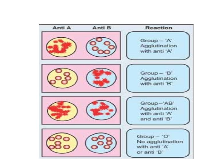
Antigen and antibody present in ABO blood groups
The blood having antigen A is called A group. This group has β antibody in the serum. The blood with antigen B and α antibody is called B group. If both the antigens are present, the blood group is called AB group and serum of this group does not contain any antibody. If both antigens are absent, the blood group is called O group and both α and β antibodies are present in the serum.
DETERMINATION OF THE ABO GROUP Determination of the ABO group is also called blood grouping, blood typing or blood matching. Principle of Blood Typing- Agglutination The blood typing is done on the basis of agglutination. Agglutination occurs if an antigen is mixed with its corresponding antibody which is called isoagglutinin. Agglutination occurs when A antigen is mixed with anti A with anti B. or when B antigen is mixed
BLOOD GROUPING
IMPORTANCE OF ABO GROUPS IN BLOOD TRANSFUSION During blood transfusion, only compatible blood must be used. The one who gives blood is called the donor and the one who receives the blood is called recipient. While transfusing the blood, antigen of the donor and the antibody of the recipient are considered. The antibody of the donor and antigen of the recipient are ignored mostly. Thus, RBC of "O" group has no antigen and so agglutination does not occur with any other group of blood. So, 'O' group blood can be given to any blood group persons and the people of this blood group called universal donors. are The plasma of AB group blood has no antibody. This does not cause agglutination of RBC from any other group of blood. The people of AB group can receive blood from any blood group persons. So, people with this blood group are called universal recipients
MATCHING AND CROSS MATCHING Blood matching (typing) is a laboratory test done to determine the blood group of a person. When the person needs blood transfusion, another test called cross matching the blood is typed. It is done to find out is done after whether the person's body will accept the donor's blood or not. For blood matching, RBC of the individual (recipient) and test sera are used. Cross matching is done by mixing the serum of the recipient and he RBCs of donor. Cross matching is always done before blood transfusion. If agglutination of RBCs from a donor occurs during cross from that person is not used for matching, the blood transfusion. Matching = Recipient's RBC + Test sera Cross matching = Recipient's serum +Donor’s RBC
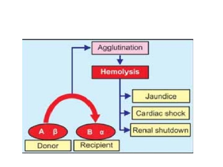
TRANSFUSION REACTIONS DUE TO ABO INCOMPATIBILITY Transfusion reactions are the adverse reactions in the body which occur due to transfusion of incompatible (mismatched) blood. The reactions may vary from fever and hives (skin disorder characterized by itching) to renal failure, shock and death. In mismatched transfusion, the transfusion reactions occur between donor's RBC and recipient's plasma. So, if the donor's plasma contains antibody against recipient's RBC, agglutination does not occur because these antibodies are diluted in recipient's blood But, if recipient’s plasma contains antibodies against donor’s RBCs, the immune system launches a response against the new blood cells. BCs are agglutinated and hemolyzed. The hemolysis of RBCs results in release of large amount of hemoglobin into the plasma. This leads to the following complications
1. Jaundice Normally, hemoglobin released from destroyed RBC is degraded and bilirubin is formed from it. When the serum bilirubin level increases above 2 mg/d. L jaundice occurs. 2. Cardiac Shock Simultaneously, the hemoglobin released into the plasma increases the viscosity of blood. This increases the workload on the heart leading to heart failure. 3. Renal Shutdown Dysfunction of kidneys is called renal shutdown. The toxic substances from hemolyzed cells cause constriction of blood vessels in kidney.
In addition, the toxic substances along with free hemoglobin are filtered through glomerular membrane and enter renal tubules. Because of poor rate of reabsorption from renal tubules, all these substances precipitate and obstruct the renal tubule. This suddenly stops formation of urine (anuria). If not treated with artificial kidney, the person dies within 10 -12 days because of jaundice, circulatory shock and more specifically due to renal shutdown and anuria
Rh FACTOR Rh factor is an antigen present in RBC. The antigen was discovered by Landsteiner and Wiener. It was first discovered in rhesus monkey and hence the name Rh factor. There are many Rh antigens but only the D is more antigenic in human. The persons having D antigen are called Rh positive and those without D antigen are called Rh negative.
Rh negative. Among Asian population, 85 percent of people are Rh positive and 15 percent are Rh negative. Rh system is different from ABO group system because, the antigen D does not have corresponding natural antibody (anti D). However, if Rh positive blood is transfused to a Rh negative person for the first time, then anti D is formed in that person. On the other hand, there is no risk of complications if Rh positive person receives Rh negative blood
TRANSFUSION REACTIONS DUE TO Rh INCOMPATIBILITY When a Rh negative person receives Rh positive blood for the first time, he is not • affected much, since the reactions do not occur immediately. But, the Rh antibodies develop within one month. The transfused RBCs, which are still present in recipient's blood are agglutinated. These agglutinated cells are lysed by macrophages. So, a delayed transfusion reaction occurs. But, it is usually mild and does not affect the recipient. However, antibodies developed in the recipient remain in the body for ever. So, when this person receives Rh positive blood for the second time, the donors RBCs are agglutinated and severe transfusion reactions occur immediately. These reactions are similar to the reactions of ABO incompatibility.
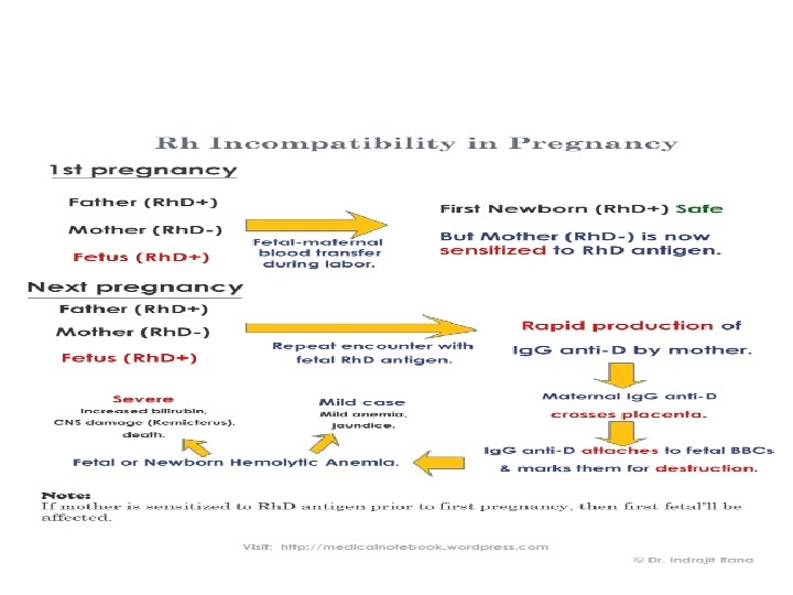
HEMOLYTIC DISEASE OF FETUS AND NEWBORN — ERYTHROBLASTOSIS FETALIS: Hemolytic disease is the disease in fetus and newborn characterized by abnormal hemolysis of RBCs. It is due to Rh incompatibility, i. e. the difference between the e mother and baby. c disease leads to erythroblastosis fetalis. Erythroblastosis fetalis is a disorder in fetus characterized by the presence of erythroblasts in blood.
The Rh agglutinins which enter the fetus cause agglutination of fetal RBCs resulting in hemolysis. The severe hemolysis in the fetus causes jaundice. To compensate the hemolysis of more and more number of RBCs, there is rapid production of RBCs, not only from bone marrow, but also from spleen and liver. Now, many large and immature cells in proerythroblastic stage are released into circulation. Because of this, the disease is called erythroblastosis fetalis. Ultimately due to excessive hemolysis severe complications develop, viz. 1. Severe anemia 2. Hydrops fetalis 3. Kernicterus.
1. Severe Anemia Excessive hemolysis results in anemia. And the infant dies when anemia becomes severe. 2. Hydrops Fetalis It is a serious condition in fetus characterized by edema. Severe hemolysis results in the development of edema, enlargement of liver and spleen and cardiac failure. When this condition becomes more severe it may lead to intrauterine death of fetus. 3. Kernicterus is the form of brain damage in infants caused by severe jaundice. If the baby survives anemia in erythroblastosis fetalis then kernicterus develops because of high bilirubin content.
Prevention or Treatment for Erythroblastosis Fetalis i. If mother is found to be Rh negative and fetus is Rh positive, anti D (antibody against D antigen) should be administered to the mother at 28 th and 34 th weeks of gestation as prophylactic measure. If Rh negative mother delivers Rh positive baby, then anti D should be administered to the mother within 48 hours of delivery. This develops passive immunity and prevents the formation of Rh antibodies in mother’s blood. So the hemolytic disease of newborn does not occur in a subsequent pregnancy. ii. If the baby is born with erythroblastosis fetalis, the treatment is given by means of transfused into the exchange transfusion (see below). Rh negative blood is infant replacing infant's own Rh positive blood. It will now take at least 6 months for the infant's new Rh positive blood to replace the transfused Rh negative blood. By this time all the molecules of Rh antibody derived from the mother get destroyed.