Heterochromatin vs Euchromatin Stains darkly highly condensed Repetitive
 Repetitive sequences Replicates later")
 • constitute ~ 10% of nuclear DNA")
 • Euchromatin: lightstaining, less condensed (complex")


 that")


- Slides: 45
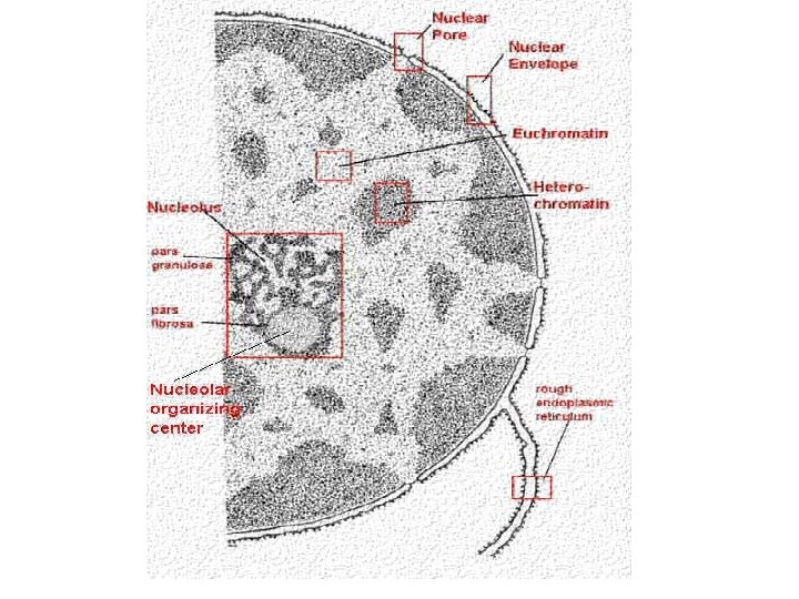
Heterochromatin vs Euchromatin • • • Stains darkly (highly condensed) Repetitive sequences Replicates later in the cell cycle Little or no recombination Transcriptionally repressive: silences gene expression • • • Stains lightly (decondensed) Single copy sequences (genes) Replicates early in the cell cycle Recombines Transcriptionally active: permissive for gene expression
heterochromatin Constitutive heterochromatin: euchromatin (and facultative heterochromatin) • constitute ~ 10% of nuclear DNA • highly compacted, transcriptionally inert, replicates late in S phase Euchromatin + facultative heterochromatin: • constitute ~ 90% of nuclear DNA • less condensed, rich in genes, replicates early in S phase however, • only small fraction of euchromatin is transcriptionally active • the rest is transcriptionally inactive/silenced (but can be activated in certain tissues or developmental stages) • these inactive regions are also known as “facultative heterochromatin”
• Heterochromatin: dark-staining, condensed (mostly simple-sequence DNA) • Euchromatin: lightstaining, less condensed (complex sequence DNA: e. g. genes)
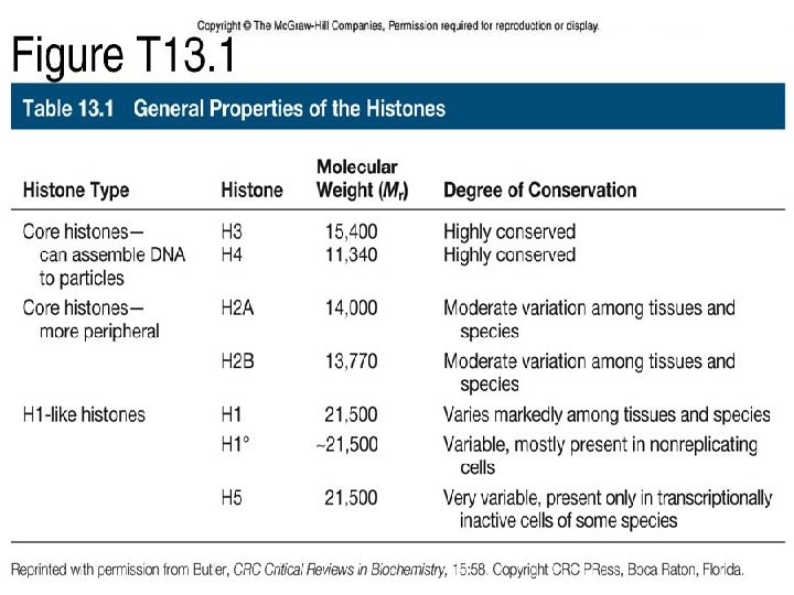
Nucleosomes are composed of histones H 1 N C • 2/3 of chromatin mass is protein • 95% of chromatin protein are histones • H 1, H 2 A, H 2 B, H 3, H 4
The difference between histone and nonhistone is simple. Both are proteins, both provide structure to DNA, and both are components of chromatin. Their chief difference is in the structure they provide. Histone proteins are the spools about which DNA winds, whereas nonhistone proteins provide the scaffolding structure. Another way to think of the difference is that nonhistone proteins are those proteins remaining after all histones have been removed from chromatin.
• Chromatin is DNA packaged with specialised proteins (and even some RNA!) that serve to control the degree to which DNA sequences are accessible for synthesis and transcription • These proteins include specialized structural proteins and enzymes
Orders of chromatin structure from naked DNA to chromatin to fully condensed chromosomes
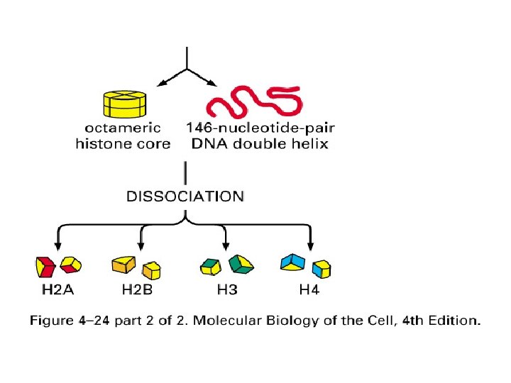
Nucleosome Structures Histone octamer 2 H 2 A 2 H 2 B 2 H 3 2 H 4
Nucleosome structure Chromatosome: octamer of histones plus ~146 bp DNA AND linker histone H 1 Nucleosome core particle: octamer of histones plus ~146 bp DNA
Level One: Building blocks of chromatin: nucleosomes • Linker can vary (8 -114 bp or more) 10 nm Fiber
Level three: nuclear scaffolding • Not well understood • Organization is not random; involved sequence elements (red dots), more non-histone chromatin proteins and tethering to the nuclear envelope and matrix
Metaphase chromatin: level 4 packaging: fully condensed
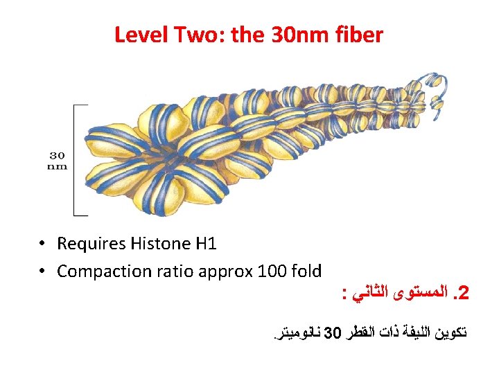
Irregularities in the 30 -nm fiber Flexible linker, DNA binding proteins Structural modulators: H 1 histone, ATP-driven Chromatin remodeling machine, covalent modification of histone tails
The function of Histone H 1
The function of Histone tails
Chromatin Remodeling
Cyclic Diagram for nucleosome formation and disruption
Human Chromosome Complex of DNA and protein is called chromatin 44 homologous chromosomes and 2 sex chromosomes Complementary DNA with different Dyes The arrangement of the full chromosome set is called karyotype
The Karyotype ﺍﻟﻄﺮﺯ ﺍﻟﻜﺮﻭﻣﻮﺳﻮﻣﻰ : It is a display of an individual’s chromosomes that arranged according to size and shapes)
Identifying chromosomes Chromosomes can be identified by: • Their size • Their shape (the position of the centromere) NB Chromosomes are flexible • Banding patterns produced by specific stains (Giemsa) Chromosomes are analysed by organising them into a KARYOTYPE
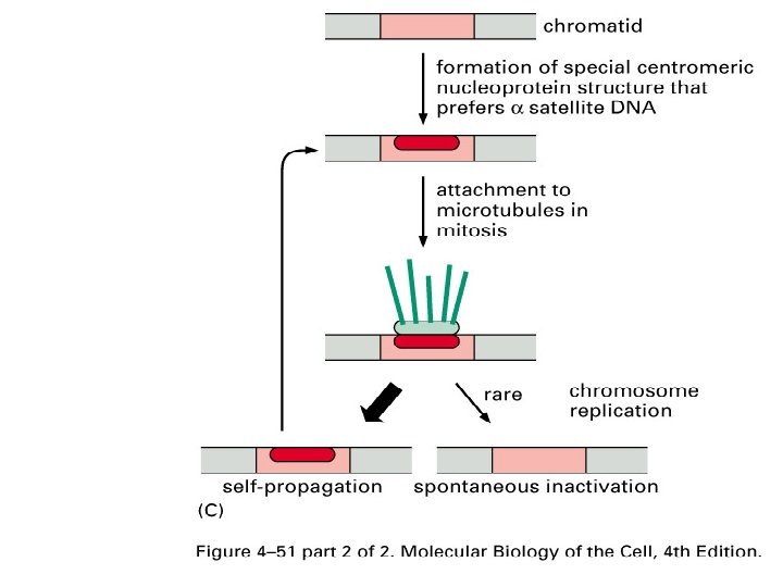
The structure of a human centromere 1. Alpha satellite DNA sequence 2. Kinetochore inner plate 3. Kinetochore outer plate 4. Spindle microtubules
The plasticity of human centromere formation
A typical mitotic chromosome at metaphase
Three important DNA sequences Telomere, replication origin, centromere
The organization of genes of a human chromosome
Chromosomes in eukaryotes and prokaryotes are different PROKARYOTES EUKARYOTES single chromosome plus plasmids many chromosomes circular chromosome linear chromosomes made only of DNA made of chromatin, a nucleoprotein (DNA coiled around histone proteins) found in cytoplasm found in a nucleus copies its chromosome and divides immediately afterwards copies chromosomes, then the cell grows, then goes through mitosis to organise chromosomes in two equal groups
A prokaryotic chromosome consists of a single molecule of DNA in the form of a closed loop. The chromosome is described as circular. A prokaryotic cell has only one chromosome. A eukaryotic chromosome is linear, not circular, in other words it has two ends, like a sausage. Each chromosome contains one molecule of DNA for the first half or so of interphase, then the DNA replicates, and the two DNA molecules remain together (as sister-chromatids) in the same chromosome for the rest of interphase. This does not happen in prokaryotic cells. Eukaryotic cells have more than one chromosome. A further difference: prokaryotic chromosomes consist only of a naked DNA molecule, but eukaryotic chromosomes also contain many molecules of proteins (mostly histones). The DNA is wound around these proteins.