Heart and Pericardium Prof Dr Selda nderolu Prof


 surface L ventricle Prof. Dr. H. Selçuk Sürücü")
 surface R atrium R ventricle L ventricle Prof. Dr. H. Selçuk Sürücü")


 (ant. +post. +septal)")
 Septomarginal trabecule (moderator band) Conus arteriosus (infundibulum)")
 Valve of foramen ovale During development")
 Papillary mm(ant+post) Chordae tendineae")


 Least cardiac vv (venae")


 Middle cervical cardiac n")
 Sup cervical cardiac brr inferior cervical cardiac brr Thoracic")


 “pericardial cavity ” “liquor")

 vagus Sympathetic trunk")

- Slides: 48
Heart and Pericardium Prof. Dr. Selda Önderoğlu Prof. Dr. H. Selçuk Sürücü
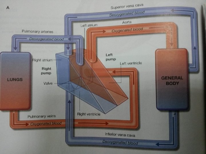
heart muscular organ Pumps blood to body. Shape: conical Apex is directed downwards
location: thoracic cavity Middle mediastinum Weight: 250 -300 gr. In the adult
relations Right and left: mediastinal surfaces of the lungs inferior: Diaphragm Anterior: : Sternum, costal cartilages, remains of thymus gland partially lungs Posterior: Oesophagus
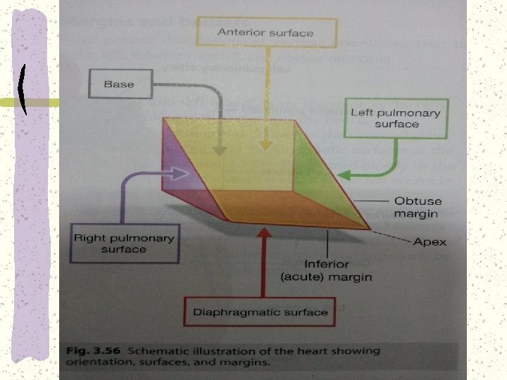
General shape Apex Base 4 surfaces Four borders Apex: directed anterior, inferior and to the left, Base: faces posteriorly and to the right
Apex L ventricle Forward, downward&to the left Prof. Dr. H. Selçuk Sürücü
Base L & R atria Faces posteriorly Prof. Dr. H. Selçuk Sürücü
Diaphragmatic (inf) surface L ventricle Prof. Dr. H. Selçuk Sürücü
Sternocostal (ant) surface R atrium R ventricle L ventricle Prof. Dr. H. Selçuk Sürücü
+ v brachiocephalica
Inf border R ventricle L ventricle
Sup border L atrium, roots of great vessels
Right and left borders Right border, Right atrium. Left border, left ventricle, left auricle.
Surface projections of borders Apex: 5 th intercostal space 9 cm from midline 2 nd left costal cartilage 1 cm from sternum 3 rd right costal cartilage 1 cm from sternum 6 th right costal cartilage 1 cm from sternum Prof. Dr. H. Selçuk Sürücü
External features “ coronary sulcus”, between atria and ventricles. “anterior interventricular sulcus”, between ventricules “posterior interventricular sulcus”, between ventricles
R atrium V cava sup&inf Coronary sinus Pectinate mm Sinus venorum (sinus of venae cavae) Terminal crest Interatrial septum Foramen ovale Fossa ovalis Limbus fossa ovalis Auricle (ant. to crest) Tricuspid valve (bw. R atr. &R vent. ) Prof. Dr. H. Selçuk Sürücü
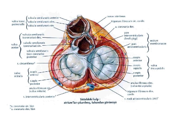
R ventricle Cusps of tricuspid valve/R atrioventricular valve (at sup wall) (ant. +post. +septal) Interventricular septum (septal wall) Pro. Membranous part muscular part Atrioventricular part ( bw. R atr&L ventr. )
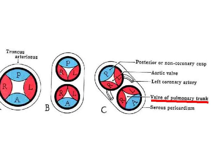
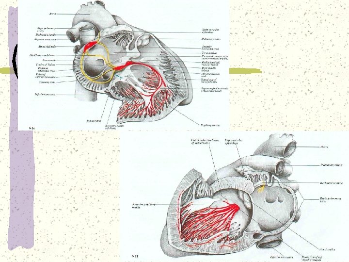
R ventricle Trabeculae carnea Papillary mm (ant+post+septal) Septomarginal trabecule (moderator band) Conus arteriosus (infundibulum) Supraventricular crest Opening of pulmonary trunk (has 3 semilunar cusps. R+L+Ant)
L atrium Pulmonary vv openings (4 in number) Valve of foramen ovale During development prevents blood from L atr. to R atr) Auricle Mitral valve/ L atrioventricular valve (Ant+post cusps) Prof. Dr. H. Selçuk Sürücü
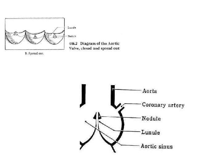
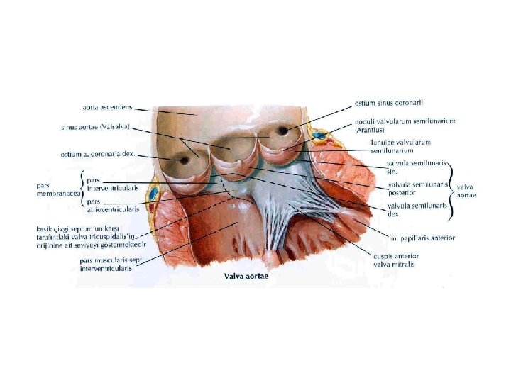
L ventricle Cusps of mitral valve/L atrioventricular valve (Ant+post cusps) Papillary mm(ant+post) Chordae tendineae Trabecula carneae Aortic opening Aortic valve ( R+L+post semilunar cusps) (each cusp has lunule, nodule, sinus)
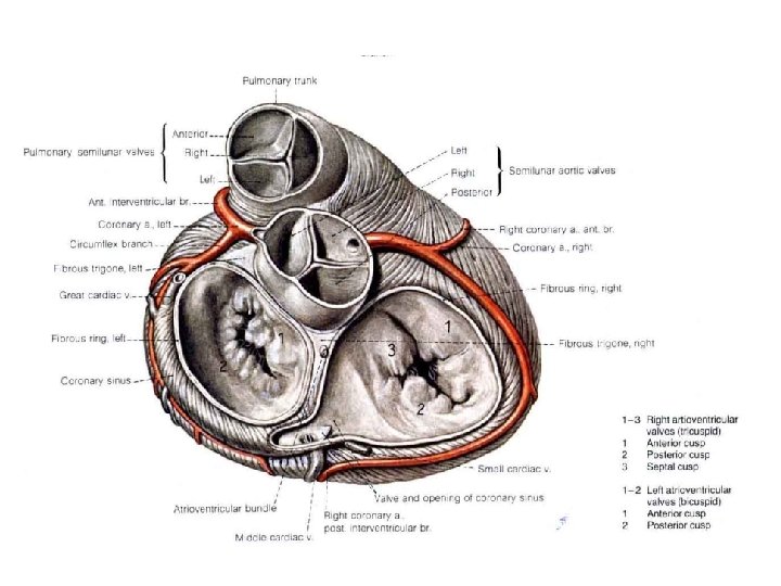
Arteries of heart R&L coronary aa- Both Exit from bulbus aorta Right coronary artery (R marginal br &Post interventricular br) Left coronary artery (Circumflex br+Ant interventricular br)
Prof. Dr. H. Selçuk Sürücü
Veins of heart coronary sinus-receives great, middle, small&post. Cardiac v) Least cardiac vv (venae cordis minimae-Thebasian vv)directly into chambers Anterior right ventricular vv Right marginal v
Great cardiac v Ant interventricular v L coronary v
Small cardiac v R coronary v
Middle cardiac v Post interventricular v
Innervation of heart Symp Psymp Together They form the cardiac plexus
Sympathetics Sup cervical cardiac n (from sup. Cerv. Gang. ) Middle cervical cardiac n br(from middle cerv. gang. ) Inf cervical cardiac n (from cervicothoracic (stellate)gang. Thoracic cardiac brrs (T 1 -4)
Parasympathetics (vagus nerve (CN X) Sup cervical cardiac brr inferior cervical cardiac brr Thoracic cardiac brr ( of vagus n. )
Lymphatics of heart R inf tracheabronchial lymph nodes
Layers of heart Endocardium: innermost layer Myocardium: middle layer, muscular layer, striated, under control of autonomic nervous system Pericardium: outermost layer, like a sac
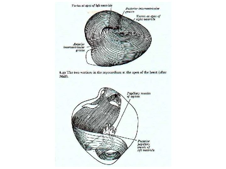
Myocardium Atrial Ventricular Conducting system Striated-special type Under autonomic nervous system control Atrial muscle fibers: 2 layers (inner-each atrium, outer- two atria together) Ventricular muscle fibers- has complex structure, no distinct layers- vortex
Myocardium Prof. Dr. H. Selçuk Sürücü
Skeleton of heart R & L fibrous rings (around mitral&tricuspid valves R & L fibrous trigones Around aortic & pulmonary Prof. Dr. H. Selçuk Sürücü valves
Conducting system of heart- specialised myocardial fibers SA node at terminal crest AV node, at interaatrial septum AV bundle, pars membranacea R & L bundle branches Purkinje fibers
Pericardium Fibrous pericardium Serous pericardium Parietal layer Visceral layer (epicardium) “pericardial cavity ” “liquor pericardii”
Pericardial sinuses-of the pericardial cavity Oblique sinus Bw. Left atrium- Pulmonary vv Like the letter “j”, inverted Transverse sinus superior v. c. &leftatriumpulmonary trunk&ascending aorta two ends open
nerves of pericardium Phrenic n –parietal pericardium (afferent) vagus Sympathetic trunk
Aorta ascending aorta Arch of aorta descending aorta