STRUCTURES AND FUNCTIONS OF THE HEART COVERINGS Pericardium


- Slides: 18

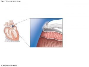
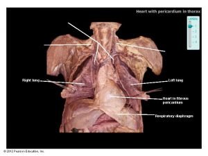
STRUCTURES AND FUNCTIONS OF THE HEART COVERINGS • Pericardium • Sac around the heart. • Fibrous pericardium: tough, loose-fitting, inextensible sac • Serous pericardium: parietal layer lies inside the fibrous pericardium; visceral layer (epicardium) covers the outside of the heart • Pericardial space: lies between visceral and parietal layers and contains 10 to 15 ml of pericardial fluid • Function of the heart coverings: to provide protection against friction 1

PERICARDIUM 2

STRUCTURE OF THE HEART • The wall of the heart is made up of three distinct layers: • Epicardium: outer layer • Myocardium: thick, contractile middle layer; compresses the heart cavities and the blood within them with great force • Endocardium: delicate inner layer of endothelial tissue 3

WALL OF THE HEART 4

CHAMBERS OF THE HEART • Atria • Two superior chambers known as “receiving chambers” because they receive blood from veins • Atria alternately relax to receive blood and then contract to push blood into ventricles • Myocardial wall of each atrium is not very thick because little pressure is needed to move blood such a short distance • Auricle: earlike flap protruding from each atrium 5

INTERIOR OF THE HEART 6

CHAMBERS AND VALVES OF THE HEART 7

CHAMBERS AND VALVES OF THE HEART (CONT. ) 8

CHAMBERS OF THE HEART • Ventricles • Two lower chambers; known as “pumping chambers” because they push blood into the large network of vessels • Ventricular myocardium is thicker (the left ventricle more than the right) than the myocardium of the atria because great force must be generated to pump the blood a large distance 9

VALVES OF THE HEART • Atrioventricular (AV) valves prevent blood from flowing back into the atria from the ventricles when the ventricles contract • Tricuspid valve (right AV valve): guards the right atrioventricular orifice; free edges of three flaps of endocardium are attached to papillary muscles by chordae tendineae • Bicuspid (mitral) valve (left AV valve): similar in structure to tricuspid valve except has only two flaps 10

VALVES OF THE HEART (CONT. ) • Semilunar (SL) valves prevent blood from flowing back into the ventricles from the aorta and pulmonary trunk • Pulmonary valve is at the entrance of the pulmonary trunk • Aortic valve is at the entrance of the aorta 11

BLOOD SUPPLY OF THE HEART TISSUE • Coronary arteries: myocardial cells receive blood from the right and left coronary arteries • First branches to come off of the aorta • Ventricles receive blood from branches of both the right and left coronary arteries • Each ventricle receives blood only from a small branch of the corresponding coronary artery • Most abundant blood supply goes to the myocardium of the left ventricle 12

CORONARY ARTERIES 13

BLOOD SUPPLY OF THE HEART TISSUE • Cardiac veins • As a rule, veins follow a course that closely parallels that of coronary arteries • After passing through the cardiac veins, blood enters the coronary sinus to drain into the right atrium 14

CORONARY VEINS 15

CONDUCTION SYSTEM OF THE HEART • Composed of four major structures: • Sinoatrial (SA) node • Atrioventricular (AV) node • AV bundle (bundle of his) • Subendocardial branches (purkinje fibers) • System generates rhythmic impulses and distributes them to regions of the myocardium 16

CONDUCTION SYSTEM OF THE HEART (CONT. ) • SA node (pacemaker) • Initiates each heartbeat and sets its pace • Specialized pacemaker cells in the node have an intrinsic rhythm • Sequence of cardiac stimulation • After generation by the SA node, each impulse travels throughout the muscle fibers of both atria and the atria begin to contract 17

CONDUCTION SYSTEM OF THE HEART 18
 Pericardial membranes
 Coronal cut of sheep heart
 Functions of pericardium
 Coverings of kidney
 Coverings of the brain
 Covering of brain
 Two kidneys
 True capsule of thyroid gland
 Scrotal sac layers
 Body coverings of animals
 Dartos muscle
 Conjoint tendon
 Example of homologous structure
 Pericardium
 Posterior region
 Nerve fibers
 Trabeculae carnae
 Parietal and visceral pericardium
 Layers of pericardium from superficial to deep
