Figure 11 2 Heart wall and coverings 2015


- Slides: 17

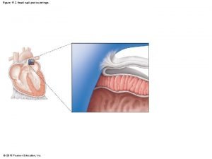
Figure 11. 2 Heart wall and coverings. © 2015 Pearson Education, Inc.

Figure 11. 2 Heart wall and coverings. Pulmonary trunk Pericardium Myocardium Fibrous pericardium Parietal layer of serous pericardium Pericardial cavity Epicardium (visceral layer of serous pericardium) Heart wall Myocardium Endocardium Heart chamber © 2015 Pearson Education, Inc.

Figure 11. 3 a Gross anatomy of the heart. © 2015 Pearson Education, Inc.

Figure 11. 3 a Gross anatomy of the heart. Brachiocephalic trunk Left common carotid artery Superior vena cava Left subclavian artery Right pulmonary artery Aortic arch Ascending aorta Pulmonary trunk Right pulmonary veins Right atrium Right coronary artery in coronary sulcus (right atrioventricular groove) Anterior cardiac vein Right ventricle Marginal artery Small cardiac vein Inferior vena cava (a) © 2015 Pearson Education, Inc. Ligamentum arteriosum Left pulmonary artery Left pulmonary veins Left atrium Auricle of left atrium Circumflex artery Left coronary artery in coronary sulcus (left atrioventricular groove) Left ventricle Great cardiac vein Anterior interventricular artery (in anterior interventricular sulcus) Apex

Figure 11. 3 b Gross anatomy of the heart. © 2015 Pearson Education, Inc.

Figure 11. 3 b Gross anatomy of the heart. Superior vena cava Aorta Left pulmonary artery Right pulmonary artery Left atrium Right atrium Left pulmonary veins Right pulmonary veins Fossa ovalis Right atrioventricular valve (tricuspid valve) Right ventricle Chordae tendineae Pulmonary semilunar valve Left atrioventricular valve (bicuspid valve) Aortic semilunar valve Left ventricle Interventricular septum Inferior vena cava Myocardium (b) Frontal section showing interior chambers and valves. © 2015 Pearson Education, Inc. Visceral pericardium (epicardium)

Figure 11. 4 The systemic and pulmonary circulations. © 2015 Pearson Education, Inc.

Figure 11. 4 The systemic and pulmonary circulations. Capillary beds of lungs where gas exchange occurs Pulmonary Circuit Pulmonary arteries Pulmonary veins Aorta and branches Venae cavae Left atrium Right atrium Left ventricle Heart Right ventricle Systemic Circuit KEY: Oxygen-rich, CO 2 -poor blood Oxygen-poor, CO 2 -rich blood © 2015 Pearson Education, Inc. Capillary beds of all body tissues where gas exchange occurs

Figure 11. 6 Operation of the heart valves. (a) Operation of the AV valves 1 Blood returning to the atria puts pressure against AV valves; the AV valves are forced open. 4 Ventricles contract, forcing blood against AV valve flaps. 2 As the ventricles fill, AV valve flaps hang limply into ventricles. 6 Chordae tendineae tighten, preventing valve flaps from everting into atria. 3 Atria contract, forcing additional blood into ventricles. 5 AV valves close. Ventricles AV valves open; atrial pressure greater than ventricular pressure AV valves closed; atrial pressure less than ventricular pressure (b) Operation of the semilunar valves Pulmonary trunk 1 As ventricles contract and intraventricular pressure rises, blood is pushed up against semilunar valves, forcing them open. Semilunar valves open © 2015 Pearson Education, Inc. Aorta 2 As ventricles relax and intraventricular pressure falls, blood flows back from arteries, filling the leaflets of semilunar valves and forcing them to close. Semilunar valves closed

Figure 11. 6 a Operation of the heart valves. (a) Operation of the AV valves 1 Blood returning to the atria puts pressure against AV valves; the AV valves are forced open. 4 Ventricles contract, forcing blood against AV valve flaps. 2 As the ventricles fill, AV valve flaps hang limply into ventricles. 6 Chordae tendineae tighten, preventing valve flaps from everting into atria. 3 Atria contract, forcing additional blood into ventricles. AV valves open; atrial pressure greater than ventricular pressure © 2015 Pearson Education, Inc. 5 AV valves close. Ventricles AV valves closed; atrial pressure less than ventricular pressure

Figure 11. 6 b Operation of the heart valves. (b) Operation of the semilunar valves Pulmonary trunk 1 As ventricles contract and intraventricular pressure rises, blood is pushed up against semilunar valves, forcing them open. Semilunar valves open © 2015 Pearson Education, Inc. Aorta 2 As ventricles relax and intraventricular pressure falls, blood flows back from arteries, filling the leaflets of semilunar valves and forcing them to close. Semilunar valves closed

Figure 11. 7 The intrinsic conduction system of the heart. © 2015 Pearson Education, Inc.

Figure 11. 7 The intrinsic conduction system of the heart. Superior vena cava Sinoatrial (SA) node (pacemaker) Left atrium Atrioventricular (AV) node Right atrium Bundle branches Atrioventricular (AV) bundle (bundle of His) Purkinje fibers © 2015 Pearson Education, Inc. Interventricular septum

Figure 11. 13 Major arteries of the systemic circulation, anterior view. © 2015 Pearson Education, Inc.

Figure 11. 13 Major arteries of the systemic circulation, anterior view. Arteries of the head and trunk Internal carotid artery External carotid artery Common carotid arteries Vertebral artery Subclavian artery Brachiocephalic trunk Aortic arch Ascending aorta Coronary artery Thoracic aorta (above diaphragm) Celiac trunk Abdominal aorta Superior mesenteric artery Renal artery Gonadal artery Inferior mesenteric artery Internal iliac artery Arteries that supply the upper limb Subclavian artery Axillary artery Brachial artery Radial artery Ulnar artery Deep palmar arch Superficial palmar arch Digital arteries Arteries that supply the lower limb Common iliac artery External iliac artery Femoral artery Popliteal artery Anterior tibial artery Posterior tibial artery Dorsalis pedis artery Arcuate artery © 2015 Pearson Education, Inc.

Figure 11. 14 Major veins of the systemic circulation, anterior view. © 2015 Pearson Education, Inc.

Figure 11. 14 Major veins of the systemic circulation, anterior view. Veins of the head and trunk Dural venous sinuses External jugular vein Vertebral vein Internal jugular vein Right and left brachiocephalic veins Superior vena cava Great cardiac vein Hepatic veins Splenic vein Hepatic portal vein Renal vein Superior mesenteric vein Inferior vena cava Common iliac vein Internal iliac vein Veins that drain the upper limb Subclavian vein Axillary vein Cephalic vein Brachial vein Basilic vein Median cubital vein Ulnar vein Radial vein Digital veins Veins that drain the lower limb External iliac vein Femoral vein Great saphenous vein Popliteal vein Posterior tibial vein Anterior tibial vein Small saphenous vein Dorsal venous arch Dorsal metatarsal veins © 2015 Pearson Education, Inc.
 Figure 11-6 arteries
 Coverings of kidney
 Dura arachnoid pia
 Coverings of the brain
 Two kidneys
 Coverings of thyroid gland
 Spermatic cord covering
 Body coverings of animals
 Epididis
 Conjoint tendon
 Sound walls vs word walls
 Chris brown wall
 One brick t junction in english bond
 Members used to carry wall loads over wall openings
 Lemon diagram
 Loss of heart muscle
 Ecg septal wall view
 Heart wall
