The Pericardium Dr S Dawar Husain The Pericardium

 is a fibroserous sac which encloses")


 at the roots of great blood")


 is responsible for the")


- Slides: 35
The Pericardium Dr. S Dawar Husain
The Pericardium • The Pericardium (G. around heart) is a fibroserous sac which encloses the heart and the roots of its great blood vessels. • The pericardium is located inside the middle mediastinum, posterior to the body of the sternum and 2 nd-6 th costal cartilages and anterior to the middle 4 thoracic vertebrae (i. e. , from T 5 to T 8).
CONTENTS • The following are the contents of the pericardium: –A. Heart with its vessels and nerves. –B. Ascending aorta. –C. Pulmonary trunk. –D. Superior vena cava (lower half). –E. Inferior vena cava (terminal part). –F. Pulmonary veins (terminal parts)
FUNCTIONS • The pericardium has many physiological roles. • Fixes the heart in the mediastinum and limits motion because of its attachment to the diaphragm, the sternum, and the tunica adventitia (outer layer) of the great vessels. • Prevents overfilling of the heart. The relatively inextensible fibrous layer of the pericardium prevents the heart from increasing in size too rapidly. • Lubrication. A thin film of fluid between the two layers of the serous pericardium reduces the friction generated by the heart as it moves within the thoracic cavity. • Protection from infection. The fibrous pericardium serves as a physical barrier against infection.
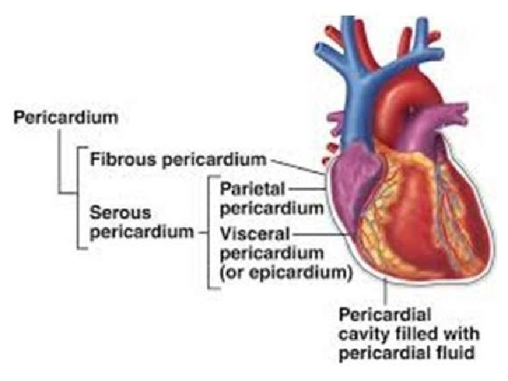
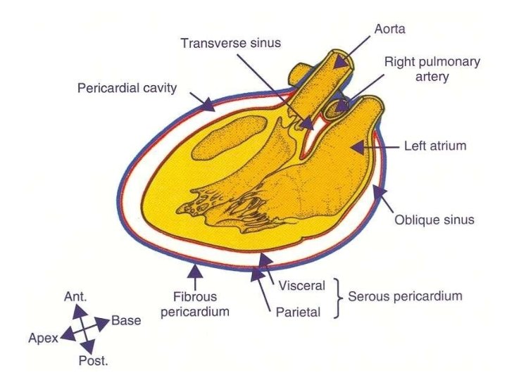
Anatomical Structure • The pericardium is made up of two main layers: • Fibrous Pericardium: • A tough, external layer known as the fibrous pericardium. • Serous Pericardium: • A thin, internal layer known as the serous pericardium. Found between the outer and inner serous layers is the pericardial cavity, which contains a small amount of lubricating serous fluid.
SUBDIVISIONS • The heart and great vessels be located within the fibrous sac and invaginate the serous sac from behind during development. • The pericardium thus is composed of: • 1) Fibrous layer of the pericardium(outside) • 2) Serous pericardium(two layers) – Parietal layer(outer). – Visceral layer also called as epicardium (inner).
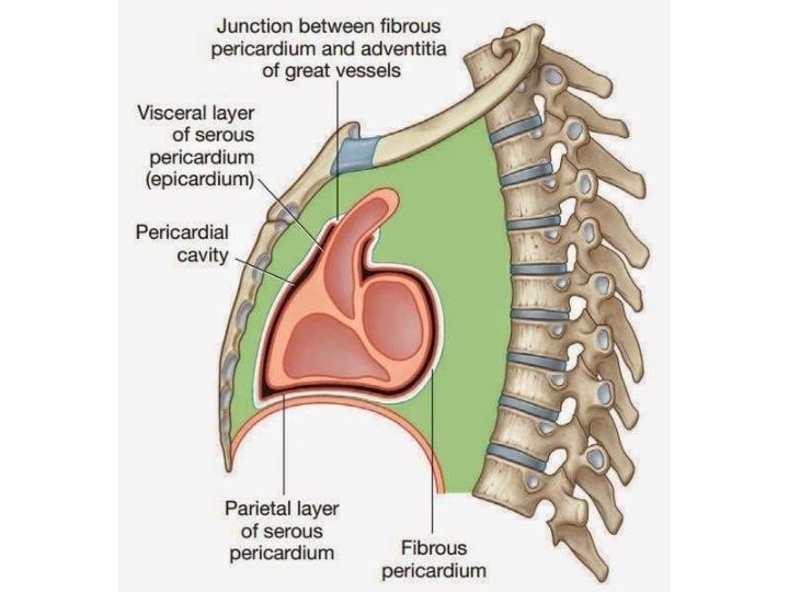
FIBROUS PERICARDIUM • • The fibrous pericardium is strong fibrous sac which supports the delicate parietal layer of the serous pericardium with which it is firmly adherent. FEATURES: • A. It is conical in shape. • B. Its apex is blunt and fused with the outer coats of the roots of great blood vessels (e. g ascending aorta, pulmonary trunk). • • C. Its base is broad and blended with the central tendon of the diaphragm. D. Anteriorly it is connected to the posterior aspect of the body of sternum by the superior and inferior sternopericardialligaments. • E. Posteriorly it is related to principal bronchi, esophagus, and descending thoracic aorta. F. Laterally, it is related to phrenic nerves and pericardiophrenic vessels. Continuous with the central tendon of the diaphragm, the fibrous pericardium is made of tough connective tissue and is relatively non-distensible. • •
SEROUS PERICARDIUM • Enclosed within the fibrous pericardium, the serous pericardium is itself divided into two layers: – The outer parietal layer that lines the internal surface of the fibrous pericardium. – The internal visceral layer that forms the outer layer of the heart (also known as the epicardium). Each layer is made up of a single sheet of epithelial cells, known as mesothelium.
SEROUS PERICARDIUM • The outer layer lines the fibrous pericardium and is reflected around the roots of great blood vessels to become continuous with the visceral layer of the pericardium. It is termed parietal pericardium. • The inner layer is closely applied to the heart with the exception of along the cardiac grooves, where it is divided from the heart by blood vessels. It is referred to as visceral pericardium or epicardium. • The 2 layers of serous pericardium are continuous with every other at the roots of great blood, and superior and inferior pulmonary veins where the pericardial sac was invaginated by the developing heart.
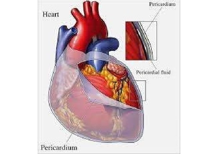
PERICARDIAL CAVITY • The slit-like potential space between the parietal and visceral layers of serous pericardium is called pericardial cavity. • Normally it includes a thin film of serous fluid (about 50 ml) named pericardial fluid which lubricates the opposed surfaces to avoid friction during the movements of the heart.
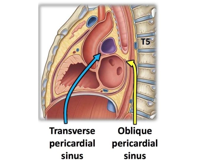
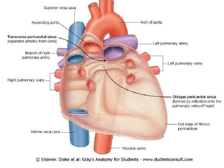
SINUSES OF PERICARDIUM • The Sinuses of Pericardium lie between the parietal and visceral layers of serous pericardium and are 2 in number: • Transverse sinus. • Oblique sinus. • They are created because of the reflection of visceral layer of serous pericardium around great vessels of the heart.
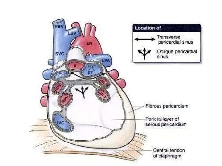
Sinuses of pericardium • The visceral pericardium (epicardium) at the roots of great blood vessels is arranged into tubes: – Arterial tube – Venous tube. • The arterial tube encloses the ascending aorta and pulmonary trunk (arterial end of the heart tube). • The venous tube encloses the superior and inferior vena cava and 4 pulmonary veins
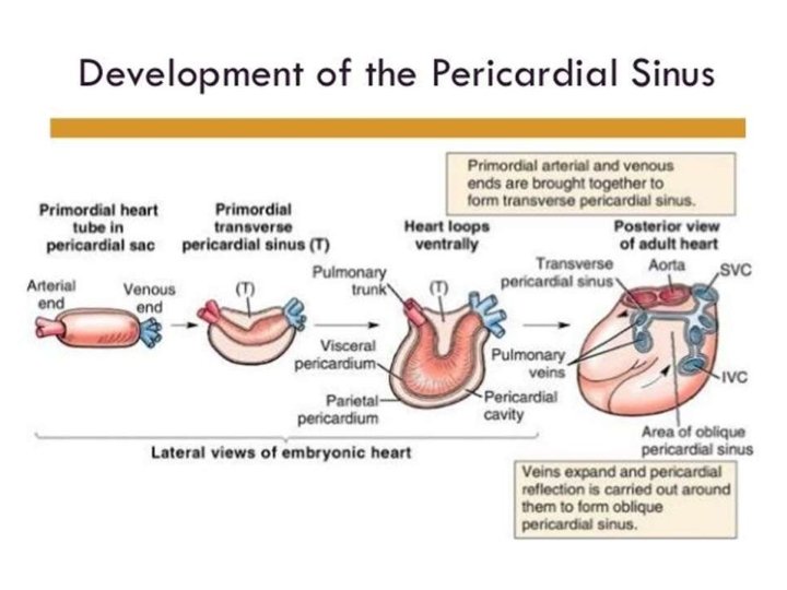
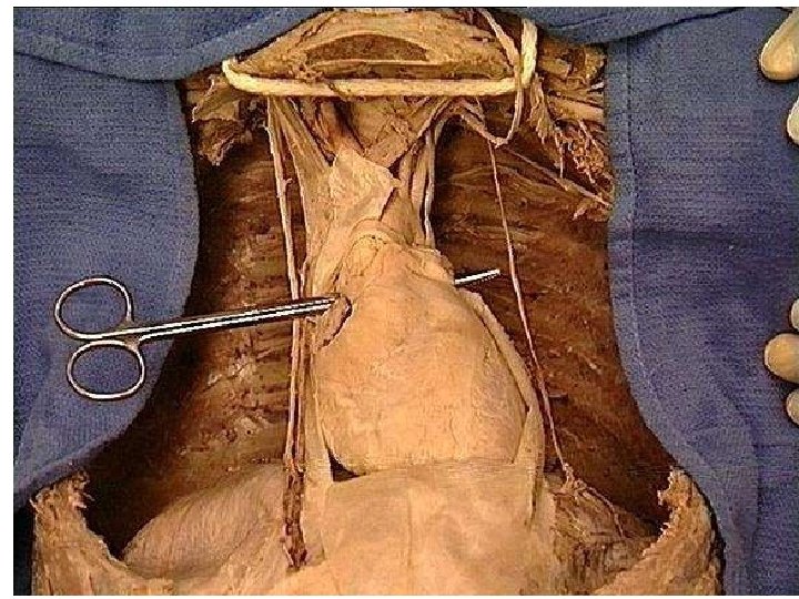
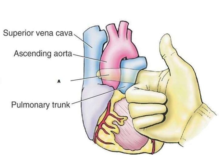
Transverse Pericardial Sinus • Formed as a result of the embryological folding of the heart tube, the transverse pericardial sinus is a passage through the pericardial cavity. • • The transverse pericardial sinus separates the heart’s arterial outflow (aorta, pulmonary trunk) from its venous inflow (superior vena cava, pulmonary veins). It is located: – Posterior to the ascending aorta and pulmonary trunk. – Anterior to the superior vena cava. – Superior to the left atrium. • The transverse pericardial sinus can be used to identify and subsequently ligate the arteries of the heart during coronary artery bypass grafting.
TRANSVERSE SINUS OF PERICARDIUM • It is a transverse recess behind the ascending aorta and pulmonary trunk and in front of superior vena cava and superior pulmonary veins. It develops because of degeneration of dorsal mesocardium. • It has a horizontal passage between the 2 pericardial tubes. On every side it interacts with all the general pericardial cavity.
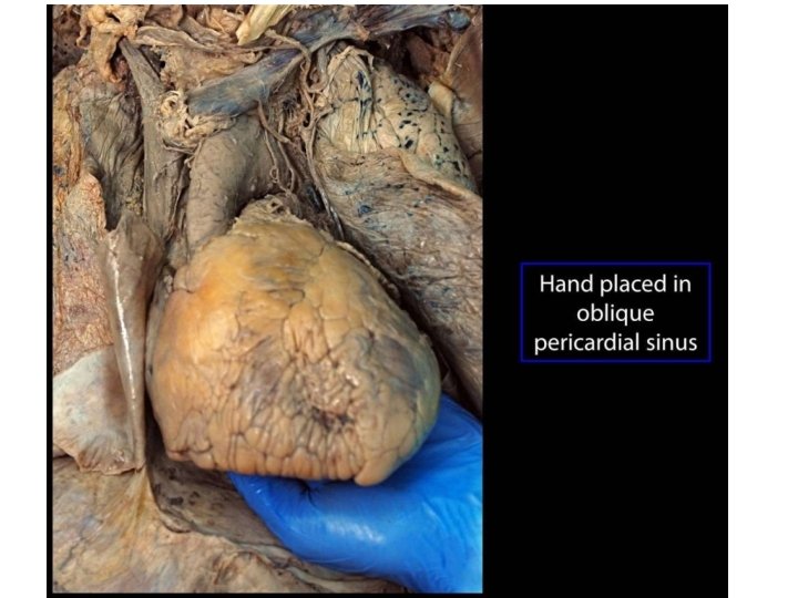
OBLIQUE SINUS OF • It is. PERICARDIUM a recess of serous pericardium supporting the base of the heart (actually left atrium). • It is enclosed by ‘J-shaped’ sheath of visceral layer of serous pericardium enclosing 6 veins (i. e. , 2 vena cavae and 4 pulmonary veins). • The oblique sinus develops as an effect of absorption of 4 pulmonary veins into the left atrium. • The oblique sinus permits the distension of left atrium during return of oxygenated blood in it
• • BOUNDARIES OBLIQUE Oblique sinus of pericardium is bounded OF in the following ways: SINUS Anteriorly: – By left atrium. • Posteriorly: – By parietal pericardium. • On right side: – By reflection of visceralpericardium alongthe right pulmonary veins and inferior vena cava. • On the left side: – By reflection of visceral pericardium along the left pulmonary veins. • Superiorly: – By reflection of visceralpericardium alongthe left and right superior pulmonary veins.
The arterial supply of the pericardium • The arterial supply of the pericardium is mainly from a slender branch of the internal thoracic artery, the pericardiacophrenic artery, that often accompanies or at least parallels the phrenic nerve to the diaphragm. • Smaller contributions of blood come from the: – Musculophrenic artery, a terminal branch of the internal thoracic artery. – Bronchial, esophageal, and superior phrenic arteries, branches of the thoracic aorta. – Coronary arteries (visceral layer of serous pericardium only), the first branches of the aorta.
The venous drainage of the pericardium • The venous drainage of the pericardium is from the: • Pericardiacophrenic veins, tributaries of the brachiocephalic (or internal thoracic) veins. • Variable tributaries of the azygos venous system
Innervatio n • The phrenic nerve (C 3 -C 5) is responsible for the somatic innervation of the pericardium, as well as providing motor and sensory innervation to the diaphragm. • Originating in the neck and travelling down through the thoracic cavity, the phrenic nerve is a common source of referred pain, with a key example being shoulder pain experienced as a result of pericarditis. • The supraclavicular nerves (descending branches) arise from the third and fourth cervical nerves.
Kehr's • Kehr's sign is asign classic example of referred pain: irritation of the diaphragm is signaled by the phrenic nerve as pain in the area above the collarbone. • This is because the supraclavicular nerves have the same cervical
SURGICAL IMPORTANCE OF TRANSVERSE • During cardiac surgery, following the PERICARDIAL SINUS pericardial sac is opened anteriorly, a finger is gone through the transverse sinus of pericardium, posterior to the aorta and pulmonary trunk. • A temporary ligature is gone through the transverse sinus around the aorta and pulmonary trunk. The tubes of heart-lung machine are inserted into these vessels and ligature is tightened.
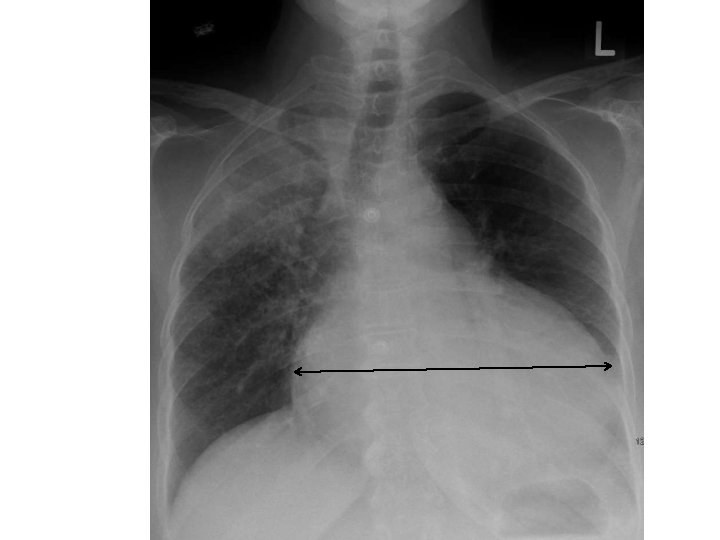
Cardiac Tamponade • The relatively inextensible fibrous pericardium can cause problems when there is an accumulation of fluid, known as pericardial effusion, within the pericardial cavity. • The rigid pericardium cannot expand, and thus the heart is subject to the resulting increased pressure. The chambers can become compressed, thus compromising cardiac output.
Pericarditis • Pericarditis, or inflammation of the pericardium, has myriad causes, including bacterial infection and myocardial infarction. • The main symptom is chest pain, and the condition cause acute cardiac tamponade due to an accumulation of fluid in the pericardial cavity.