HEART TUBE PERICARDIUM Dr Mujahid Khan Early Development

HEART TUBE & PERICARDIUM Dr. Mujahid Khan

Early Development of Heart Ø The earliest sign of heart is the appearance of paired endothelial strands called angioblastic cords Ø They develop in the cardiogenic mesoderm during the third week Ø These cords canalize to form two heart tubes Ø These cords fuse together to form the tubular heart late in the third week


Early Development of Heart Ø Primordium of heart is first evident at 18 days in the cardiogenic area Ø The heart begins to beat at 22 -23 days Ø Blood flow begins during the fourth week and can be visualized by Doppler ultrasonography

Development of Heart Ø The endocardial heart tubes approach each other and fuse to form a single heart tube after lateral folding Ø Fusion of tubes begins at the cranial end of the developing heart and extends caudally
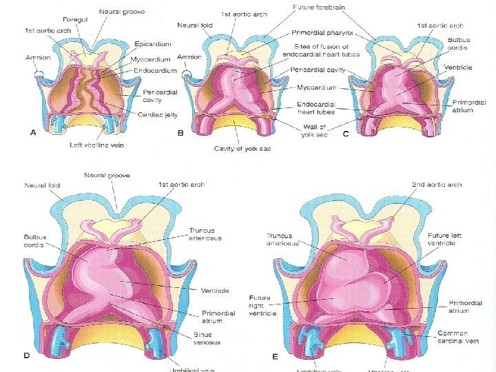

Primordial Myocardium Ø As the heart tubes fuse, an external layer of the embryonic heart, the primordial myocardium is formed from splanchnic mesoderm around pericardial coelom Ø At this stage the developing heart is composed of a thin endothelial tube, separated from thick muscular tube by gelatinous connective tissue, cardiac jelly

Endocardium Ø The endothelial tube becomes the internal endothelial lining of the heart, called endocardium Ø The primordial myocardium becomes the muscular wall of the heart or myocardium Ø The visceral pericardium or epicardium is derived from mesothelial cells and spread over the myocardium

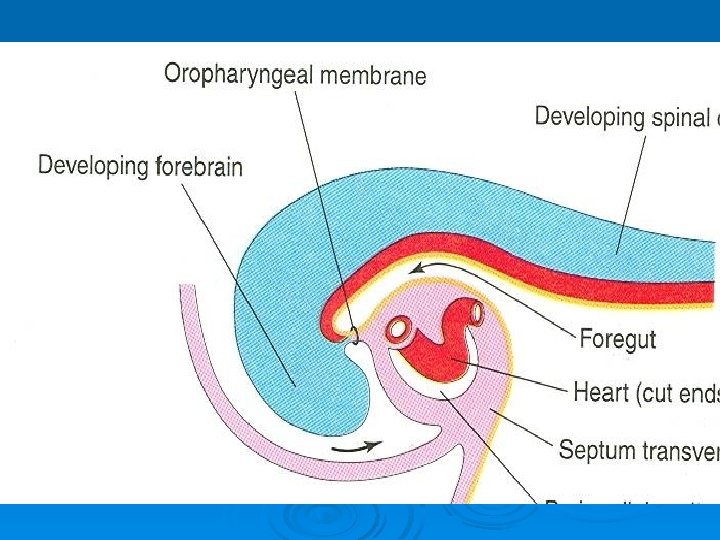
After Folding Ø As folding of head region occurs Ø The heart and pericardial cavity come to lie ventral to the foregut and caudal to the oropharyngeal membrane

Fate of Heart Tubes Ø The tubular heart elongates and develops alternate dilations and constrictions: Ø Truncus Arteriosus Ø Bulbus Cordis Ø Ventricle Ø Atrium Ø Sinus venosus
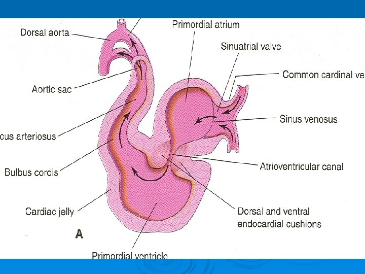

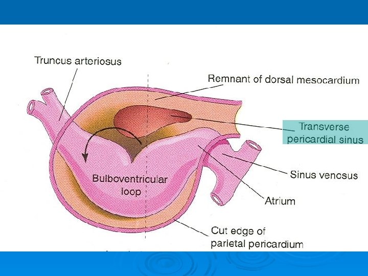
Fate of Heart Tubes Ø As the developing heart elongates and bends, it gradually invaginates into the pericardial cavity Ø Initially suspended from the dorsal wall by a mesentery, the dorsal mesocardium Ø Central part of this mesentery soon degenerates Ø Heart is now attached only at its cranial and caudal ends

Truncus Arteriosus Ø Is continuous cranially with the aortic sac, from which the aortic arches arise Ø The sinus venosus receives umbilical, vitelline, and common cardinal veins from the chorion, yolk sac, and embryo respectively Ø Bulbus cordis and ventricle grow faster than other regions, the heart bends upon itself, forming bulboventricular loop
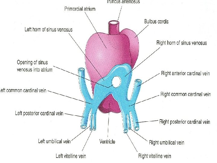

Truncus Arteriosus Ø As the primordial heart bends, the atrium and sinus venosus come to lie dorsal to the truncus arteriosus, bulbus cordis, and ventricle Ø By this stage the sinus venosus has developed lateral expansions, the right and left horns of the sinus venosus


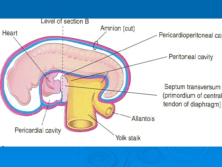
Pericardial Cavity Ø As the heart elongates and bends, it gradually invaginates into the pericardial cavity Ø The heart is initially suspended from the dorsal wall by a mesentery, the dorsal mesocardium Ø The central part of the mesentery soon degenerates Ø Forms a communication, the transverse pericardial sinus between the right and left sides of the pericardial cavity


Pericardial Cavity Ø During the fourth week three well defined body cavities are formed: Ø Pericardial cavity Ø 2 pericardioperitoneal canals Ø Peritoneal cavity

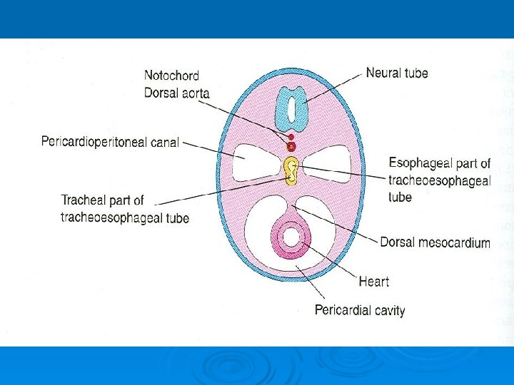
Division of Body Cavities Ø Each pericardioperitoneal canal lies lateral to the foregut and dorsal to the septum transversum Ø Partitions of pleuropericardial fold form in each pericardioperitoneal canal Ø This separates pericardial cavity from pleural cavities Ø Also pleural cavities from peritoneal cavity

Pleuropericardial Membranes Ø As the pleuropericardial folds enlarge, they form partitions that separate the pericardial cavity from the pleural cavities Ø These partitions are called pleuropericardial membranes Ø They contain the common cardinal veins

Pleuropericardial Membranes Ø As the primordial pleural cavities expand ventrally around the heart, they extend into the body wall, splitting the mesenchyme into: Ø An outer layer that becomes the thoracic wall Ø An inner layer becomes the fibrous pericardium, the outer layer of the pericardial sac enclosing the heart
- Slides: 27