GLUTEAL REGION GLUTEAL REGION The gluteal region hip

 is the prominent area posterior to")


and")


- Slides: 30
GLUTEAL REGION
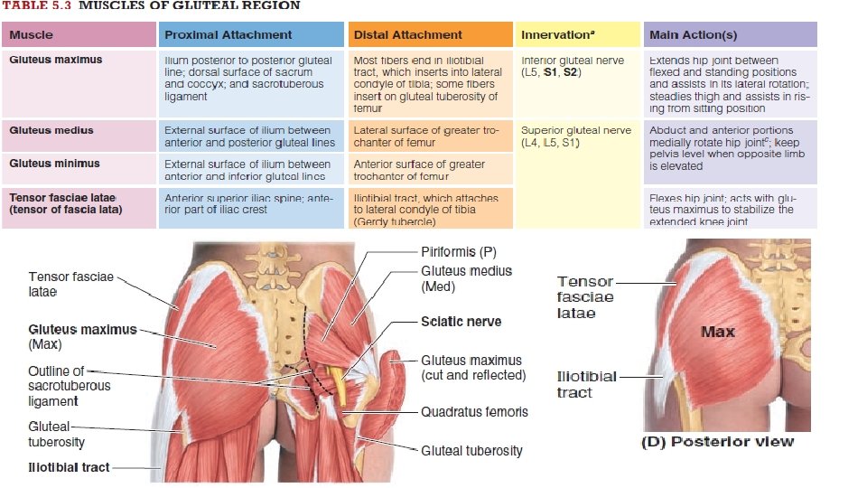
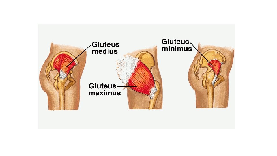
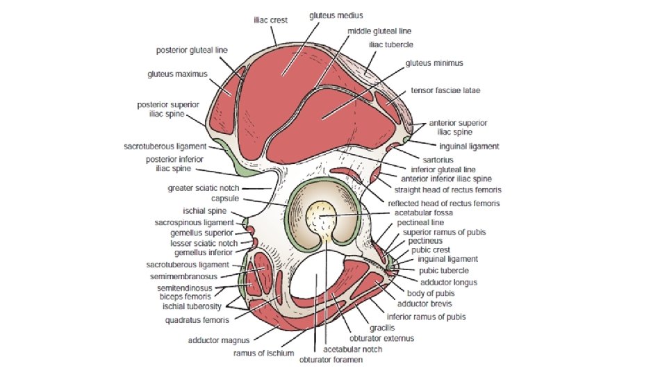
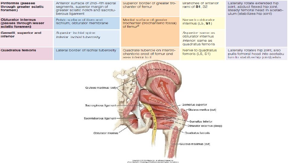
GLUTEAL REGION The gluteal region (hip and buttocks) is the prominent area posterior to the pelvis. It is bounded superiorly by the iliac crest, and ASIS (anterior superior iliac spine) and inferiorly by the gluteal fold. The gluteal folds demarcate the inferior border of the buttocks and the superior boundary of the thigh. Gluteal Muscles The gluteal muscles are organized into two layers: superficial and deep. The superficial layer consists of the three glutei (maximus, medius, and minimus) and the tensor fasciae latae The main actions of the gluteus maximus are extension and lateral rotation of the hip joint. The gluteus medius and minimus are abductors and medial rotators of the thigh. The tensor fasciae latae lies on the lateral side of the hip, enclosed between two layers of fascia lata. The tensor fasciae latae is an abductor and medial rotator of the hip joint. The deep layer consists of smaller muscles: the piriformis, obturator internus, superior and inferior gemelli, and quadratus femoris. These muscles, covered by the inferior half of the gluteus maximus, are lateral rotators of the thigh, but they also stabilize the hip joint
HIP JOINT
The ilium, the superior and largest part of the hip bone, contributes to the superior part of the acetabulum, the cup-like cavity (socket) on the lateral aspect of the hip bone for articulation with the head of the femur.
-The ilium consists of a body, which joins the pubis and ischium to the acetabulum, and an ala (wing), which is bordered superiorly by the iliac crest. it extends in front at the anterior superior iliac spine and behind at the posterior superior iliac spine. The iliac tubercle lies about 2 in. (5 cm) behind the anterior superior spine. -Below the anterior superior iliac spine is a prominence, the anterior inferior iliac spine; a similar prominence, the posterior inferior iliac spine, is located below the posterior superior iliac spine. Above and behind the acetabulum, the ilium possesses a large notch, the greater sciatic notch.
The proximal end of the femur consists of a head, neck, and greater and lesser trochanters. The head of the femur is covered with articular cartilage, except for a medially placed depression or pit, the fovea for the ligament of the head. The neck of the femur is trapezoidal; the narrow end supports the head and its broader base is continuous with the shaft.
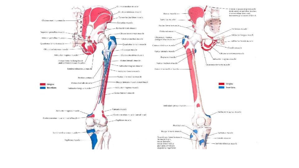
Where the neck joins the shaft are two large, blunt elevations—the trochanters. The conical lesser trochanter, with its rounded tip, extends medially from the posteromedial part of the junction of the femoral neck and shaft. Pectineal line just below lesser trochanter. The greater trochanter is a large, laterally placed mass that projects superomedially where the neck joins the shaft. The intertrochanteric line is a roughened ridge running from the greater to the lesser trochanter. A similar but smoother ridge, the intertrochanteric crest, joins the trochanters posteriorly. On the posterior surface of the shaft below the greater trochanter is the gluteal tuberosity for the attachment of the gluteus maximus muscle.
Hip Joint The hip joint forms the connection between the lower limb and the pelvic girdle. It is a strong, stable multiaxial ball and socket type of synovial joint. The femoral head is the ball, and the acetabulum is the socket. This joint is designed for stability over a wide range of movement. During standing, the weight of the upper body is transferred through the hip bones to the heads of the femurs.
ARTICULAR SURFACES The round head of the femur articulates with the cup-like acetabulum of the hip bone. The head is covered with articular cartilage, except for the pit or fovea for the ligament of the head of femur. The rim of the acetabulum consists of a semilunar articular part covered with articular cartilage, the lunate surface of the acetabulum.
Because the depth of the acetabulum is increased by the fibrocartilaginous acetabular labrum (lip)and the transverse acetabular ligament (bridging the acetabular notch), more than half of the head fits within the acetabulum. Centrally, a deep nonarticular part, the acetabular fossa, is formed mainly by the ischium.
JOINT CAPSULE The external fibrous layer of the joint capsule attaches proximally on the hip bone to the bony rim of the acetabulum and the transverse acetabular ligament. Distally, it attaches to the femoral neck only anteriorly at the intertrochanteric line and at the root of the greater trochanter. Posteriorly, the fibrous layer has an arched border that crosses the neck proximal to the intertrochanteric crest but is not attached to it. The joint capsule covers approximately the proximal two thirds of the neck of the femur posteriorly.
Anteriorly and superiorly, the capsule supports by the strong inverted Yshaped iliofemoral ligament , which attaches to the anterior inferior iliac spine and acetabular rim proximally and the intertrochanteric line distally. The iliofemoral ligament prevents hyperextension of the hip joint during standing.
Inferiorly and anteriorly by the pubofemoral ligament, which arises from the obturator crest of the pubic bone ( which forms part of the circumference of the obturator foramen superiorly and affords attachment to the obturator membrane) and passes laterally and inferiorly to merge with the fibrous layer of the joint capsule. This ligament blends with the medial part of the iliofemoral ligament and tightens during extension and abduction of the hip joint. The pubofemoral ligament resists excessive abduction of the hip joint.
Posteriorly by the weak ischiofemoral ligament, which arises from the ischial part of the acetabular rim and spirals superolaterally to the neck of the femur, medial to the base of the greater trochanter
The ligament of head of femur, primarily a synovial fold conducting a blood vessel, is weak and of little importance in strengthening the hip joint. Its wide end attaches to the margins of the acetabular notch and the transverse acetabular ligament; its narrow end attaches to the femur at the fovea for the ligament of the head of femur. Usually, the ligament contains a small artery to the head of the femur.
The transverse acetabular ligament is formed by the acetabular labrum as it bridges the acetabular notch. The ligament converts the notch into a tunnel through which the blood vessels and nerves enter the joint.
Synovial Membrane -The synovial membrane lines the capsule and is attached to the margins of the articular surfaces. -It covers the portion of the neck of the femur that lies within the joint capsule. -It ensheathes the ligament of the head of the femur and covers the pad of fat contained in the acetabular fossa. Bursae related to hip joint: The hip joint may communicate with the psoas bursa through a circular aperture between the pubofemoral and iliofemoral ligaments. Lymphatic drainage of hip joint: The lymph vessels from front aspect drain to the deep inguinal nodes, while those from the posterior and medial aspects run with the gluteal and obturator arteries repectively to reach the internal iliac nodes
HIP MOVEMENTS Hip movements are flexion–extension, abduction– adduction, medial & lateral rotation, and circumduction. ■■ Flexion is performed by the iliopsoas, rectus femoris, and sartorius and also by the adductor muscles. ■■ Extension (a backward movement of the flexed thigh) is performed by the gluteus maximus and the hamstring muscles. ■■ Abduction is performed by the gluteus medius and minimus, assisted by the sartorius, tensor fasciae latae, and piriformis. ■■ Adduction is performed by the adductor longus and brevis and the adductor fibers of the adductor magnus. These muscles are assisted by the pectineus and the gracilis.
■■ Lateral rotation is performed by the piriformis, obturator internus and externus, superior and inferior gemelli, and quadratus femoris, assisted by the gluteus maximus. ■■ Medial rotation is performed by the anterior fibers of the gluteus medius and gluteus minimus and the tensor fasciae latae. ■■ Circumduction is a combination of the previous movements. The extensor group of muscles is more powerful than the flexor group, and the lateral rotators are more powerful than the medial rotators.
flexion is limited by the tension of the hamstring group of muscles. Extension, is limited by the tension of the iliofemoral, pubofemoral, and ischiofemoral ligaments. Abduction is limited by the tension of the pubofemoral ligament, and adduction is limited by contact with the opposite limb. Lateral rotation is limited by the tension in the iliofemoral and pubofemoral ligaments, and medial rotation is limited by the ischiofemoral ligament.
BLOOD SUPPLY The arteries supplying the hip joint are : • Medial and lateral circumflex femoral arteries, which are usually branches of the profunda femoris artery but are occasionally branches of the femoral artery. The main blood supply is from the retinacular arteries arising as branches from the circumflex femoral arteries (especially the medial circumflex femoral artery) • Artery to the head of femur, a branch of the obturator artery that traverses the ligament of the head.
NERVE SUPPLY -The nerve supplying the muscles extending directly across and acting at a given joint also innervate the joint. Therefore, the nerve supply of the hip joint is from the following: • Femoral nerve or its muscular branches, anteriorly. • Obturator nerve, inferiorly. • Superior gluteal nerve, superiorly. • Nerve to quadratus femoris, posteriorly. • Siatic nerve , posteriorly.
Important Relations ■■ Anteriorly: Iliopsoas, pectineus, and rectus femoris muscles. The iliopsoas and pectineus separate the femoral vessels and nerve from the joint. ■■ Posteriorly: The obturator internus, the gemelli, and the quadratus femoris muscles separate the joint from the sciatic nerve. ■■ Superiorly: Piriformis and gluteus minimus. ■■ Inferiorly: Obturator externus tendon.
Thank You