Gas Exchangetopic 6 4 and H 6 Ventilation


")


, a globular protein, is the primary vehicle for transporting oxygen")


 • the majority of carbon dioxide is")


- Slides: 45
Gas Exchange-topic 6. 4 and -H 6
Ventilation • Ventilation: The flow of air in and out of the alveoli is called ventilation • two stages: • inspiration (or inhalation) and expiration (or exhalation).
Why do we need it ? • to maintain concentration gradients in the alveoli. • Diffusing O 2 and CO 2
Gas exchange occurs across specialized respiratory surfaces • Gas exchange-diffusion – Supplies oxygen for cellular respiration and disposes of carbon dioxide Respiratory medium (air of water) O 2 CO 2 Respiratory surface Organismal level Circulatory system Cellular level Energy-rich molecules from food Cellular respiration ATP
• Animals require large, moist respiratory surfaces for the adequate diffusion of respiratory gases – Between their cells and the respiratory medium, either air or water
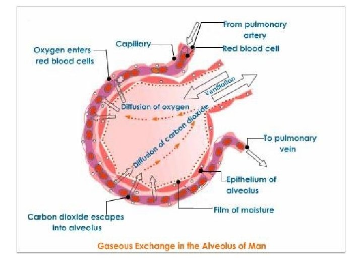
Mammalian Respiratory Systems: A Closer Look • A system of branching ducts – Conveys air to the lungs Branch from the pulmonary artery (oxygen-poor blood) Branch from the pulmonary vein (oxygen-rich blood) Terminal bronchiole Nasal cavity Pharynx Left lung Alveoli 50 µm Larynx Esophagus Trachea 50 µm Right lung Bronchus Bronchiole Diaphragm Heart SEM Colorized SEM
• In mammals, air inhaled through the nostrils – Passes through the pharynx into the trachea, bronchioles, and deadend alveoli, where gas exchange occurs
• Draw and label a diagram of the ventilation system, including trachea, lungs, bronchioles and alveoli, diaphragm
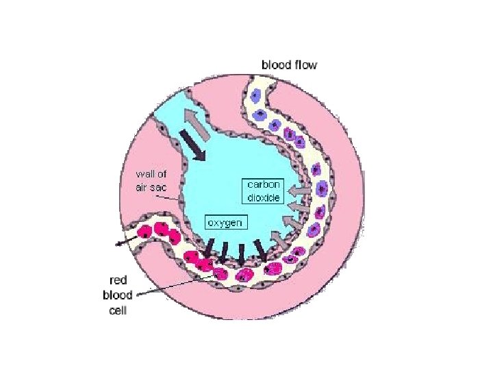
Features of Alveoli • Large surface area (600 million alveoli = 80 m 2) • Flattened epithelial cells of alveoli and close association with network of capillaries • Short diffusion distance from alveoli to blood (0. 5 -1. 0 um) • Moist surface for the solution of gases • Warm as blood supply
Alveoli
• Breathing ventilates the lungs • The process that ventilates the lungs is breathing – The alternate inhalation and exhalation of air – Is needed to maintain high concentration gradient in the alveoli.
How a Mammal Breathes • Mammals ventilate their lungs – By negative pressure breathing, which pulls air into the lungs Rib cage expands as rib muscles contract Air inhaled Rib cage gets smaller as rib muscles relax Air exhaled Lung Diaphragm INHALATION Diaphragm contracts (moves down) EXHALATION Diaphragm relaxes (moves up)
Inhalation • • Diaphragm contracts- flattens-downwards External intercostal muscles contract. Ribs move up and out Volume in chest/thoracic cavity/lung increases • Pressure decrease • Air rushes in
Exhalation • • • Diaphragm relaxes-moves up-curved up External muscle relaxes Decrease in lung volume Increase in pressure Air is rushed out
During exercise • Abdominal muscles, • Intercoastal muscles contracts • Faster exhalation
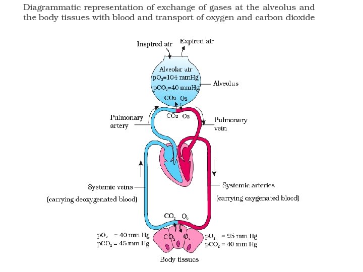
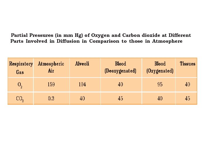
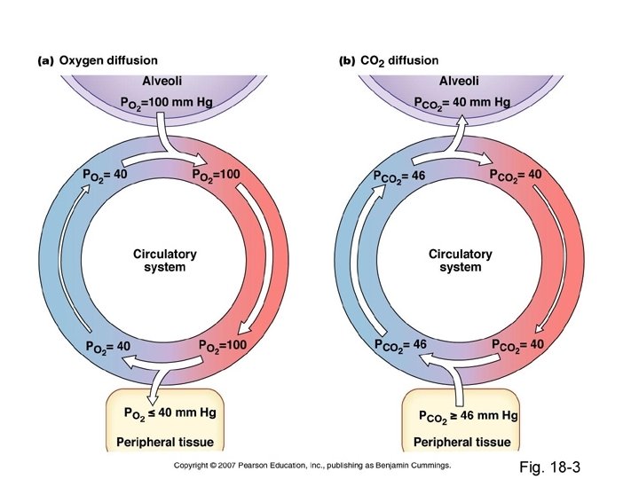
• Partial pressure-each gas has a partial pressure which is the pressure which the gas would have if it alone occupied the volume. • Units –k Pa-(kilo Pascal), mm Hg
• No. of membrane it has to cross
Control of Breathing in Humans • The main breathing control centers – Are located in two regions of the brain, the medulla oblongata and the pons Cerebrospinal fluid 1 The control center in the medulla sets the basic rhythm, and a control center in the pons moderates it, smoothing out the transitions between inhalations and exhalations. Pons 2 Nerve impulses trigger muscle contraction. Nerves from a breathing control center in the medulla oblongata of the brain send impulses to the diaphragm and rib muscles, stimulating them to contract and causing inhalation. Breathing control centers Medulla oblongata The medulla’s control center also helps regulate blood CO 2 level. Sensors in the medulla detect changes in the p. H (reflecting CO 2 concentration) of the blood and cerebrospinal fluid bathing the surface of the brain. 5 Nerve impulses relay changes in CO 2 and O 2 concentrations. Other sensors in the walls of the aorta and carotid arteries in the neck detect changes in blood p. H and send nerve impulses to the medulla. In response, the medulla’s breathing control center alters the rate and depth of breathing, increasing both to dispose of excess CO 2 or decreasing both if CO 2 levels are depressed. Carotid arteries In a person at rest, these nerve impulses result in about 10 to 14 inhalations per minute. Between inhalations, the muscles relax and the person exhales. Aorta Diaphragm Rib muscles The sensors in the aorta and 6 carotid arteries also detect changes in O 2 levels in the blood and signal the medulla to increase the breathing rate when levels become very low.
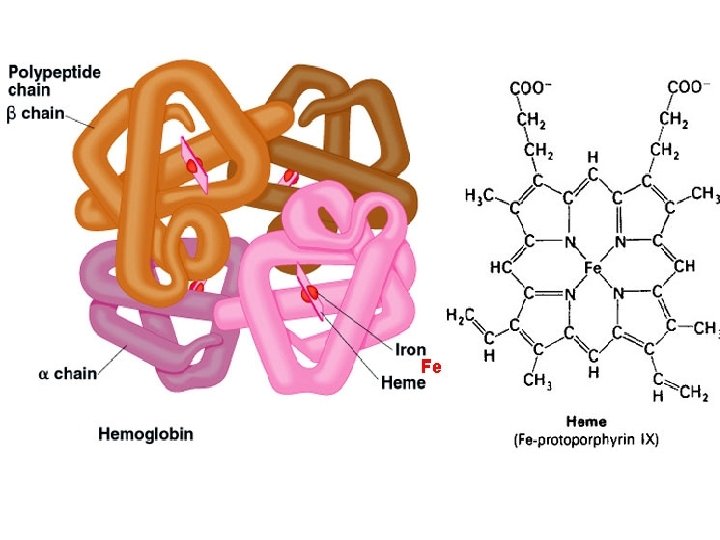
Hemoglobin
• Hemoglobin (Hb), a globular protein, is the primary vehicle for transporting oxygen in the blood. • To a much lesser degree-blood's plasma • Each Hb molecule has the capacity to carry four oxygen molecules.
Each Hb molecule can join with 4 oxygen molecules • The increasing affinity for oxygen allows the haemoglobin to rapidly saturate in the high partial pressures of oxygen of the alveoil capillaries. • Color changes-red---dark red—reddish purple
The normal range of values for hemoglobin content • Female: 11. 5 -15. 5 g /dl • Male: 13. 5 -17. 5 g/dl
• The amount of oxygen bound to the Hb at any time is related, • to the partial pressure of oxygen to which the Hb is exposed. • In the lungs, at the alveolar–capillary interface, the partial pressure of O 2 is typically high, and therefore the oxygen binds readily to Hb that is present.
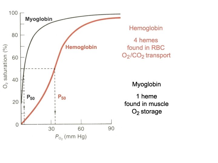
oxygen saturation. • How much of that capacity is filled by oxygen at any time is called the oxygen saturation. • Expressed as a percentage
Oxygen dissociation curves. • Important tool for understanding how our blood carries and releases oxygen. • relates oxygen saturation and partial pressure of oxygen in the blood (p. O 2), and is determined by what is called “Hb's affinity for oxygen”; • how readily Hb acquires and releases oxygen molecules into the fluid that surrounds it.
• As the blood circulates to other body tissue in which the partial pressure of O 2 is less, the Hb releases the O 2 into the tissue because the Hb cannot maintain its full bound capacity of O 2 in the presence of lower O 2 partial pressures
O 2 dissociation curve
The effect of carbon dioxide in the blood Hb can also bind carbon dioxide, but to a lesser extent. Carbaminohaemoglobin forms. Some carbon dioxide is carried in this form to the lungs from respiring tissues.
The presence of carbon dioxide helps the release of oxygen from Hb, this is known as the Bohr effect.
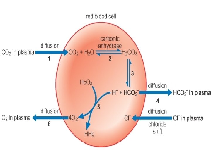
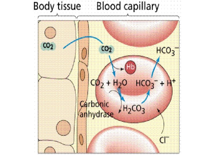
There are 3 ways in which carbon dioxide is transported in the blood: • 1. DISSOLVED CO 2 • more soluble in blood than oxygen • About 5 % of carbon dioxide is transported unchanged, simply dissolved in the plasma
2. BOUND TO HAEMOGLOBIN AND PLASMA PROTEINS • Carbon dioxide combines reversibly with Hb to form carbaminohaemoglobin. • Carbon dioxide does not bind to iron, as oxygen does, but to amino groups on the polypeptide chains of Hb. • 10 % of carbon dioxide is transported bound to Hb and plasma proteins
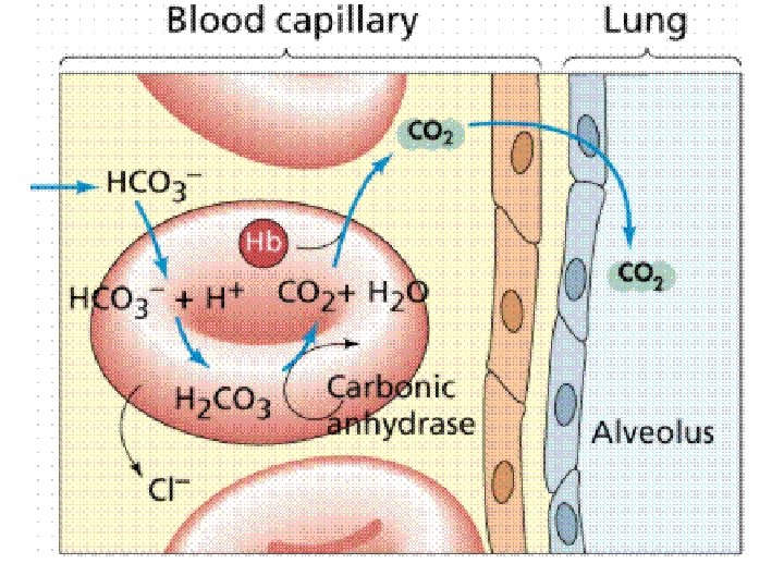
3. BICARBONATE IONS (HCO 3 - ) • the majority of carbon dioxide is transported in this way • CA-Carbonic anhydrase