
New Orleans EMS Airway Lecture Series Lecture 4
- Slides: 40
New Orleans EMS Airway Lecture Series: Lecture 4 The Pediatric Airway Jeffrey M. Elder, M. D. Deputy Medical Director
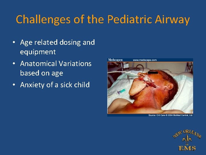
Challenges of the Pediatric Airway • Age related dosing and equipment • Anatomical Variations based on age • Anxiety of a sick child
Pediatric Airway Anatomy
Tongue • Located completely in the oral cavity until 2 years old – No portion makes up the upper/anterior pharyngeal wall • Potential site of airway obstruction – Difficult ventilation
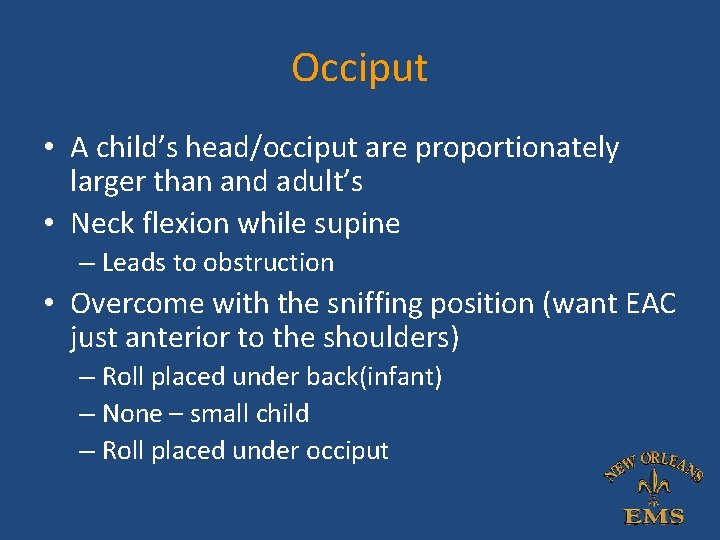
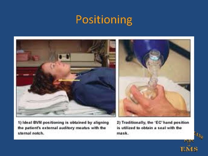
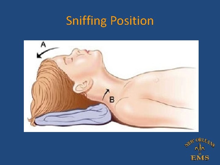
Occiput • A child’s head/occiput are proportionately larger than and adult’s • Neck flexion while supine – Leads to obstruction • Overcome with the sniffing position (want EAC just anterior to the shoulders) – Roll placed under back(infant) – None – small child – Roll placed under occiput
Positioning
Sniffing Position
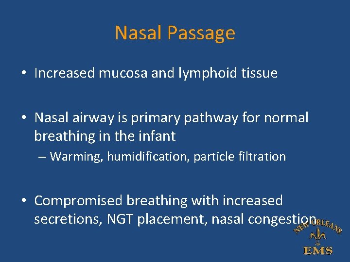
Nasal Passage • Increased mucosa and lymphoid tissue • Nasal airway is primary pathway for normal breathing in the infant – Warming, humidification, particle filtration • Compromised breathing with increased secretions, NGT placement, nasal congestion
Larynx • Newborns – Larynx at the base of the occiput/C 1 to C 4 • Enables epiglottis to lock the larynx into the nasopharynx by passing up behind the soft palate • Provides a direct air channel from the nares to the lungs, allowing liquids to pass on the sides into the esophagus
Larynx • Two separate anatomic pathways – Respiratory tract from the nose to the lungs – Digestive tract from the mouth to the stomach • Large Tongue – Entirely within the oral cavity • High Glottis • Difficult line of vision from mouth to the larynx during laryngoscopy – Anterior Airway
Anatomic Changes in Childhood • Occurs after the second year of life • Posterior 1/3 of tongue descends into the neck, forming upper anterior pharyngeal wall • By 7 years, the larynx lies between C 3 and C 6 • In adulthood, the larynx lies between C 4 – C 7 • Now loose the two separate pathways
Anatomy • In adults, the vocal cords and trachea are of equal dimensions • In newborns, the narrowest portion of the airway is the cricoid ring – Tight ET tubes may lead to cricoid damage, subglottic stenosis
Functional Issues • Children easily obstruct the airway – Racemic epinephrine can have dramatic results in the smallest areas of the airway (croup –cricoid ring) – Larger airway calibers do not see such dramatic results (epiglotitis) – forced nebs can lead to dynamic upper airway obstruction • Noxious stimuli can lead to dynamic obstruction and respiratory arrest – Crying child increases work of breathing 32 -fold – “leave them alone” • BMV may bridge through an obstruction – i. e. Epiglotitis – Increased inspiratory effort may collapse the airway – (extrathoracic trachea) – PPV can stent the airway open are relieve the obstruction
Physiology • Basal Oxygen consumption is approximately twice that of adults • Children have a decreased functional residual capacity (FRC) to body weight ratio • Desaturate much more quickly!! – Even given equivalent duration of preoxygenation • Be prepared to provide supplemental oxygen by BMV if oxygen saturation drops below 90%
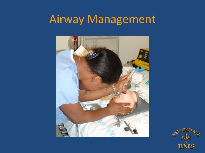
Airway Management
Evaluation • History – PMHx, Prematurity, Previous Intubations • Observation – Tachypnea – Accessory Muscle Use – Nasal Flaring – Tripoding
Evaluation • • • Position of comfort Grunting Cyanosis Drooling Wheezing Rales
Signs of Respiratory Failure • Decreased level of consciousness • Grunting and increased work of breathing • Poor Air Entry / Decreased breath sounds • Bradycardia • Apnea/Slow Respiratory Rate
Reasons to Intubate • Failure to Oxygenate • Failure to Ventilate • Expected Clinical Course
Airway Management
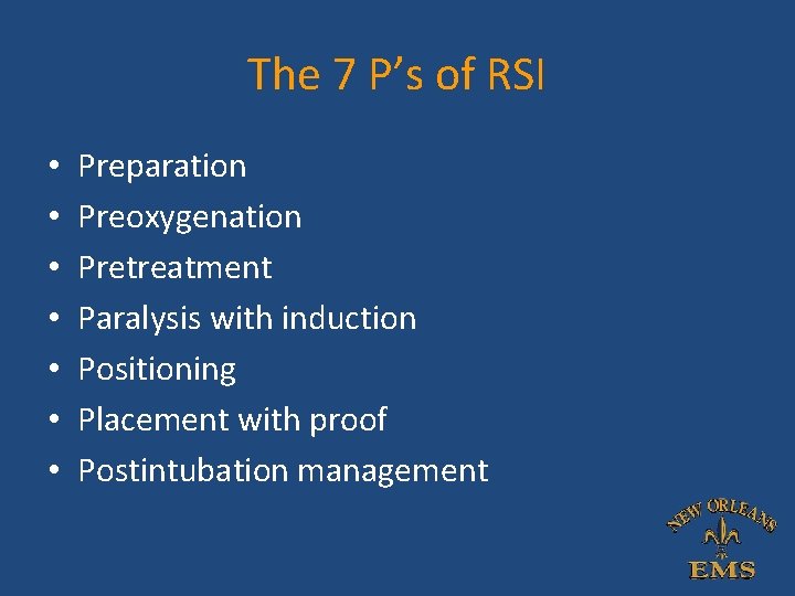
The 7 P’s of RSI • • Preparation Preoxygenation Pretreatment Paralysis with induction Positioning Placement with proof Postintubation management
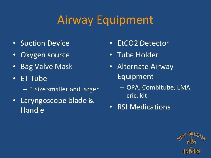
Airway Equipment • • Suction Device Oxygen source Bag Valve Mask ET Tube – 1 size smaller and larger • Laryngoscope blade & Handle • Et. CO 2 Detector • Tube Holder • Alternate Airway Equipment – OPA, Combitube, LMA, cric. kit • RSI Medications
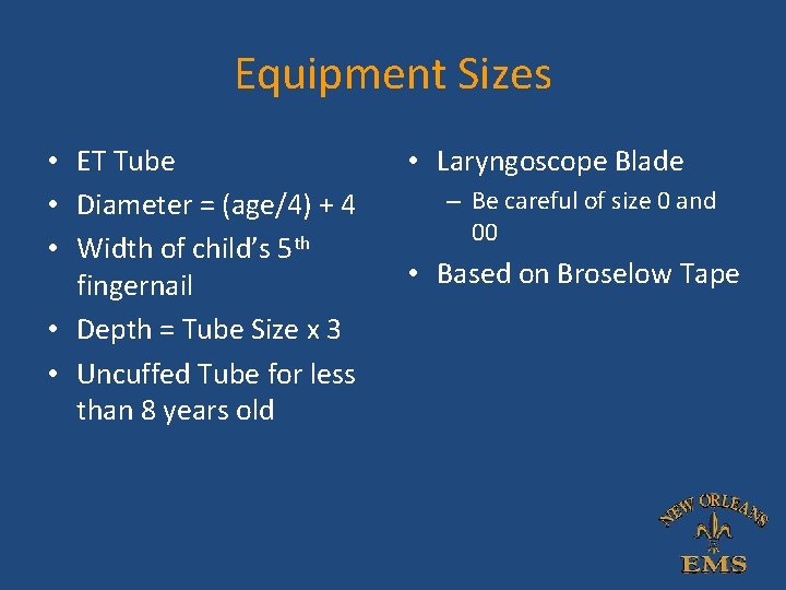
Equipment Sizes • ET Tube • Diameter = (age/4) + 4 • Width of child’s 5 th fingernail • Depth = Tube Size x 3 • Uncuffed Tube for less than 8 years old • Laryngoscope Blade – Be careful of size 0 and 00 • Based on Broselow Tape
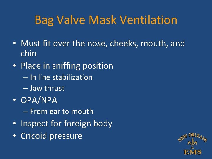
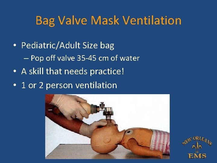
Bag Valve Mask Ventilation • Must fit over the nose, cheeks, mouth, and chin • Place in sniffing position – In line stabilization – Jaw thrust • OPA/NPA – From ear to mouth • Inspect foreign body • Cricoid pressure
Bag Valve Mask Ventilation • Pediatric/Adult Size bag – Pop off valve 35 -45 cm of water • A skill that needs practice! • 1 or 2 person ventilation
Rapid Sequence Induction – Etomidate 0. 3 mg/kg – Succinylcholine 2 mg/kg • faster metabolism than adults • Still first line for full-stomach or emergency intubation – Rocuronium 1 mg/kg – Atropine 0. 02 mg/kg min. 0. 1 mg – Lidocaine 1 mg/kg
Endotracheal Intubation • Usually after airway control and ventilation/oxygenation • Preoxygenation – Don’t bag – 3 minutes of 100% oxygen via BVM • Pick the right equipment! • Most effective and reliable airway management/protection • Always a clinical decision
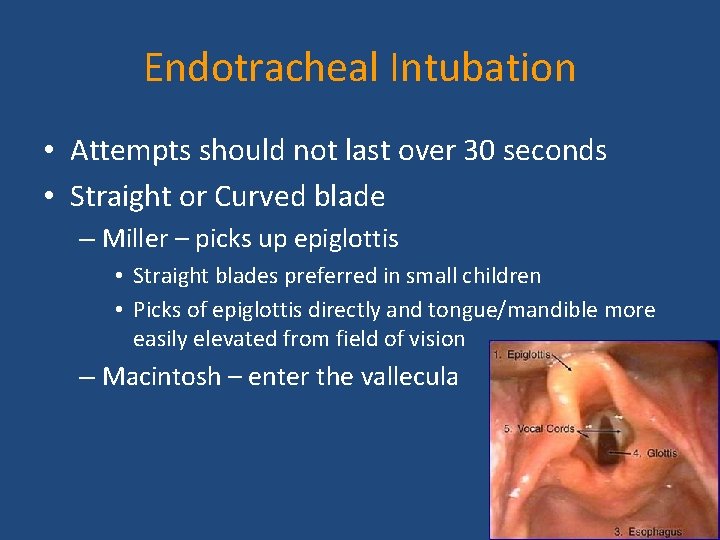
Endotracheal Intubation • Attempts should not last over 30 seconds • Straight or Curved blade – Miller – picks up epiglottis • Straight blades preferred in small children • Picks of epiglottis directly and tongue/mandible more easily elevated from field of vision – Macintosh – enter the vallecula
Post Intubation Management • Verification of Tube Placement – Visualization – ETC 02 – Auscultation • Secure the tube with tape or commercial device – Head/neck immobilization in small children to avoid neck movement and dislodgement
Post Intubation Management • Place NG tube after ETI – Decompress the stomach – Avoid micro aspiration in mechanically ventilated patients
Contraindications to RSI • Major Laryngeal trauma • Upper Airway Obstructions • Distorted Facial/Airway Anatomy • Operator Concern of Difficult Airway
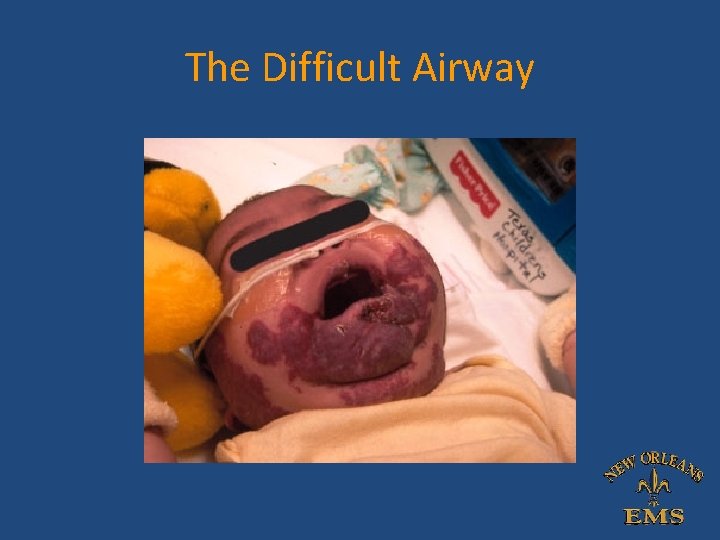
The Difficult Airway
The Difficult Airway • Direct Examination – Mental-Hyoid Distance – Upper-Lower Incisor Distance • Large Tongue • Blood, swelling, secretions • Limited C spine mobility/scoliosis • Limited mandibular ROM • Maxillofacial/ Larynx trauma • *Angoiedema • *Anaphylaxis • *Epiglottitis • *Croup • Morbid Obesity • Micrognathia • Burns • Foreign Body
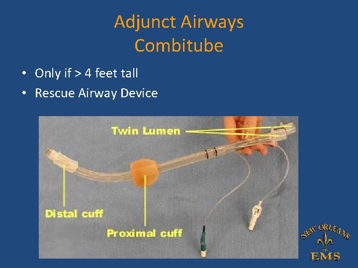
Adjunct Airways Combitube • Only if > 4 feet tall • Rescue Airway Device
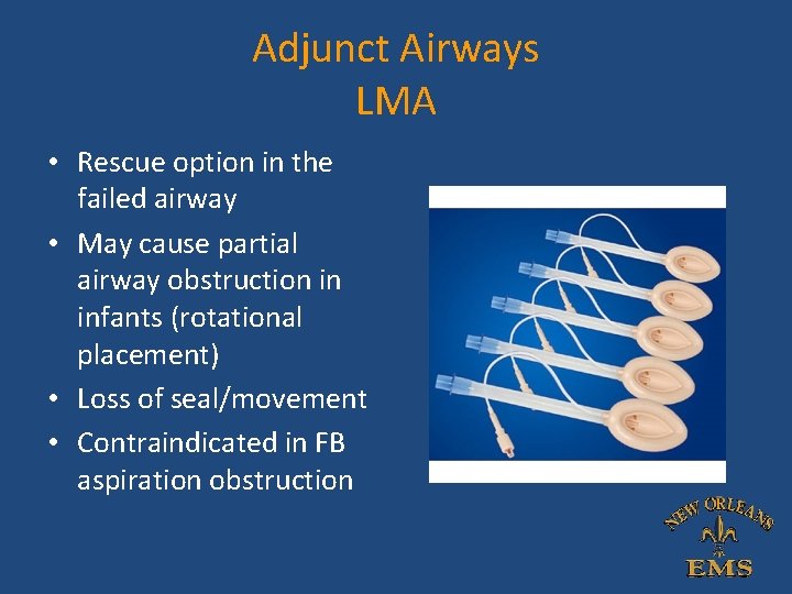
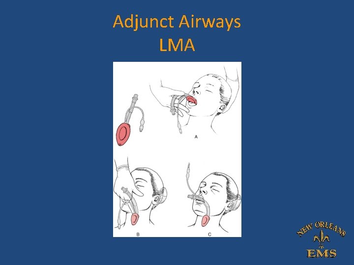
Adjunct Airways LMA • Rescue option in the failed airway • May cause partial airway obstruction in infants (rotational placement) • Loss of seal/movement • Contraindicated in FB aspiration obstruction
Adjunct Airways LMA
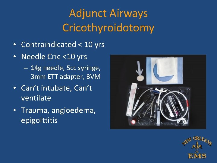
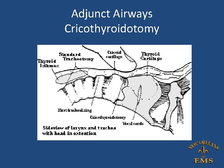
Adjunct Airways Cricothyroidotomy • Contraindicated < 10 yrs • Needle Cric <10 yrs – 14 g needle, 5 cc syringe, 3 mm ETT adapter, BVM • Can’t intubate, Can’t ventilate • Trauma, angioedema, epigolttitis
Adjunct Airways Cricothyroidotomy
References • Trauma Reports Volume 8, No. 1. Jan/Feb 2007. AHC Media, LLC. • Managing the Pediatric Airway in the ED, Pediatric Emergency Medicine Practice. Volume 3, No. 1. January 2006 • Manual of Emergency Airway Management, 3 rd Edition. Walls, R. and Murphy, M. 2008.