Fluid and Hemodynamic Disorders Wheres my water Intracellular


- Slides: 57
Fluid and Hemodynamic Disorders
Where’s my water? • Intracellular • Ions • Ion specific gates in cell membrane • Cellular proteins • Extracellular • Interstitial (between the cells) Lymph • Intravascular • Blood • Lymphatic fluid
Movement of water in the vascular system • Hydrostatic, the pumping pressure • Heart • Skeletal muscle action • Oncotic or osmotic, holds fluid in. • Proteins such as albumin • Cellular elements such as RBCs
Intracellular & Extracellular Water
Things can go wrong • • • Heart failure Kidney failure Myocardial infarction Pulmonary emobolus Tissue congestion Edema
Edema • Too much extracellular fluid. – Swelling • tumor – Localized or – Generalized – Dependent • action of gravity
Tansudate or Exudate? • Exudate – Inflammatory water – Part of the inflammatory reaction • Rubor, dolor, calor, tumor – Purposeful and intentional – Localized
Tansudate or Exudate? • Transudate – Leakage, not part of healing – Increased hydrostatic pressure • Heart failure • Lymphatic obstruction – Decreased oncotic pressure • Decreased albumin
Congestive Heart Failure
Congestive Heart Failure
Passive Congestion
Chronic Passive Congestion, Nutmeg Liver
Chronic Passive Congestion
Pulmonary Edema
Pulmonary Edema
Pulmonary Edema
Pitting Edema
Lymphedema
Papilledema
Water in Hollow Spaces • Hydrothorax • Hydropericardium • Hydroperitoneum – Ascites
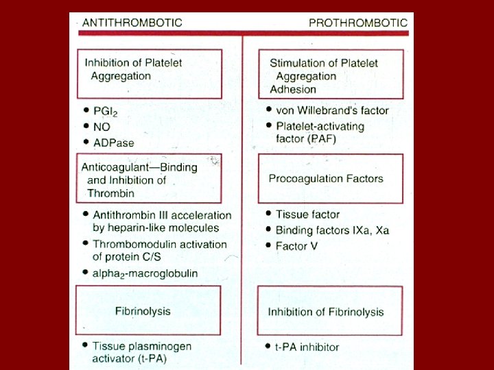
Healthy Blood Clotting • Platelets • Vessels • Clotting Proteins
Healthy Clotting
Clotting Factors
Factor Activation
Hematoma
Petechiae
Thrombosis • A pathological clot • A clot forming in the fixed vascular system.
Thrombosis 1. Endothelial damage 2. Stasis and clotting factor activation 3. Clotting factor abnormalities – Too many clotting proteins • • Pregnancy Cancers – Too little inhibition – Abnormal factors • Leiden Factor (abnormal V)
Thrombosis
Thrombosis • Arterial Side Thrombi – Platelet activation – Endothelial cell injury • Venous Side Thrombi – Stasis – Clotting factor activation – Endothelial cell injury
Coronary Artery Thombosis • Angiogram
Acute Myocardial Infarction
Mural Thrombus
Aneurysm with Thrombus
Deep Leg Vein Thrombosis
Airplane Travel • Gunner turret
Outcomes of a DVT
Embolus • Space occupying mass moving in the fixed vascular system • • Blood clot Bone Fragments Amniotic Fluid Air
Pulmonary Embolus
Pulmonary Embolus
Infarction • Anemic – End artery supply – No blood – White • Hemorrhagic – Venous occlusion – Loose tissues – Dual blood supply – Red
Anemic Infarct
Anemic Infarct
Cerebral Infarction
Hemorrhagic Infarct
Shock • • Poor perfusion Tissue hypoxia Tissue acidosis Many causes – – Poor pumping by heart Low blood volume Loss of fluid Overwhelming infections
Types of Shock • Cardiogenic – Decreased output • Hypovolemic – Blood loss – Fluid loss • Anaphylaxis – Ig. E and histamine • Septic – Gram negative rods – Toxins
What Happens Next? • Compensated – Fluid shifts • Decompensated – Progression possible • Irreversible – No recovery
The Shock Spiral
Summary • Fluid shifts • Oncotic & Hydrostatic Pressures • Excessive tissue water • Exudate vs. Transudate • Clot formation • Vessels, platelets & proteins • Thrombosis • Pathological clot • Arterial = endothelial damage & platelet activation. • Venous = stasis and factor activation
Summary • Infarction • Ischemic = end artery organ • Hemorrhagic = venous or dual blood supply • Tissue vulnerability – Brain – Kidney – muscle