Emergency Medical Responder First on Scene Eleventh Edition



- Slides: 81
Emergency Medical Responder: First on Scene Eleventh Edition Chapter 22 Caring for Chest and Abnominal Emergencies Slides in this Presentation Contain Hyperlinks. JAWS users should be able to get a list of links by using “INSERT + F 7 Copyright © 2019, 2016, 2011 Pearson Education, Inc. All Rights Reserved
Objectives (1 of 7) 22. 1 Review the anatomy of the chest and abdomen from Chapter 4. 22. 2 Define the following terms: – Closed chest injury – Crepitus – Diaphragm – Distention – Evisceration – Flail chest Copyright © 2019, 2016, 2011 Pearson Education, Inc. All Rights Reserved
Objectives (2 of 7) 22. 2 Define the following terms: – Guarding – Hemothorax – Mediastinum – Occlusive dressing – Open chest injury – Paradoxical movement – Penetrating injury – Pleura Copyright © 2019, 2016, 2011 Pearson Education, Inc. All Rights Reserved
Objectives (3 of 7) 22. 2 Define the following terms: – Pleural space – Pneumothorax – Quadrant – Retroperitoneal cavity – Spontaneous pneumothorax – Sucking chest wound – Tension pneumothorax Copyright © 2019, 2016, 2011 Pearson Education, Inc. All Rights Reserved
Objectives (4 of 7) 22. 3 Describe the major structures of the thoracic cavity. 22. 4 Explain the relationship between chest injury and perfusion. 22. 5 Differentiate between an open and closed chest injury. 22. 6 Describe the signs and symptoms of a closed chest injury. Copyright © 2019, 2016, 2011 Pearson Education, Inc. All Rights Reserved
Objectives (5 of 7) 22. 7 Explain the appropriate assessment of a patient with a chest injury. 22. 8 Explain the appropriate care of a patient with a closed chest injury. 22. 9 Explain the appropriate care of a patient with an open chest injury. 22. 10 Describe the major structures of the abdominal and pelvic cavities. Copyright © 2019, 2016, 2011 Pearson Education, Inc. All Rights Reserved
Objectives (6 of 7) 22. 11 Describe the signs and symptoms of internal bleeding. 22. 12 Explain the appropriate assessment and care of a patient with abdominal pain. 22. 13 Explain the appropriate assessment and care of a patient with an open abdominal injury. Copyright © 2019, 2016, 2011 Pearson Education, Inc. All Rights Reserved
Objectives (7 of 7) 22. 14 Demonstrate the appropriate assessment and care of a patient with a chest injury. 22. 15 Demonstrate the appropriate assessment and care of a patient with abdominal pain. 22. 16 Demonstrate the appropriate assessment and care of an open abdominal injury. 22. 17 Value the importance of proper body substance isolation (BSI) precautions when assisting with chest and abdominal injuries. Copyright © 2019, 2016, 2011 Pearson Education, Inc. All Rights Reserved
SECTION: ANATOMY OF THE CHEST Copyright © 2019, 2016, 2011 Pearson Education, Inc. All Rights Reserved
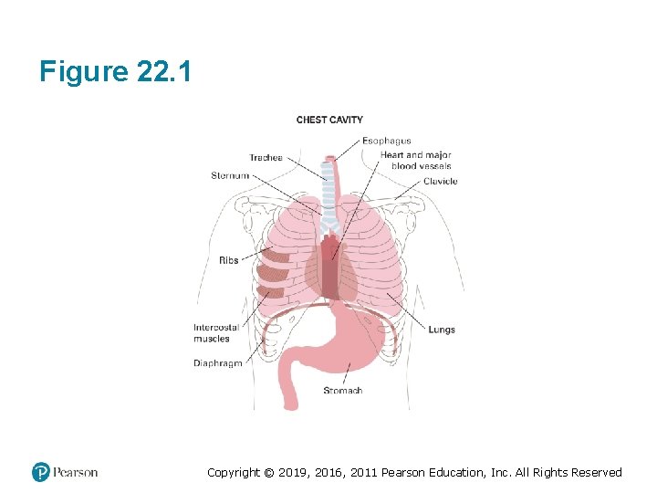
Anatomy of the Chest (1 of 3) • Chest cavity or thoracic cavity makes up approximately half of torso. • 12 pairs of ribs make the shape of the chest. • Major organs contained within chest are the heart and lungs. Copyright © 2019, 2016, 2011 Pearson Education, Inc. All Rights Reserved
Anatomy of the Chest (2 of 3) • Mediastinum houses the following: – Trachea – Esophagus – Heart – Vena cava – Aorta Copyright © 2019, 2016, 2011 Pearson Education, Inc. All Rights Reserved
Figure 22. 1 Copyright © 2019, 2016, 2011 Pearson Education, Inc. All Rights Reserved
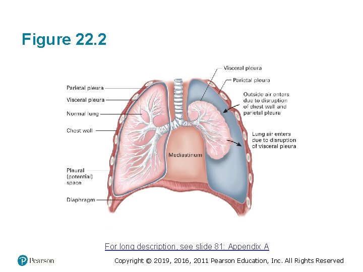
Anatomy of the Chest (3 of 3) • Left and right sides of chest are occupied by the lungs. – Pleura ▪ Thin saclike structure that surrounds each lung – Pleural space ▪ Potential space that exists between visceral and parietal pleura in chest Copyright © 2019, 2016, 2011 Pearson Education, Inc. All Rights Reserved
Figure 22. 2 For long description, see slide 81: Appendix A Copyright © 2019, 2016, 2011 Pearson Education, Inc. All Rights Reserved
Anatomy of the Chest: Think About It • What might happen if a chest injury disrupts the parietal pleura? • What might happen if a lung injury disrupts the visceral pleura? • What if there is a problem with the fluid that provides lubrication between the pleural layers? Copyright © 2019, 2016, 2011 Pearson Education, Inc. All Rights Reserved
SECTION: CHEST INJURIES Copyright © 2019, 2016, 2011 Pearson Education, Inc. All Rights Reserved
Chest Injuries (1 of 12) • Closed Chest Injuries – Blunt force trauma ▪ Blow to chest can fracture ribs, sternum, and rib cartilages. –Falls –Contact sports –Vehicle collisions –Blasts Copyright © 2019, 2016, 2011 Pearson Education, Inc. All Rights Reserved
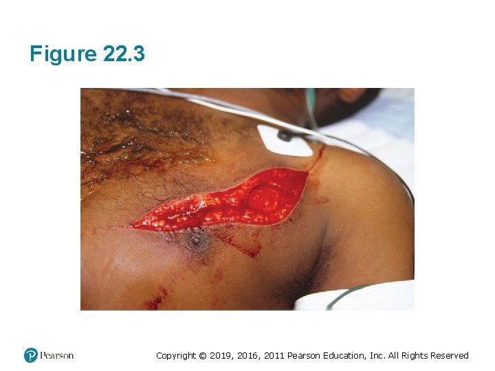
Chest Injuries (2 of 12) • Open Chest Injuries – Penetrating injury ▪ Penetrate chest wall, damaging internal organs and impairing respiration. –Bullet –Knife –Glass –Steel rods –Pipes Copyright © 2019, 2016, 2011 Pearson Education, Inc. All Rights Reserved
Figure 22. 3 Copyright © 2019, 2016, 2011 Pearson Education, Inc. All Rights Reserved
Chest Injuries (3 of 12) • Compression – Results from severe blunt trauma in which chest is rapidly compressed ▪ Driver in motor-vehicle collision strikes chest on steering column. Copyright © 2019, 2016, 2011 Pearson Education, Inc. All Rights Reserved
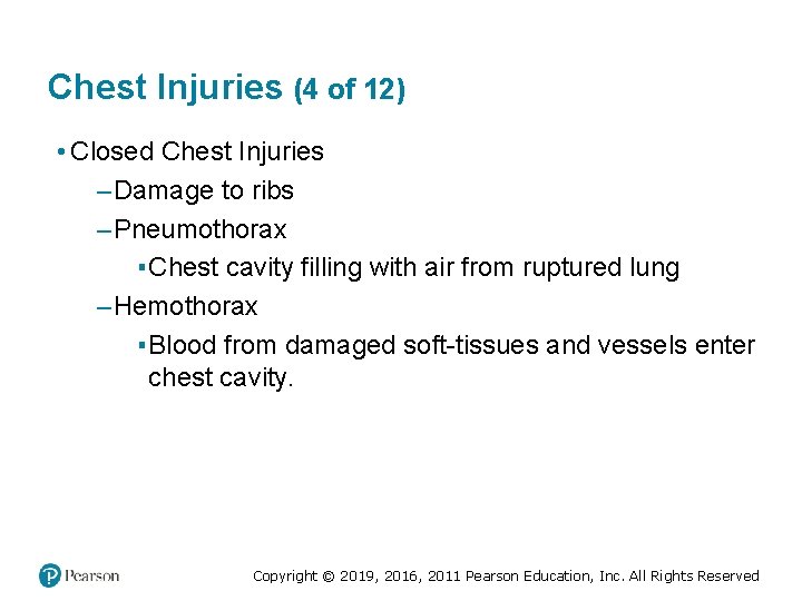
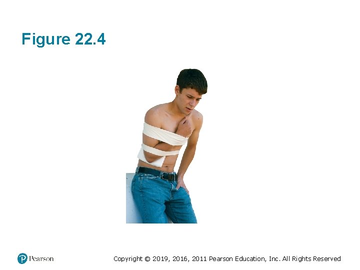
Chest Injuries (4 of 12) • Closed Chest Injuries – Damage to ribs – Pneumothorax ▪ Chest cavity filling with air from ruptured lung – Hemothorax ▪ Blood from damaged soft-tissues and vessels enter chest cavity. Copyright © 2019, 2016, 2011 Pearson Education, Inc. All Rights Reserved
Figure 22. 4 Copyright © 2019, 2016, 2011 Pearson Education, Inc. All Rights Reserved
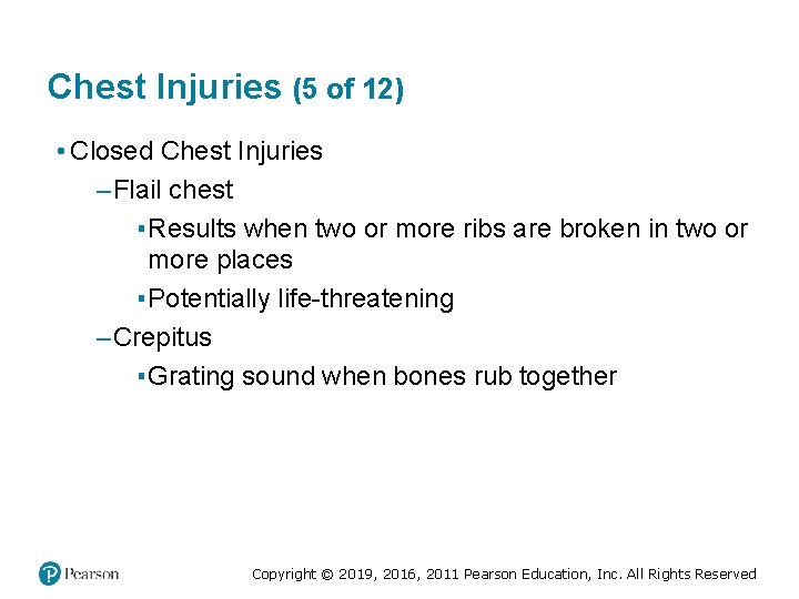
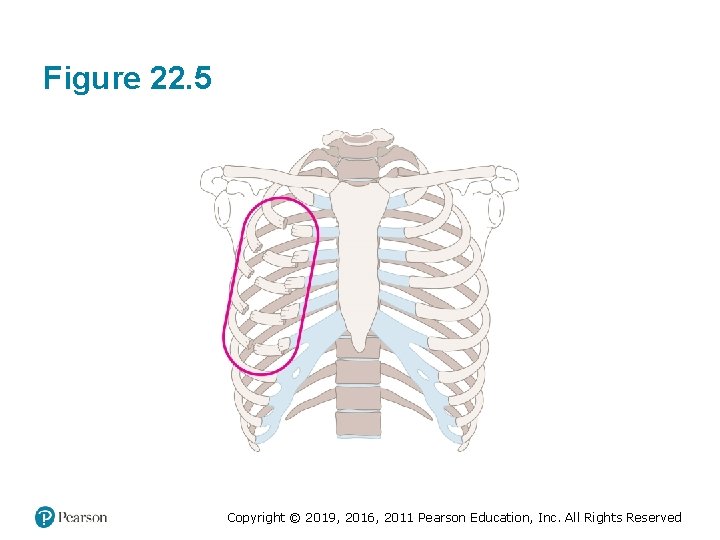
Chest Injuries (5 of 12) • Closed Chest Injuries – Flail chest ▪ Results when two or more ribs are broken in two or more places ▪ Potentially life-threatening – Crepitus ▪ Grating sound when bones rub together Copyright © 2019, 2016, 2011 Pearson Education, Inc. All Rights Reserved
Figure 22. 5 Copyright © 2019, 2016, 2011 Pearson Education, Inc. All Rights Reserved
Chest Injuries (6 of 12) • Closed Chest Injury Signs/Symptoms – Pain on breathing – Ecchymosis – Increased difficulty breathing – Accessory muscle use – Uneven chest wall movement during breathing – Signs and symptoms of shock Copyright © 2019, 2016, 2011 Pearson Education, Inc. All Rights Reserved
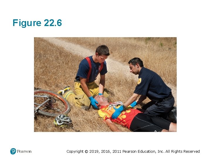
Chest Injuries (7 of 12) • Closed Chest Injury Management – Perform primary assessment. – Ensure ABCs are intact. – Provide positive pressure ventilations, if breathing is inadequate. – Remove clothing over area where there is complaint of pain. – Observe and palpate for signs of deformity. Copyright © 2019, 2016, 2011 Pearson Education, Inc. All Rights Reserved
Chest Injuries (8 of 12) • Closed Chest Injury Management – Administer oxygen per local protocols. – Splint chest using bulky dressings or towels. – Place patient in a position of comfort, if no suspected spine injury. – Care for shock. – Transport patient. Copyright © 2019, 2016, 2011 Pearson Education, Inc. All Rights Reserved
Figure 22. 6 Copyright © 2019, 2016, 2011 Pearson Education, Inc. All Rights Reserved
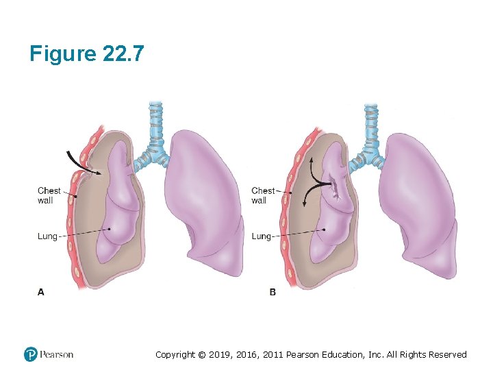
Chest Injuries (9 of 12) • Open Chest Injuries – Sucking chest wound ▪ Open chest wound characterized by sucking sound each time patient inhales – Tension pneumothorax ▪ Air builds up inside chest cavity, causing excessive pressure on one side of chest. Copyright © 2019, 2016, 2011 Pearson Education, Inc. All Rights Reserved
Figure 22. 7 Copyright © 2019, 2016, 2011 Pearson Education, Inc. All Rights Reserved
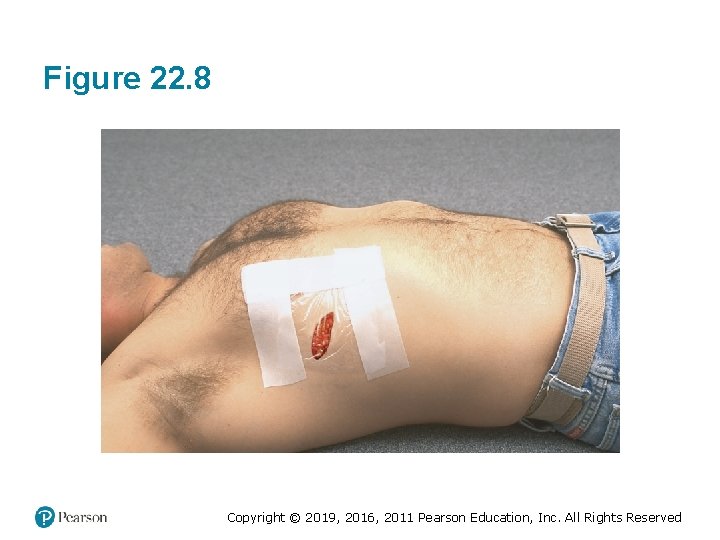
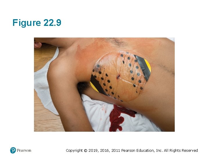
Chest Injuries (10 of 12) • Open Chest Injuries Management – Immediately seal with something that prevents air from entering wound, like gloved hand. – Take appropriate Standard Precautions. – Place an occlusive dressing directly over wound and hold it in place. – Provide high-flow oxygen. – Care for shock. Copyright © 2019, 2016, 2011 Pearson Education, Inc. All Rights Reserved
Figure 22. 8 Copyright © 2019, 2016, 2011 Pearson Education, Inc. All Rights Reserved
Figure 22. 9 Copyright © 2019, 2016, 2011 Pearson Education, Inc. All Rights Reserved
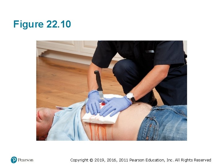
Chest Injuries (11 of 12) • Impaled Chest Wounds – Impaled objects must be stabilized and left in place. – Take appropriate Standard Precautions. – Perform primary assessment. – Ensure ABCs are intact. – Assist ventilations as appropriate. Copyright © 2019, 2016, 2011 Pearson Education, Inc. All Rights Reserved
Chest Injuries (12 of 12) • Impaled Chest Wounds – Provide high-flow oxygen per local protocol. – Provide care for shock. – Initiate immediate transport. Copyright © 2019, 2016, 2011 Pearson Education, Inc. All Rights Reserved
Figure 22. 10 Copyright © 2019, 2016, 2011 Pearson Education, Inc. All Rights Reserved
Chest Injuries: Think About It • You respond to a residential home to find a 26 -year-old female who was struck by a felled tree which impacted her chest. • What are your concerns? • How do you proceed? Copyright © 2019, 2016, 2011 Pearson Education, Inc. All Rights Reserved
SECTION: ABDOMINAL EMERGENCIES Copyright © 2019, 2016, 2011 Pearson Education, Inc. All Rights Reserved
Abdominal Emergencies (1 of 10) • Anatomy of the Abdomen and the Pelvis – Diaphragm ▪ Primary muscle of respiration ▪ Divides chest cavity from abdominal cavity – Epigastric ▪ Area below the ribs to just above the naval Copyright © 2019, 2016, 2011 Pearson Education, Inc. All Rights Reserved
Abdominal Emergencies (2 of 10) • Anatomy of the Abdomen and the Pelvis – Periumbilical ▪ Area around the naval – Superpubic ▪ Area below the naval to the pubic region Copyright © 2019, 2016, 2011 Pearson Education, Inc. All Rights Reserved
Abdominal Emergencies (3 of 10) • Anatomy of the Abdomen and the Pelvis – Parietal peritoneum ▪ Thin layer of tissue lining the wall of the abdominal cavity – Visceral peritoneum ▪ Thin layer of tissue lining most of the abdominal organs Copyright © 2019, 2016, 2011 Pearson Education, Inc. All Rights Reserved
Abdominal Emergencies (4 of 10) • Anatomy of the Abdomen and the Pelvis – Retroperitoneal cavity ▪ Area behind abdominal cavity that contains kidneys and ureters Copyright © 2019, 2016, 2011 Pearson Education, Inc. All Rights Reserved
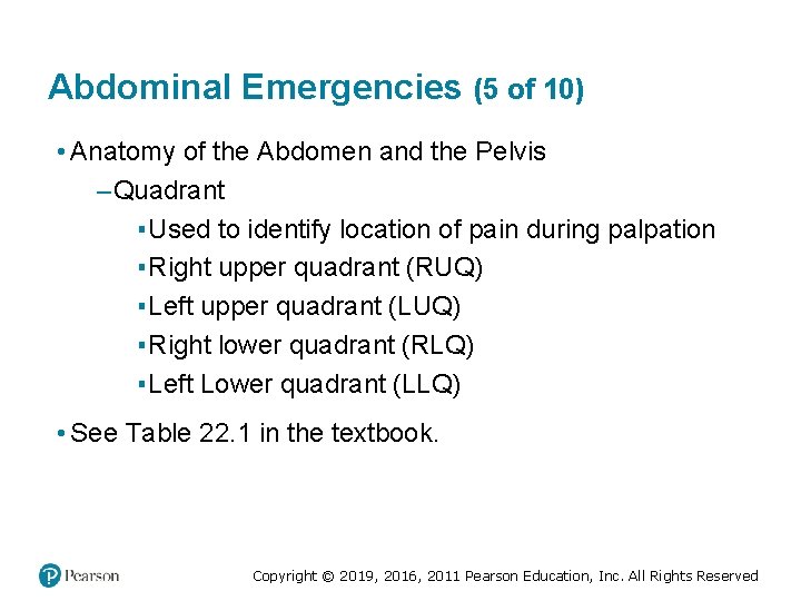
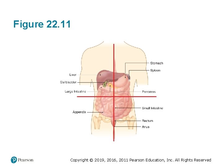
Abdominal Emergencies (5 of 10) • Anatomy of the Abdomen and the Pelvis – Quadrant ▪ Used to identify location of pain during palpation ▪ Right upper quadrant (RUQ) ▪ Left upper quadrant (LUQ) ▪ Right lower quadrant (RLQ) ▪ Left Lower quadrant (LLQ) • See Table 22. 1 in the textbook. Copyright © 2019, 2016, 2011 Pearson Education, Inc. All Rights Reserved
Figure 22. 11 Copyright © 2019, 2016, 2011 Pearson Education, Inc. All Rights Reserved
Abdominal Emergencies (6 of 10) • Generalized Abdominal Pain – Bleeding – Infection – Ulcers – Indigestion – Constipation – Food poisoning – Menstrual cramps Copyright © 2019, 2016, 2011 Pearson Education, Inc. All Rights Reserved
Abdominal Emergencies (7 of 10) • Generalized Abdominal Pain – Diabetic emergencies – Kidney stones – Gallstones – Appendicitis – Ectopic pregnancy Copyright © 2019, 2016, 2011 Pearson Education, Inc. All Rights Reserved
Abdominal Emergencies (8 of 10) • Acute Abdominal Pain Signs/Symptoms – Pain that is either sharp or dull – Pain on palpation – Rigid or tight abdomen – Bloating (distention) Copyright © 2019, 2016, 2011 Pearson Education, Inc. All Rights Reserved
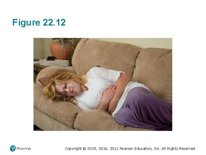
Abdominal Emergencies (9 of 10) • Acute Abdominal Pain Signs/Symptoms – Nausea/vomiting – Cramping – Pain that radiates to other areas – Guarding ▪ Protecting abdomen Copyright © 2019, 2016, 2011 Pearson Education, Inc. All Rights Reserved
Figure 22. 12 Copyright © 2019, 2016, 2011 Pearson Education, Inc. All Rights Reserved
Abdominal Emergencies (10 of 10) • Acute Abdominal Pain Assessment – Rule out history of trauma. – Injuries to abdomen cause bleeding that is very slow. – Signs and symptoms can be delayed for hours, sometimes days. – Obtain a thorough medical history. Copyright © 2019, 2016, 2011 Pearson Education, Inc. All Rights Reserved
Abdominal Emergencies: Think About It (1 of 3) • You respond to the home of a 14 -year-old female. Her mother reports that she dismissed her from school because of severe abdominal pain. She attempted to take the girl to the pediatrician, but the pain had intensified and she is unable to walk. Copyright © 2019, 2016, 2011 Pearson Education, Inc. All Rights Reserved
Abdominal Emergencies: Think About It (2 of 3) • The patient has no other medical history, takes no medication, and has no allergies. She has a pulse of 100, blood pressure of 90/60, and respiratory rate of 24. You palpate the abdomen and find the patient guarding her RLQ, so you palpate that quadrant last (gently). She screams out in pain. Copyright © 2019, 2016, 2011 Pearson Education, Inc. All Rights Reserved
Abdominal Emergencies: Think About It (3 of 3) 1. What are your suspicions? 2. How will you proceed? 3. What other questions should you be sure to include in your assessment of this patient? Copyright © 2019, 2016, 2011 Pearson Education, Inc. All Rights Reserved
SECTION: ABDOMINAL INJURIES Copyright © 2019, 2016, 2011 Pearson Education, Inc. All Rights Reserved
Abdominal Injuries (1 of 4) • Abdominal injuries can produce life-threatening emergencies. • Signs and Symptoms – Deep cut or puncture wound to abdomen, pelvis, or lower back – Blunt trauma to abdomen or pelvis – Pain or cramps in abdominal or pelvic region Copyright © 2019, 2016, 2011 Pearson Education, Inc. All Rights Reserved
Abdominal Injuries (2 of 4) • Signs and Symptoms – Guarding – Lying still with legs drawn up – Rapid, shallow breathing – Rapid pulse – Rigid, distended, and/or tender abdomen Copyright © 2019, 2016, 2011 Pearson Education, Inc. All Rights Reserved
Abdominal Injuries (3 of 4) • Caring for Closed Abdominal Injury – Perform thorough assessment of abdomen. – Palpate all quadrants. – Expose abdomen to observe for signs of injury. – Allow patient to maintain position of comfort. Copyright © 2019, 2016, 2011 Pearson Education, Inc. All Rights Reserved
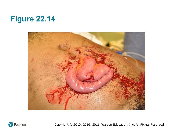
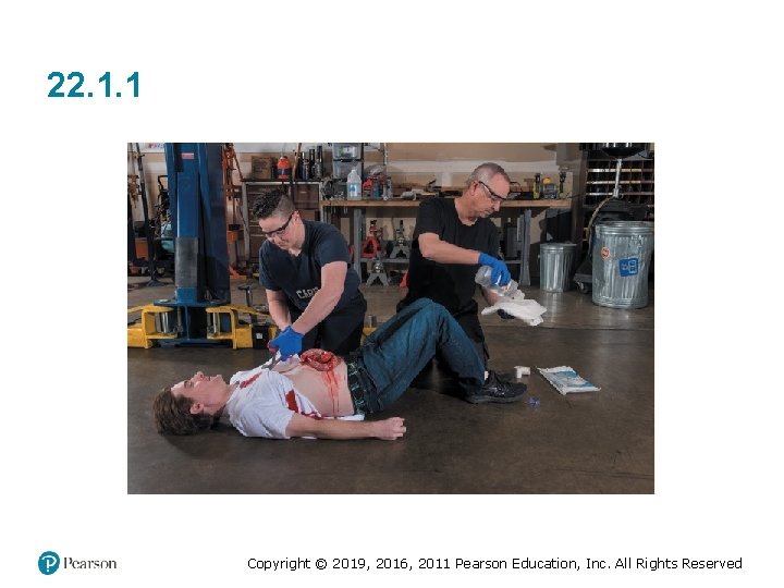
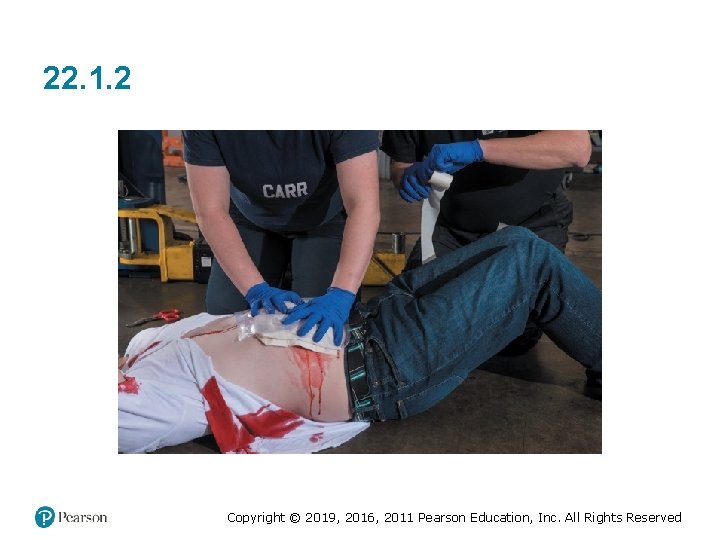
Abdominal Injuries (4 of 4) • Abdominal Evisceration – Open wound of abdomen characterized by protrusion of intestines through abdominal wall ▪ Never attempt to place spilled abdominal contents back into open wound. Copyright © 2019, 2016, 2011 Pearson Education, Inc. All Rights Reserved
Figure 22. 14 Copyright © 2019, 2016, 2011 Pearson Education, Inc. All Rights Reserved
22. 1. 1 Copyright © 2019, 2016, 2011 Pearson Education, Inc. All Rights Reserved
22. 1. 2 Copyright © 2019, 2016, 2011 Pearson Education, Inc. All Rights Reserved
22. 1. 3 Copyright © 2019, 2016, 2011 Pearson Education, Inc. All Rights Reserved
Abdominal Injuries: Think About It (1 of 3) • You respond to a skateboard park where a 17 -year-old boy has fallen onto concrete from a 20 foot height following a failed ramp jump attempt. He's complaining of persistent LUQ abdominal pain and nausea. His friends tell you he tried to get up but felt lightheaded. Copyright © 2019, 2016, 2011 Pearson Education, Inc. All Rights Reserved
Abdominal Injuries: Think About It (2 of 3) • Your patient is left lateral recumbent, guarding his abdomen, and moaning in pain. He has no other medical history, takes no medication, and is allergic to penicillin. His pulse is 100, blood pressure 100/70, and respirations are 28. Copyright © 2019, 2016, 2011 Pearson Education, Inc. All Rights Reserved
Abdominal Injuries: Think About It (3 of 3) 1. What are your suspicions? 2. How will you proceed? 3. What else could be going on? Copyright © 2019, 2016, 2011 Pearson Education, Inc. All Rights Reserved
SECTION: SUMMARY Copyright © 2019, 2016, 2011 Pearson Education, Inc. All Rights Reserved
Summary (1 of 9) • Injuries to chest can affect ability of patient to breathe adequately and cause damage to internal organs. • Injuries to chest classified as either open or closed. • Types of closed injuries – Broken ribs – Collapse of a lung, pneumothorax – Blood in chest cavity, hemothorax Copyright © 2019, 2016, 2011 Pearson Education, Inc. All Rights Reserved
Summary (2 of 9) • Signs and Symptoms of Closed Chest Injuries – Pain on breathing – Difficulty breathing – Discoloration – Deformity – Paradoxical movement Copyright © 2019, 2016, 2011 Pearson Education, Inc. All Rights Reserved
Summary (3 of 9) • Flail Chest – Caused when two or more ribs are broken in two or more places ▪ Compromises integrity of chest wall ▪ Makes it very difficult for patient to breathe adequately Copyright © 2019, 2016, 2011 Pearson Education, Inc. All Rights Reserved
Summary (4 of 9) • Be sure to expose all areas and palpate thoroughly with both hands. • Consider high-flow oxygen when local protocols indicate. Copyright © 2019, 2016, 2011 Pearson Education, Inc. All Rights Reserved
Summary (5 of 9) • Open chest injuries result in sucking chest wound – Must be covered immediately with occlusive dressing • Stabilize all impaled objects and secure object in place. • Do not attempt to remove an impaled object. Copyright © 2019, 2016, 2011 Pearson Education, Inc. All Rights Reserved
Summary (6 of 9) • Abdomen and pelvis contain organs that are both hollow and solid. • Injuries to organs can cause organ failure and severe internal bleeding. Copyright © 2019, 2016, 2011 Pearson Education, Inc. All Rights Reserved
Summary (7 of 9) • Signs and Symptoms of Abdominal Emergency – Pain – Rigidity – Distention – Blood in vomit or feces Copyright © 2019, 2016, 2011 Pearson Education, Inc. All Rights Reserved
Summary (8 of 9) • Abdominal injuries classified as closed or open. • Closed injuries result in organ damage and/or severe bleeding. Copyright © 2019, 2016, 2011 Pearson Education, Inc. All Rights Reserved
Summary (9 of 9) • Abdominal evisceration is open wound that has allowed abdominal contents to spill out. – Cover with a sterile moist dressing and cover with plastic to minimize chances that exposed organs will dry out. Copyright © 2019, 2016, 2011 Pearson Education, Inc. All Rights Reserved
SECTION: REVIEW QUESTIONS Copyright © 2019, 2016, 2011 Pearson Education, Inc. All Rights Reserved
Review Questions (1 of 3) 1. What are the major structures of the thoracic cavity? 2. What is the relationship between chest injury and perfusion? 3. What is the difference between an open and closed chest injury? 4. What are the signs and symptoms of a closed chest injury? Copyright © 2019, 2016, 2011 Pearson Education, Inc. All Rights Reserved
Review Questions (2 of 3) 5. What is the appropriate assessment of a patient with a chest injury? 6. What is the appropriate care of a patient with a closed chest injury? 7. What is the appropriate care of a patient with an open chest injury? 8. What are the major structures of the abdominal and pelvic cavities? Copyright © 2019, 2016, 2011 Pearson Education, Inc. All Rights Reserved
Review Questions (3 of 3) 9. What are the signs and symptoms of internal bleeding? 10. What is the appropriate assessment and care of a patient with abdominal pain? 11. What is the appropriate assessment and care of a patient with an open abdominal injury? Copyright © 2019, 2016, 2011 Pearson Education, Inc. All Rights Reserved
Copyright This work is protected by United States copyright laws and is provided solely for the use of instructors in teaching their courses and assessing student learning. Dissemination or sale of any part of this work (including on the World Wide Web) will destroy the integrity of the work and is not permitted. The work and materials from it should never be made available to students except by instructors using the accompanying text in their classes. All recipients of this work are expected to abide by these restrictions and to honor the intended pedagogical purposes and the needs of other instructors who rely on these materials. Copyright © 2019, 2016, 2011 Pearson Education, Inc. All Rights Reserved
Appendix A Long Description for Figure 22. 2 The parts labeled on the left are parietal pleura, visceral pleura, chest wall, Pleural (potential) space, and Diaphragm. One of the two lungs is shown smaller in size than the normal lung. Outside air enters the region between the lung and chest wall due to disruption of chest wall and parietal pleura. Lung air enters the region between the lung and chest wall due to disruption of visceral pleura. Return to presentation Copyright © 2019, 2016, 2011 Pearson Education, Inc. All Rights Reserved