
Wound Management UNC Emergency Medicine Medical Student Lecture






- Slides: 36
Wound Management UNC Emergency Medicine Medical Student Lecture Series
2 Principles of Wound Management Goals of Wound Care Facilitate hemostasis n Decrease tissue loss n Promote wound healing n Minimize scar formation n
3 Principles of Wound Management Mechanism of Injury n Wounds are caused by three different types of forces – – – Shear Compressive Tensile
4 Principles of Wound Management Shear Forces n Result from sharp objects – – Low energy Minimal cell damage Result in straight edges, little contamination Heals with a good result
5 Principles of Wound Management Compressive Forces n Result from blunt objects impacting the skin at a right angle – – – Results in stellate or complex laceration Ragged or shredded edges More prone to infection
6 Principles of Wound Management Tensile Forces n Result from blunt objects impacting the skin at an oblique angle – – – Results in triangular wound Sometimes produces a flap More prone to infection
7 Principles of Wound Management Evaluation of Wounds ABC’s first Always! n Ensure hemostasis n – – n Remove obstructions – n Saline gauze dressing Compression Rings, clothing, other jewelry History
8 Principles of Wound Management History n n n n Symptoms Type of Force Contamination Event Potential foreign body Function Non-accidental trauma n n n Tetanus status Allergies Medications Comorbidities Previous scar formation
9 Principles of Wound Management Wound Examination n n n Location Size Shape Margins Depth Alignment with skin lines Neuro function n n Vascular function Tendon function Underlying structures Wound contamination Foreign bodies
10 Principles of Wound Management Wound Consultation n n n n Tarsal plate or lacrimal duct Open fracture or joint space Extensive facial wounds Associated with amputation Associated with loss of function Involves tendons, nerves, or vessels Involves significant loss of epidermis Any wound that you are uncertain about
11 Principles of Wound Management Wound Preparation - Anesthesia n Topical – – – n Local – – – n Solution or paste LET EMLA Direct infiltration 1% lidocaine with or without epinephrine Bupivicaine or sensorcaine for longer acting anesthesia Regional Block – – Local infiltration proximally in order to avoid tissue disruption Smaller amount of anesthesia required
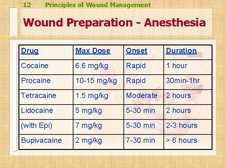
12 Principles of Wound Management Wound Preparation - Anesthesia Drug Max Dose Onset Duration Cocaine 6. 6 mg/kg Rapid 1 hour Procaine 10 -15 mg/kg Rapid 30 min-1 hr Tetracaine 1. 5 mg/kg Moderate 2 hours Lidocaine 5 mg/kg 5 -30 min 2 hours (with Epi) 7 mg/kg 5 -30 min 2 -3 hours Bupivacaine 2 mg/kg 7 -30 min > 6 hours
13 Principles of Wound Management Minimize the Pain of Injection Use sodium bicarbonate mixed with the anesthetic (1 ml/10 ml solution) n Use smallest needle possible n Inject slowly n Insert needle through open wound edge and skin that has already been anesthetized n
14 Principles of Wound Management Wound Preparation - Hemostasis n Physical vs. chemical – – n Direct pressure Epinephrine Gelfoam Cautery Refractory – Use a tourniquet
15 Principles of Wound Management Wound Preparation – Foreign Body Removal Visual inspection n Imaging n – – n Glass, metal, gravel fragments >1 mm should be visible on plain radiographs Organic substances and plastics are usually radiolucent Always discuss and document possibility of retained foreign body
16 Principles of Wound Management Wound Preparation – Irrigation Local anesthesia prior to irrigation n Do not soak the wound n Use normal saline n Large syringe (60 m. L) with Zerowet attachment n Do not use iodine, chlorhexidine, peroxide or detergents n
17 Principles of Wound Management Wound Preparation – Debridement Removes foreign matter & devitalized tissue n Creates sharp wound edge n Excision with elliptical shape n Respect skin lines n
18 Principles of Wound Management Wound Preparation – Antibiotics n n Infections occur in ~3 -5% of traumatic wounds seen in the ED Factors that increase risk – – n Heavily contaminated wound, especially with soil Immunocompromised patients Diabetics Human bites > animal bites Most important prevention adequate irrigation & debridement
19 Principles of Wound Management Wound Preparation – Antibiotics n Dog & cat bites – – n Human bites – – n Cover pasteurella Augmentin Cover eikenella Augmentin Puncture wounds – – Cover pseudomonas Cipro, levaquin
20 Principles of Wound Management Wound Preparation – Tetanus Prophylaxis n Clean wounds – – n Incomplete immunization toxoid >10 years, then give toxoid Tetanus prone wound – Incomplete immunization n Toxoid – n & immune globulin > 5 years, give toxoid Remember to think about rabies!
21 Principles of Wound Management Wound Closure n Primary closure – – n Secondary closure – – n Suture, staple, adhesive, or tape Performed on recently sustained lacerations: <12 hours generally and <24 hours on face Secondary intent Allowed to granulate Tertiary closure – Delayed primary (observed for 4 -5 days)
22 Principles of Wound Management Suture Material n Absorbable – – – n Non-Absorbable – – – n Chromic gut Vicryl PDS II Silk Prolene Dermalon Monofilament vs. braided
23 Principles of Wound Management Staples, Adhesives & Tape n Staples – n Adhesives – n Quick, poor aesthetic result Dermabond- painless, petroleum dissolves Tape – Steri-strips
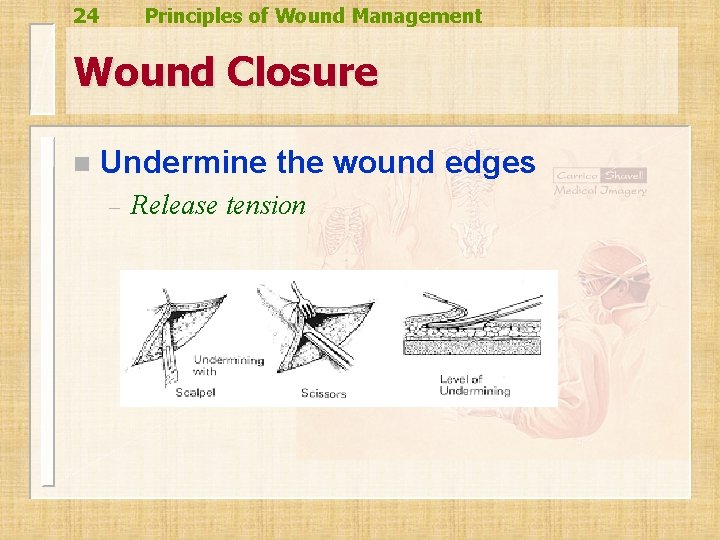
24 Principles of Wound Management Wound Closure n Undermine the wound edges – Release tension
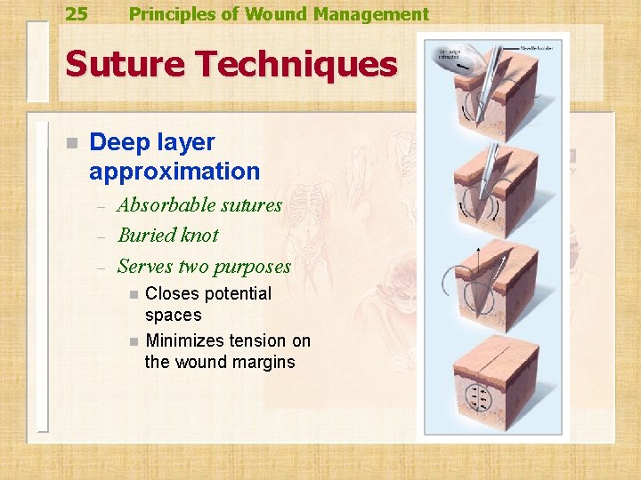
25 Principles of Wound Management Suture Techniques n Deep layer approximation – – – Absorbable sutures Buried knot Serves two purposes n n Closes potential spaces Minimizes tension on the wound margins
26 Principles of Wound Management Skin Closure Key – wound edge eversion n “Approximate, don’t strangulate” n Anticipate wound edema n Choose appropriate size of suture for location of laceration n
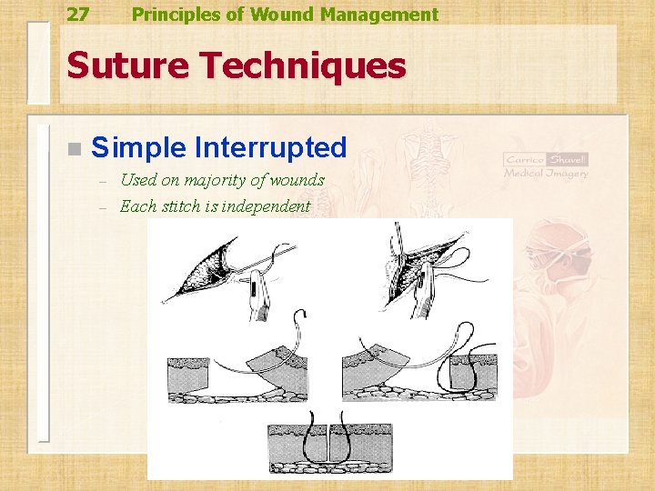
27 Principles of Wound Management Suture Techniques n Simple Interrupted – – Used on majority of wounds Each stitch is independent
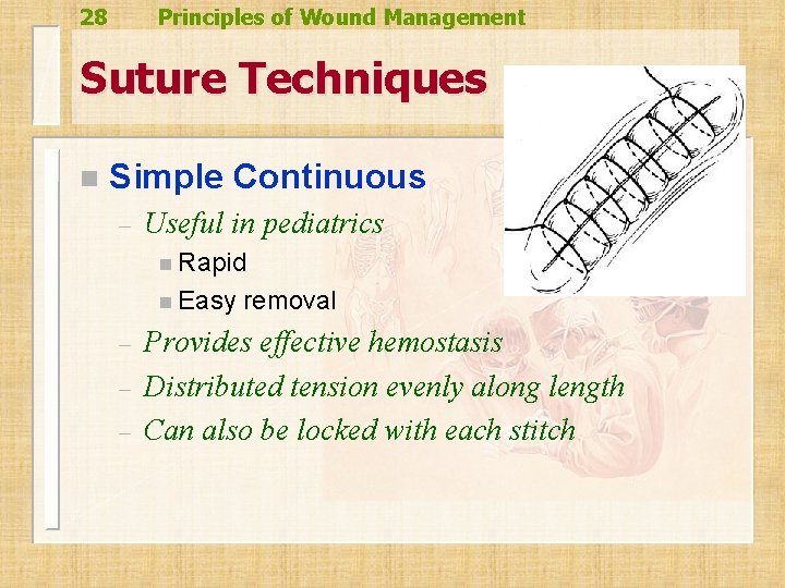
28 Principles of Wound Management Suture Techniques n Simple Continuous – Useful in pediatrics n Rapid n Easy – – – removal Provides effective hemostasis Distributed tension evenly along length Can also be locked with each stitch
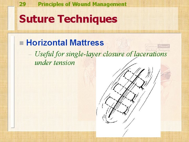
29 Principles of Wound Management Suture Techniques n Horizontal Mattress – Useful for single-layer closure of lacerations under tension
30 Principles of Wound Management Horizontal Mattress
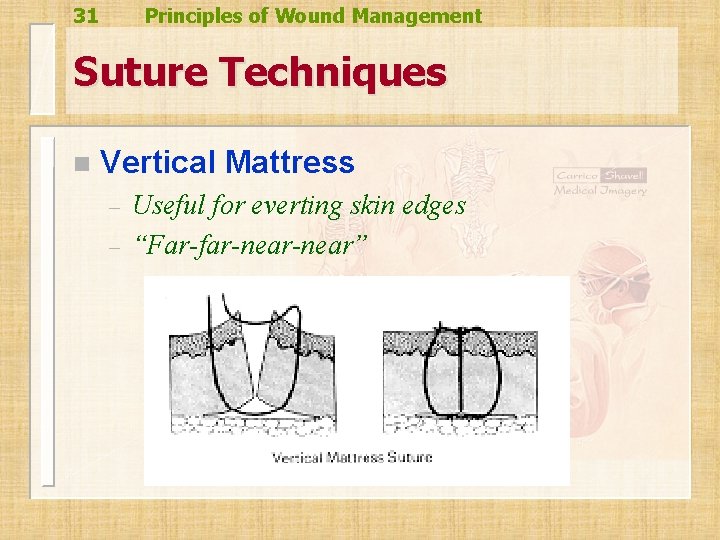
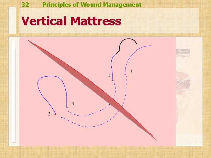
31 Principles of Wound Management Suture Techniques n Vertical Mattress – – Useful for everting skin edges “Far-far-near”
32 Principles of Wound Management Vertical Mattress
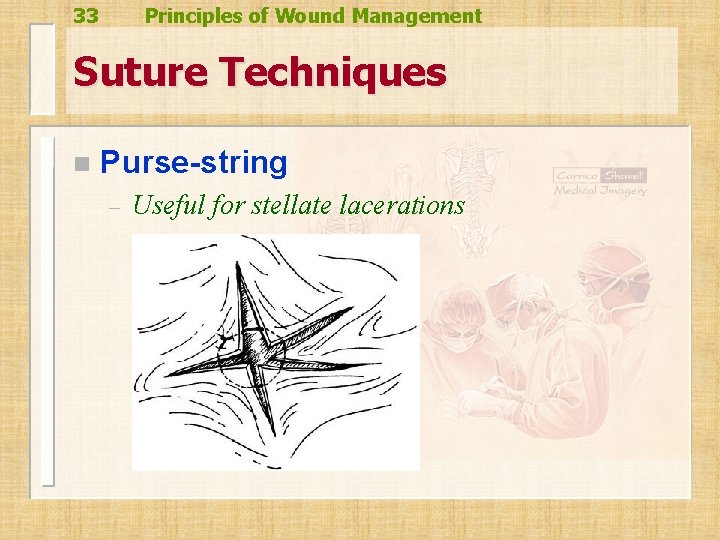
33 Principles of Wound Management Suture Techniques n Purse-string – Useful for stellate lacerations
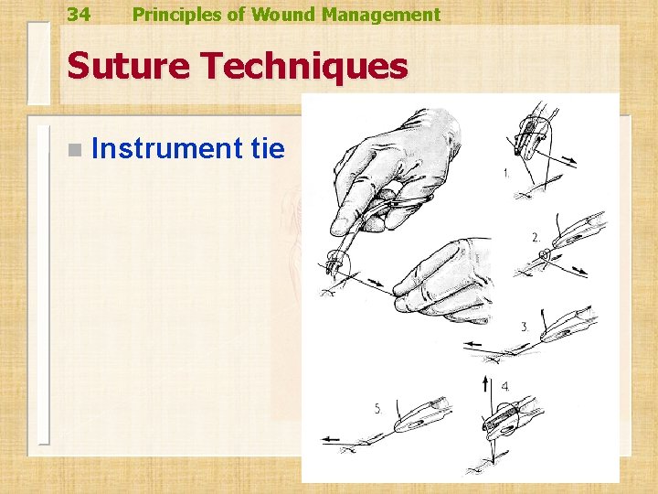
34 Principles of Wound Management Suture Techniques n Instrument tie
35 Principles of Wound Management Wound Care n Dressing – – – Maintain dry for 24 -48 hours Use antibiotic to maintain moist environment If overlying a joint, splint in a position of function Sun protection to prevent scar hyperpigmentation Suture removal instructions!
36 Principles of Wound Management Practice Time! n