Current Radiology diagnostic radiology interventional radiology nonvascular and


 radiography, fluoroscopy • conventional tomography • •")
 • contrast studies • barium studies GIT •")
")
 – spectra – color coded (CDI)")

 CT • - volumetric imaging • no gaps")


 endocarditis, peripheral")
 • Positive contrast = more absorbing substances (elements with high proton")


")
- Slides: 31
Current Radiology • diagnostic radiology • interventional radiology –nonvascular and vascular IR • background - imaging –diagnosis –imaging guided miniinvasive procedures • biopsies, fine needle aspirations • therapeutical procedures: drainages, angioplasty, stenting, thrombolysis
Wilhelm Conrad Roentgen Discovery of x-rays 8. 11. 1895
Imaging methods • conventional radiology • (digital) radiography, fluoroscopy • conventional tomography • • ultrasound (US, 2 D, Doppler) computed tomography (CT) magnetic resonance (MRI) angiography (DSA) • hybrid imaging (PET/CT, SPECT/CT) • (nuclear medicine imaging: planar, SPECT, PET)
Conventional radiology • Plain films (native) • contrast studies • barium studies GIT • double contrast: air+barium, enteroclysis: metylcelulosis+barium • iodinated CA: • • • intravenous pyelography (IVP) fistulography ERCP, PTC
Visualization of roentgen image • Fluoroscopy – image is generated on fluoroscopic screen (shield) - excitation – image is positive (bones are dark) • Radiography – record of image on film - photochemical reaction – image is negative (bones are light)
Categories of US imaging • morphological (2 D) – spectra – color coded (CDI) – power (CDE, CPA) triplex • Doppler duplex – B mode
What is 3 D and 4 D ultrasonography? 2 D 3 D 4 D
Advances in CT • spiral (helical) CT • - volumetric imaging • no gaps due to breathing • faster scanning, CTA feasibility • multidetector systems • HRCT - (lung, bones – petrous bone etc) • thin collimation of sections • high resolution image recostruction algorithm
Conventional x spiral CT inkrementální sken spirální sken
Postprocessing-2 D rekonstrukce MPR=multiplanární rekonstrukce sagitální MPR
MDCT – coronal sections
MDCT CTCA – 16 rows of detektorů J. Ferda, FN Plzeň - Lochotín
Example: Case 1 10. 3. 2000 31. 10. 2000
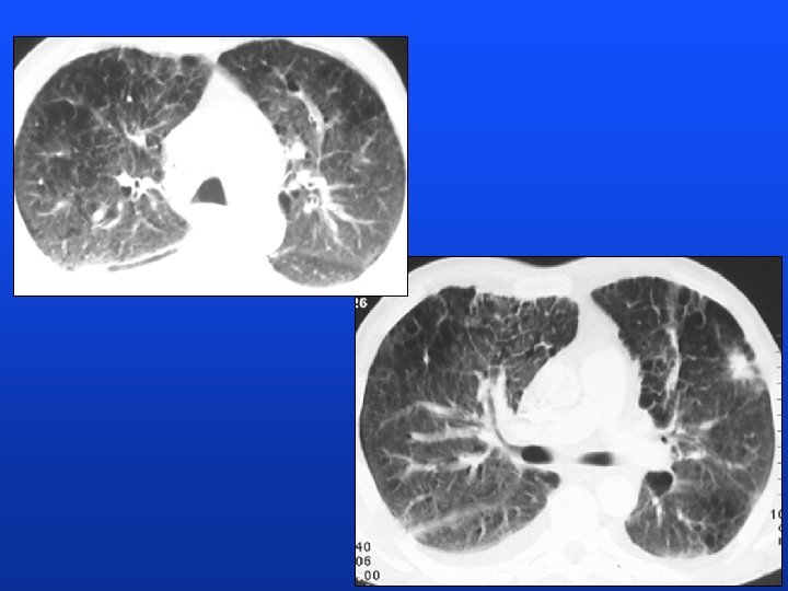
HRCT
CT 2. 11. 2000
CT, brain 3. 11. 2000 Autopsy: lung CA, carcinomatous lymphangiopathy, non-infectious (marantic) endocarditis, peripheral embolization
Contrast agents (substances) • Positive contrast = more absorbing substances (elements with high proton number Z) – barium - barium sulfate suspension (GIT) – iodine substances water soluble iodinated CA: angiocardiography, bronchography lipiodol: lymhography • Negative contrast = gases of low density – air, carbon dioxide, nitrogen (pneumoencephalography) • Double contrast = combination of both contrasts – gastrointestinal tract • • • CT - diluted barium (GIT), iodinated CM (GIT, i. v. ) MR - paramagnetic CM (gadolinium) US - microbubbles (work in progress)
Example: case 2
Diagnosis: non-occlusive mesenteric ischemia
Diagnostic algorithms Case 3: acute biliary pancreatitis Ultrasound 1. day: etiology
ERCP 1. day, papilotomy: treatment CT with CA, 4. day: staging AP
Clinical value of MRI • Commonest indications • neuroradiology – brain – spinal canal • • musculoskeletal system heart, vessels - MRA, abdomen, pelvis (prenatal MR) • advances • Faster acquisitions • MR angiography - TOF, PC, with contrast agent - gadolinium
Kongenitální PNET
Congenital PNET
Subarachnoid bleeding
CTA - MDCT
Saccular aneurysm on AB MPR MIP
MR of tle heart - cine mode
Trends in radiology: less radiation, less invasivity Typical field: angiography • Doppler ultrasonography • CT angiography • MR angiography
Enjoy your radiology lessons! (we will enjoy your exam)