
Interventional Radiology Useful for All and Always Dr








- Slides: 44
Interventional Radiology : Useful for All and Always ? Dr S. Murgo CHU Tivoli, La Louvière, Belgique Hôpital Erasme, Bruxelles, Belgique
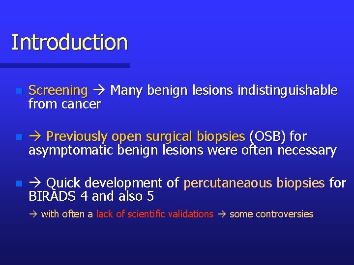
Introduction n Screening Many benign lesions indistinguishable from cancer n Previously open surgical biopsies (OSB) for asymptomatic benign lesions were often necessary n Quick development of percutaneaous biopsies for BIRADS 4 and also 5 with often a lack of scientific validations some controversies
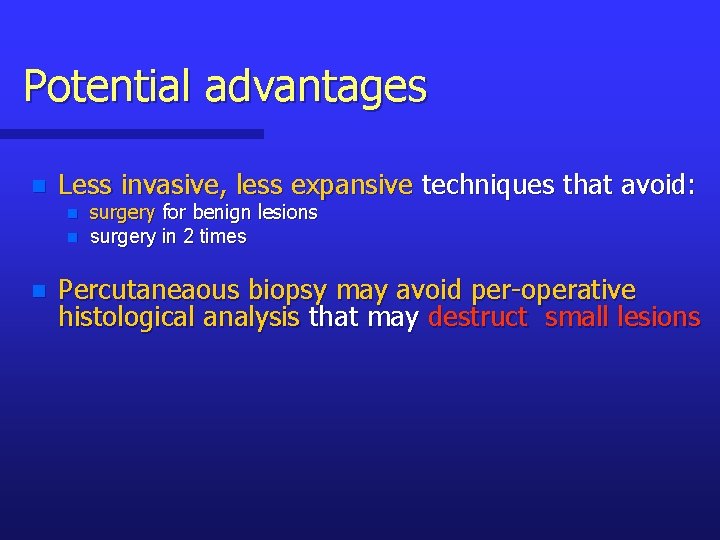
Potential advantages n Less invasive, less expansive techniques that avoid: n n n surgery for benign lesions surgery in 2 times Percutaneaous biopsy may avoid per-operative histological analysis that may destruct small lesions
Potential drawbacks n Epithelial displacement (FNA, CNB > VACB): n n No evidence of biologic significance No of the recurrence rate after BCS But some displaced cells associated with DCIS can sometime mimic IDC for pathologist. Risk of missed cancers good knowledge of limitations
Interventional Radiology Includes: Guidewire Localization Radio. Frequency Fine Needle Aspiration Core Needle Biopsy Large Core Needle Biopsy
Main Mammographic Signs Mass Architectural Distorsion Microcalcifications
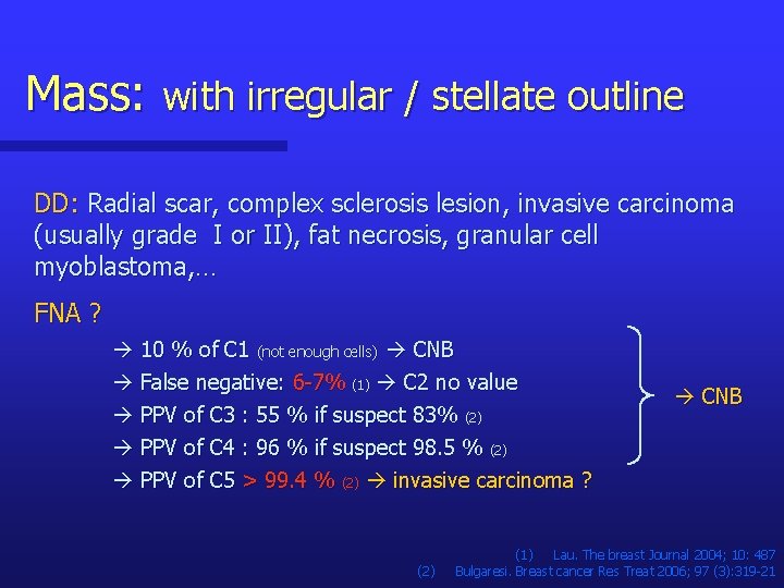
Mass: with irregular / stellate outline DD: Radial scar, complex sclerosis lesion, invasive carcinoma (usually grade I or II), fat necrosis, granular cell myoblastoma, … FNA ? 10 % of C 1 (not enough cells) CNB False negative: 6 -7% (1) C 2 no value PPV of C 3 : 55 % if suspect 83% (2) PPV of C 4 : 96 % if suspect 98. 5 % (2) CNB PPV of C 5 > 99. 4 % (2) invasive carcinoma ? (2) (1) Lau. The breast Journal 2004; 10: 487 Bulgaresi. Breast cancer Res Treat 2006; 97 (3): 319 -21
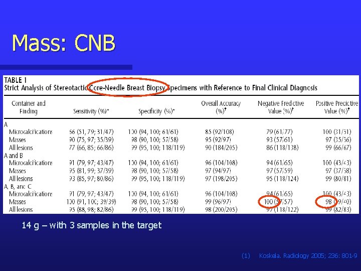
Mass: CNB 14 g – with 3 samples in the target (1) Koskela. Radiology 2005; 236: 801 -9
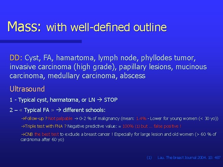
Mass: with well-defined outline DD: Cyst, FA, hamartoma, lymph node, phyllodes tumor, invasive carcinoma (high grade), papillary lesions, mucinous carcinoma, medullary carcinoma, abscess Ultrasound 1 - Typical cyst, harmatoma, or LN STOP 2 – « Typical FA » different schools: Follow-up ? Not palpable 0 -2 % of malignancy (mean: 1. 4% - Lower for young women (< 30 yo)) Triple test with FNA ? Negative predictive value: 100% (1) but … false positive ! CNB the best to exclude a breast cancer ! Especially for large lesion and old women (> 60 % of carcinoma after 60 yo) (1) Lau. The breast Journal 2004. 10: 487
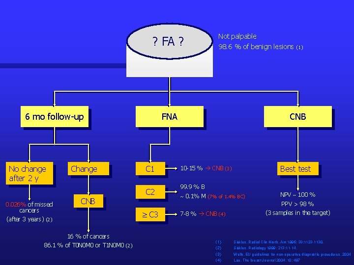
? FA ? 6 mo follow-up No change after 2 y 0. 026% of missed cancers Change CNB FNA C 1 C 2 16 % of cancers 86. 1 % of T 0 N 0 M 0 or T 1 N 0 M 0 (2) CNB Best test 10 -15 % CNB (3) 99. 9 % B 0. 1% M (7% of 1. 4% BC) NPV 100 % PPV > 98 % C 3 (after 3 years) (2) Not palpable 98. 6 % of benign lesions (1) 7 -8 % CNB (4) (1) (2) (3) (4) (3 samples in the target) Sickles. Radiol Clin North. Am 1995; 33: 1123 -1130. Sickles. Radiology 1999; 213: 11 -14. Wells. EU guidelines for non-operative diagnostic procedures. 2004 Lau. The breast Journal 2004; 10: 487
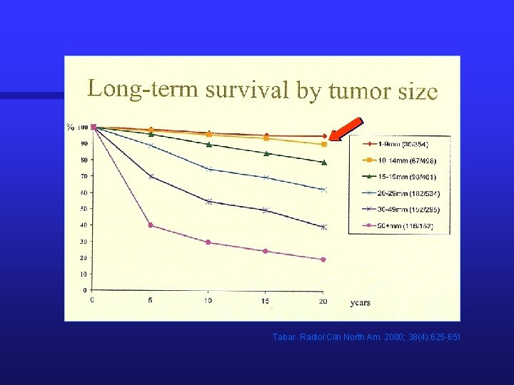
Tabar. Radiol Ciln North Am. 2000; 38(4): 625 -651
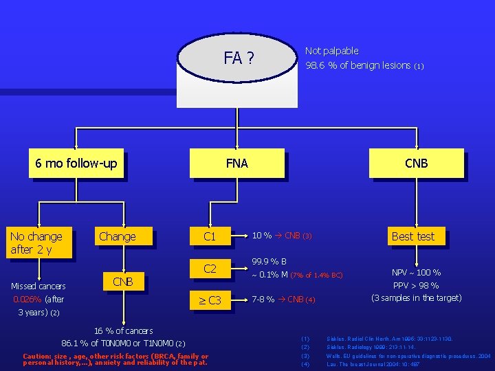
FA ? 6 mo follow-up No change after 2 y Missed cancers Change CNB FNA C 1 C 2 CNB Best test 10 % CNB (3) 99. 9 % B 0. 1% M (7% of 1. 4% BC) NPV 100 % PPV > 98 % C 3 0. 026% (after Not palpable 98. 6 % of benign lesions (1) 7 -8 % CNB (4) (3 samples in the target) 3 years) (2) 16 % of cancers 86. 1 % of T 0 N 0 M 0 or T 1 N 0 M 0 (2) Caution: size , age, other risk factors (BRCA, family or personal history, . . . ), anxiety and reliability of the pat. (1) (2) (3) (4) Sickles. Radiol Clin North. Am 1995; 33: 1123 -1130. Sickles. Radiology 1999; 213: 11 -14. Wells. EU guidelines for non-operative diagnostic procedures. 2004 Lau. The breast Journal 2004; 10: 487
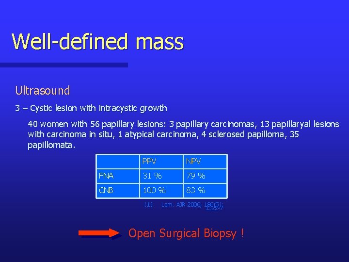
Well-defined mass Ultrasound 3 – Cystic lesion with intracystic growth 40 women with 56 papillary lesions: 3 papillary carcinomas, 13 papillaryal lesions with carcinoma in situ, 1 atypical carcinoma, 4 sclerosed papilloma, 35 papillomata. PPV NPV FNA 31 % 79 % CNB 100 % 83 % (1) Lam. AJR 2006; 186(5): 1322 -7 Open Surgical Biopsy !
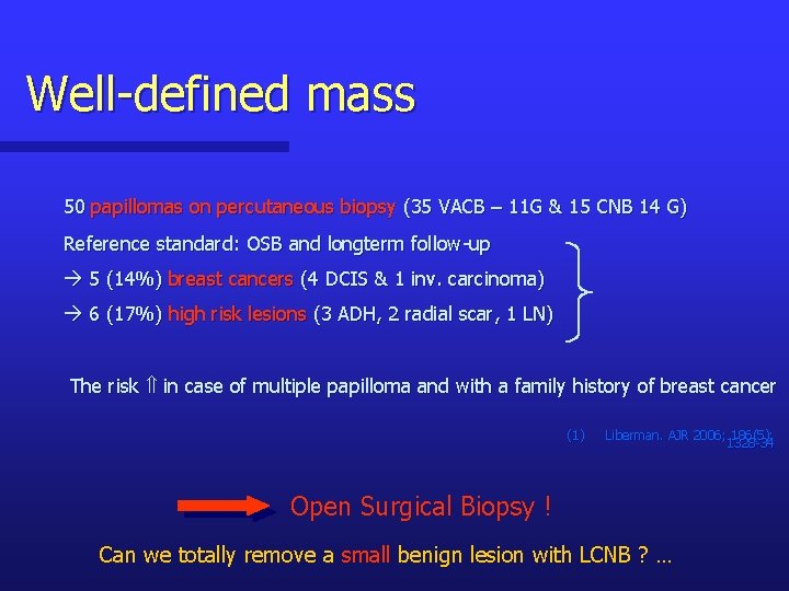
Well-defined mass 50 papillomas on percutaneous biopsy (35 VACB – 11 G & 15 CNB 14 G) Reference standard: OSB and longterm follow-up 5 (14%) breast cancers (4 DCIS & 1 inv. carcinoma) 6 (17%) high risk lesions (3 ADH, 2 radial scar, 1 LN) The risk in case of multiple papilloma and with a family history of breast cancer (1) Liberman. AJR 2006; 186(5): 1328 -34 Open Surgical Biopsy ! Can we totally remove a small benign lesion with LCNB ? …
Architectural distortion DD: Involution, radial scar, invasive lobular carcinoma, DCIS(rarely), … Radial scar: n Fibroelastic center with pseudo-infiltrative tubular structure (DD: tub car). n In the crown of the RS +/- ADH, ALH, DCIS, LN, … the risk with the age and the size (1) - None < 40 yo, rare between 40 & 50 yo, > 50 yo - Rare if < 6 -7 mm Open Surgical Biopsy ! (1) Andersen JA, Cancer 1984; 53: 2557 -2560.
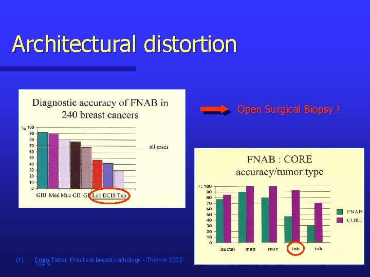
Architectural distortion Open Surgical Biopsy ! (1) From Tabar. Practical breast pathology - Thieme 2002: 104 -5
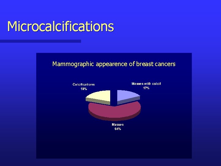
Microcalcifications Mammographic appearence of breast cancers
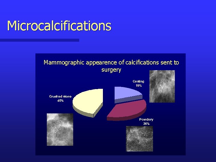
Microcalcifications Mammographic appearence of calcifications sent to surgery
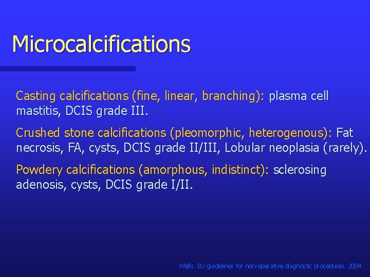
Microcalcifications Casting calcifications (fine, linear, branching): plasma cell mastitis, DCIS grade III. Crushed stone calcifications (pleomorphic, heterogenous): Fat necrosis, FA, cysts, DCIS grade II/III, Lobular neoplasia (rarely). Powdery calcifications (amorphous, indistinct): sclerosing adenosis, cysts, DCIS grade I/II. Wells. EU guidelines for non-operative diagnostic procedures. 2004
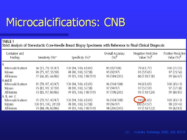
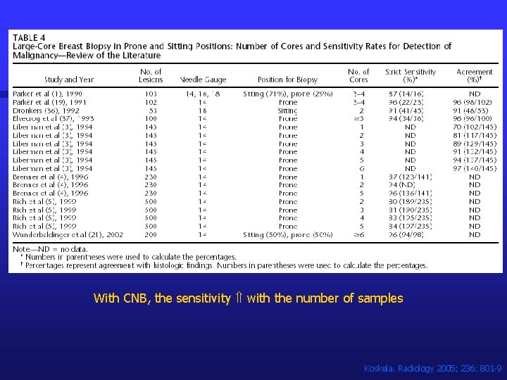
Microcalcifications: CNB (1) Koskela. Radiology 2005; 236: 801 -9
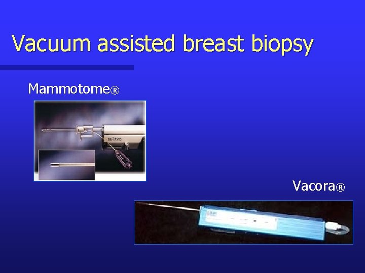
Vacuum assisted breast biopsy Mammotome® Vacora®
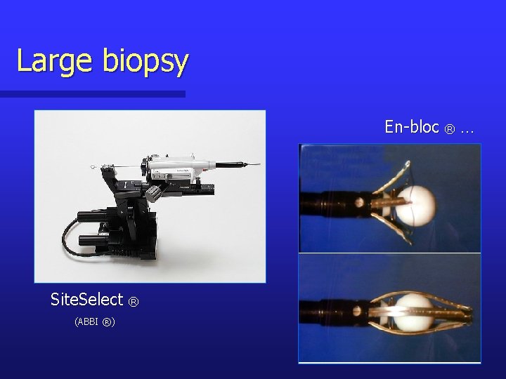
Large biopsy En-bloc ® … Site. Select ® (ABBI ®)
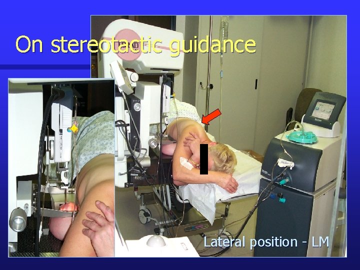
On stereotactic guidance Lateral position - LM
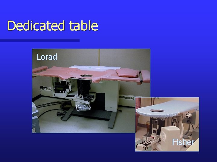
Dedicated table Lorad Fisher
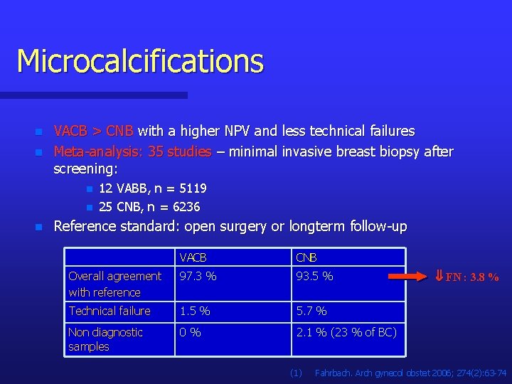
Microcalcifications n n VACB > CNB with a higher NPV and less technical failures Meta-analysis: 35 studies – minimal invasive breast biopsy after screening: n n n 12 VABB, n = 5119 25 CNB, n = 6236 Reference standard: open surgery or longterm follow-up VACB CNB Overall agreement with reference 97. 3 % 93. 5 % Technical failure 1. 5 % 5. 7 % Non diagnostic samples 0 % 2. 1 % (23 % of BC) (1) FN : 3. 8 % Fahrbach. Arch gynecol obstet 2006; 274(2): 63 -74
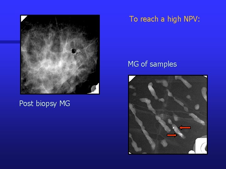
To reach a high NPV: MG of samples Post biopsy MG
With CNB, the sensitivity with the number of samples Koskela. Radiology 2005; 236: 801 -9
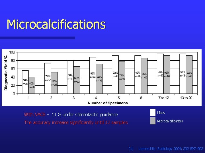
Microcalcifications With VACB - 11 G under stereotactic guidance Mass The accuracy increase significantly until 12 samples Microcalcification (1) Lomoschitz. Radiology 2004; 232: 897– 903
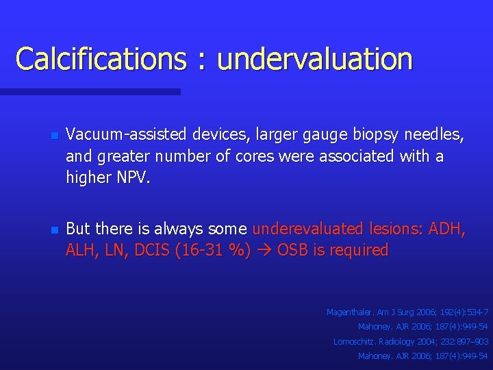
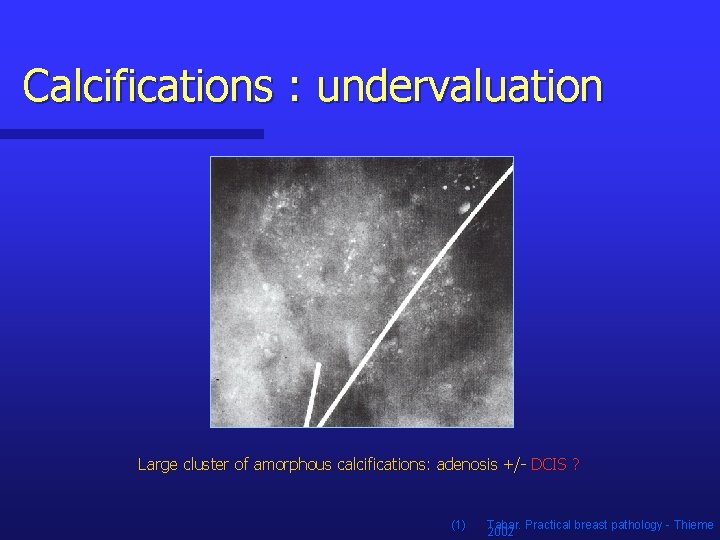
Calcifications : undervaluation n Vacuum-assisted devices, larger gauge biopsy needles, and greater number of cores were associated with a higher NPV. n But there is always some underevaluated lesions: ADH, ALH, LN, DCIS (16 -31 %) OSB is required Magenthaler. Am J Surg 2006; 192(4): 534 -7 Mahoney. AJR 2006; 187(4): 949 -54 Lomoschitz. Radiology 2004; 232: 897– 903 Mahoney. AJR 2006; 187(4): 949 -54
Calcifications : undervaluation Large cluster of amorphous calcifications: adenosis +/- DCIS ? (1) Tabar. Practical breast pathology - Thieme 2002
Microcalcifications n To avoid missed cancer, a open surgical biopsy is required after percutaneous biopsy: n n n When none or a small number of calcifications are removed For large cluster of amorphous calcifications (adenosis +/- DCIS ? ) For an histological diagnosis of ADH, ALH, and LN
Conclusions n IR is very useful and efficient BUT not for all and always ! n The knowledge of the limitations of each techniques nb of missed cancers n Confrontation of the cytological and histological results with the PE and medical imaging studies in a multidisciplinary approach ! n n Repeat biopsy is necessary if histological and imaging finding are discordant Surgical excision is necessary for some histological benign lesions: ADH, ALH, LN, radial scar, papillary lesions, possible phyllode tumor
Conclusions n Further work is necessary to optimize criteria for patient selection, to develop and define the role of new technologies. n Complete removal of the mammographic target does not ensure complete excision of the histological process Further investigation is necessary to determinate in which lesion, complete removal of the target is advantageous.