CORNEAL DEGENERATIONS DR P PRADEEP PROFESSOR OF OPHTHALMOLOGY


![B. DEPENDING UPON ETIOLOGY A] Age related degeneration: Arcus senilis, Vogt’s white limbal girdle,](https://slidetodoc.com/presentation_image_h2/3ad0bedb92c0e33afbed49e0cceb57bc/image-5.jpg "B. DEPENDING UPON ETIOLOGY A] Age related degeneration: Arcus senilis, Vogt’s white limbal girdle,")


 Band shape keratopathy Calcific degeneration (BSK) Band shape")


 and pseudopterygium may develop.")
, CL � an option though difficult")


- Slides: 45
CORNEAL DEGENERATIONS DR P. PRADEEP PROFESSOR OF OPHTHALMOLOGY
WHAT IS CORNEAL DEGENERATION corneal degeneration refers to the condition in which the normal cells undergo some degenerative changes under the influence of age or some pathological condition.
CLASSIFICATIONS A. Depending upon locations I. Axial corneal degeneration 1. 2. 3. 4. 5. Fatty degeneration Hyaline degeneration Amyloidosis Calcific degeneration(Band keratopathy) Salzmann’s nodular degeneration
II. PERIPHERAL DEGENERATION 1. Arcus senilis 2. Vogt’s white limbal girdle Hassa-Henle bodies Terrien’s marginal degeneration Morren’s ulcer pellucid marginal degeneration Furrow degeneration(senile marginal degeneration) 3. 4. 5. 6. 7.
B. DEPENDING UPON ETIOLOGY A] Age related degeneration: Arcus senilis, Vogt’s white limbal girdle, Hassal-Henle bodies, Mosaic degeneration. B] Pathological degeneration: Fatty degeneration, Amyloidosis, Calcific degeneration, Salzmann’s degeneration, Furrow degeneration, Spheroid degeneration, Pellucid marginal degeneration, Terrien’s marginal degeneration
AGE RELATED CORNEAL DEGENERATIONS
ARCUS SENILIS Arcus senilis refers to an annular lipid infiltration of corneal periphery. Sometimes, similarly changes may or may not be associated with hyperlipidemia.
CLINICAL FEATURES The arcus starts in the superior and inferior quadrants and then progresses circumferentially to form a ring which is about 1 mm wide. Peripheral border of this ring opacity is sharp while central border is diffuse This ring of opacity is separated from the limbus by a clear zone.
VOGT’S WHITE LIMBAL GRIDLE It consists of whitish cresentric limbal bands composed of chalk like flecks centred at 9 and 3 o’clock TYPE 1 : variant of band shaped. keratopathy featuring a swiss cheese hole pattern and a clear area separating the lesion from the scleral margin. TYPE 2 : more prevalent and. distinguished by absence of holes and typically also of a juxtalimbal clear zone.
HASSAL-HENLE BODIES Hassal-henle bodies are drop like excrescences of hyaline materal projectng into the anterior chamber around the corneal periphery. These form the commonest senile change seen in the cornea. In pathological conditions they become larger and invade the central area and the condition is called CORNEAL GUTTATA
• Tiny dark spots on central endothelium • Similar peripheral lesions are Hassell-Henle bodies
• Cornea farinata: visually insignificant condition characterized by bilateral, minute, flour like deposits in deep stroma, most prominent centrally
CROCODILE SHAGREEN: • Asymptomatic • Greyish white polygonal stromal opacities separated by relatively clear spaces • Frequently involves anterior to thirds of the stroma occasionally posterior one third.
PATHOLOGICAL CORNEAL DEGENERATION
FATTY DEGENERATIONS It is characterized by whitish or yellowish deposits. Initially fat deposits are intracellular but some becomes extracellular with necrosis of stromal cells. Lipid keratopathy can be primary or secondary
• Primary : charecterized by white or yellowish often with crystalline element, stromal deposits consisting of cholestrol , fats and phospholipids and not associated with vascularization. • Secondary : common associated with previous ocular injury or disease resulted in corneal vascularization. Most common cause herpes simplex and zoster.
• Treatment : primarily medical control of underlying inflammatory disease: • Photocoagulation or needle cautery of feeding vessels. • Penetrating keratoplasty in advanced but quiescent disease.
HYALINE DEGENERATION Primary Hyaline degeneration association with granular dystrophy. Secondary Hyaline degeneration is unilateral and associated with various types of corneal disease including old keratitis, long-standing Glaucoma, trachomatous pannus. It may be complicated by recurrent corneal erosion.
TREATMENT Treatment of the condition when it causes visual disturbance is keratoplasty
AMYLOID DEGENERATION Amyloid degeneration of cornea is characterized by deposition of Amyloid material underneath epithelium. It is very rare condition and occurs in primary (in a healthy cornea) and secondary forms (in a disease cornea).
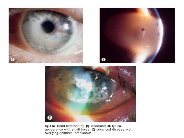
CALCIFIC DEGENERATION (BAND SHAPE KERATOPATHY ) Band shape keratopathy Calcific degeneration (BSK) Band shape keratopathy (BSK) is essentially a degenerative change associated with deposition of calcium salts in Bowman’s membrane, most superficial part of stroma and in deeper layers of epithelium.
ETIOLOGY Ocular disease complicated by band keratopathy include chronic uveitis in adults, children with still’s disease, phthisis bulbi, chronic Glaucoma, chronic keratitis and ocular trauma. Age related BSK is common and affects otherwise healthy cornea. Metabolic conditions rarely associate with BSK included hypercalcaemia
CLINICAL FEATURES It typically presents as a band shaped opacity In the interpalpebral zone with a clear interval between the ends of the band the limbus. The condition begins at the periphery and gradually progresses towards the centre.
TREATMENT • Chelation is simple and effective for mild cases. • Corneal epithelium overlying the opacity and a solid layer of calcification are first scraped of with forceps and scalpel blade. • The cornea is then rubbed with cotton tipped applicator dipped in solution of EDTA 1. 5 -3. 0 % for 15 -20 minutes. • Other modes : diamond burr , excimer laser keratectomy and lamellar keratoplasty.
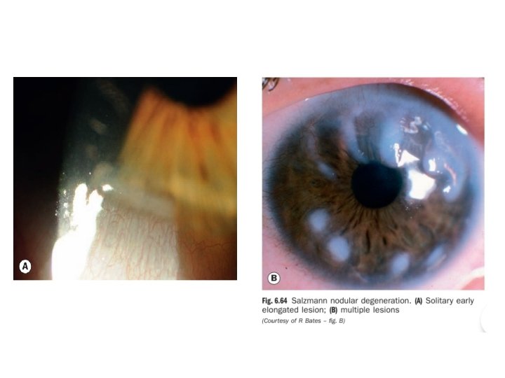
SALZMANN’S NODULAR DEGENERATION Salzmann’s nodular degeneration SND is a slowly progressive condition in which graywhite to bluish nodules measuring 1 -3 mm are seen anterior to Bowman’s layer of the cornea. They are classically round and located in the mid-periphery. SND is classically round and located in the mid-peripher
PATHOGENESIS In Salzmann's nodular degeneration raised hyaline plaque are deposited between epithelium and bowman’s membrane There is associated destruction of bowman’s membrane and the adjacent stroma
ETIOLOGY It can occur in any form of chronic corneal irritation or inflammation such as trachoma, dry eye, chronic blepharitis and chronic allergic keratoconjunctivitis. The condition occurs more commonly in women and is usually unilateral
CLINICAL FEATURES Patient may experience discomfort due to loss of epithelium from the surface of the nodules impinge on the central zone. Signs : • superficial stromal opacities progressing to elevated whitish or blue grey nodular lesions that may be round or elongated. • Base of the nodule may be associated with pannus or epithelial iron deposits.
TREATMENT To improve visual acuity and to decrease the irregular astigmatism a superficial keratectomy was recommended to remove the Salzmann’s nodules. Treatment is essential by keratoplasty
TERRIEN’S MARGINAL DEGENERATION Terrien’s Marginal Degeneration is non-ulcerative thinning of the marginal cornea.
ETIOLOGY Predominantly affects males usually after 40 years. Mostly involves superior peripheral cornea. Initial lesions asymptomatic corneal opacities separated from limbus by clear zone. Lesion progresses slowly with thinning and superficial vascularization.
COMPLICATION Complication such as perforation (due to mild trauma) and pseudopterygium may develop.
TREATMENT Early refractive treatment includes: spectacles � (polycarbonate), CL � an option though difficult to fit due to irregular astigmatism (RGP over piggyback), And � when vision uncorrectable surgical intervention includes PK.
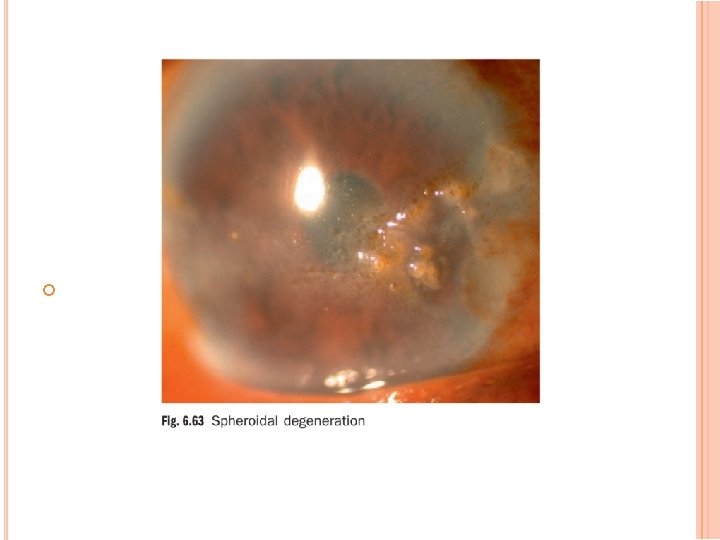
SPHEROID DEGENERATION Climatic droplet keratopathy /Labradorkeratopathy/corneal elastosis. Etiology: • Typically occurs in men who work outdoors. • Has also been related to exposure to UV rays or ageing or cornel disease
CLINICAL FEATURES In this condition amber-coloured spheroidal granules accumulate at the level of bowman’s membrane and anterior stroma in the interpalpebral zone. In marked degeneration the vision is affected
TREATMENT Treatment in advance cases is by corneal transplantation
PELLUCID MARGINAL CORNEAL DEGENERATION Acute hydrops maybe seen in the area of inferior thinning. Commonly manifests b/w ages of 20 -40 with no apparent hereditary transmission and equal gender distribution
PELLUCID MARGINAL CORNEAL DEGENERATION
SIGNS Inferior corneal thinning Severely reduced uncorrected visual acuity that typically cannot be improved with spherocylinder lens Practically normal pinhole visual acuity
DIAGNOSTIC PROCEDURES Corneal topography Pachymetry: Used to measure for inferior corneal thinning, which is a reversal of the typical pattern in which the cornea thickens from centre to periphery Orbscan: Shows a classic "kissing birds" appearance with PMD
TREATMENT Because of extremely abnormal corneal topography, the treatment of PMD is difficult. Therapeutic options are limited by the degree of corneal protrusion. A recent study has found that 88% of PMD cases were managed nonsurgically with spectacles (36%) or contacts (52%), whereas 12% underwent penetrating keratoplasty.
THANK YOU