Chronic Sinusitis Diagnosis and Managment Dr N A
- Slides: 56
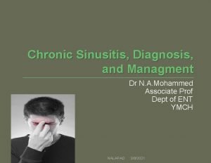
Chronic Sinusitis, Diagnosis, and Managment Dr N. A. Mohammed Associate Prof Dept of ENT YMCH NALAPAD 3/8/2021
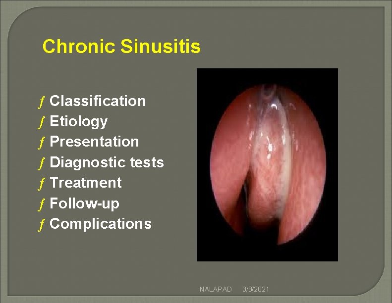
Chronic Sinusitis ƒ Classification ƒ Etiology ƒ Presentation ƒ Diagnostic tests ƒ Treatment ƒ Follow-up ƒ Complications NALAPAD 3/8/2021
Sinusitis Classification Definitions –Chronic > 60 days of sx & signs Or, 4 episodes of acute sinusitis each > 10 days in a single year NALAPAD 3/8/2021
General Contributors to Chronic Sinusitis Resistant infectious organisms Underlying systemic illness (esp. diabetes) Immunodeficiency Irreversible mucosal changes Anatomic abnormality NALAPAD 3/8/2021
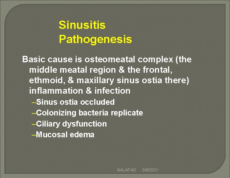
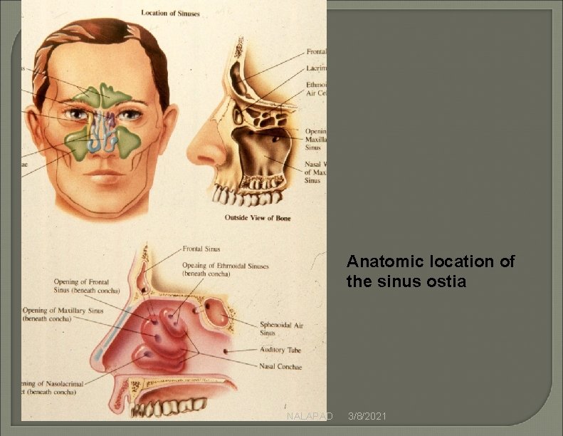
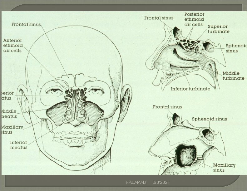
Sinusitis Pathogenesis Basic cause is osteomeatal complex (the middle meatal region & the frontal, ethmoid, & maxillary sinus ostia there) inflammation & infection –Sinus ostia occluded –Colonizing bacteria replicate –Ciliary dysfunction –Mucosal edema NALAPAD 3/8/2021
NALAPAD 3/8/2021
NALAPAD 3/8/2021
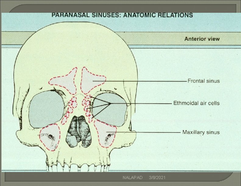
Anatomic location of the sinus ostia NALAPAD 3/8/2021
NALAPAD 3/8/2021
NALAPAD 3/8/2021
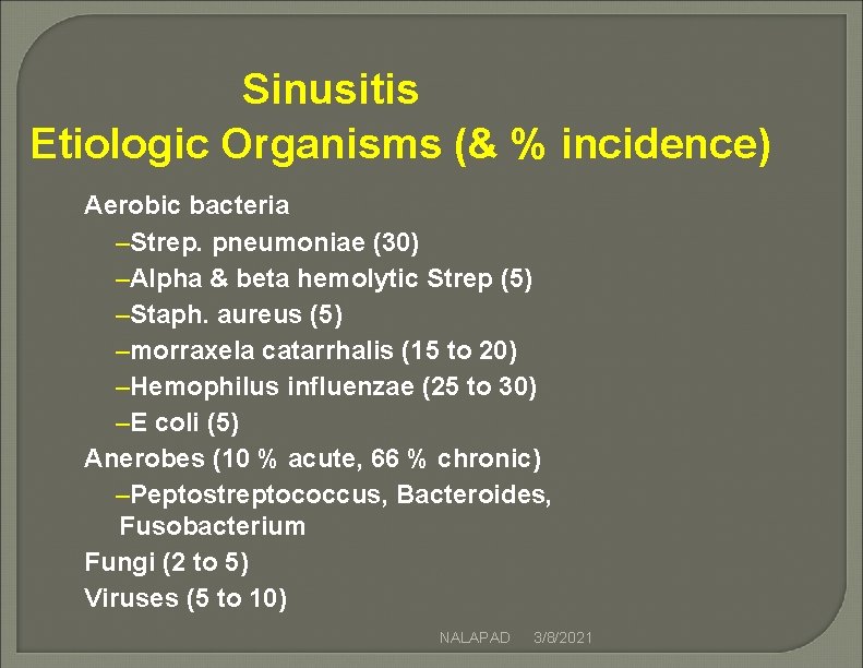
Sinusitis Etiologic Organisms (& % incidence) Aerobic bacteria –Strep. pneumoniae (30) –Alpha & beta hemolytic Strep (5) –Staph. aureus (5) –morraxela catarrhalis (15 to 20) –Hemophilus influenzae (25 to 30) –E coli (5) Anerobes (10 % acute, 66 % chronic) –Peptostreptococcus, Bacteroides, Fusobacterium Fungi (2 to 5) Viruses (5 to 10) NALAPAD 3/8/2021
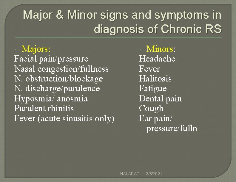
Major & Minor signs and symptoms in diagnosis of Chronic RS Majors: Facial pain/pressure Nasal congestion/fullness N. obstruction/blockage N. discharge/purulence Hyposmia/ anosmia Purulent rhinitis Fever (acute sinusitis only) Minors: Headache Fever Halitosis Fatigue Dental pain Cough Ear pain/ pressure/fulln NALAPAD 3/8/2021
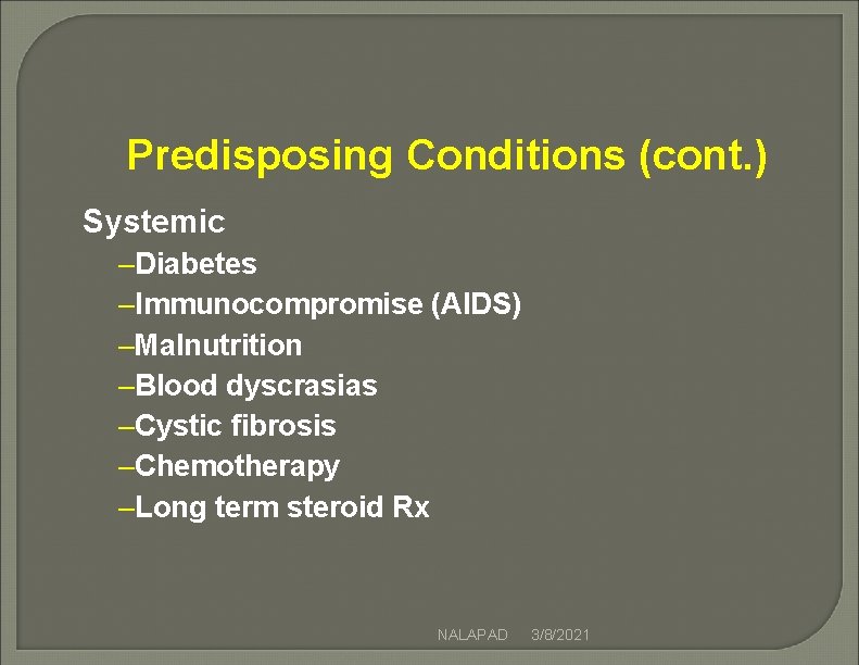
Predisposing Conditions (cont. ) Systemic –Diabetes –Immunocompromise (AIDS) –Malnutrition –Blood dyscrasias –Cystic fibrosis –Chemotherapy –Long term steroid Rx NALAPAD 3/8/2021
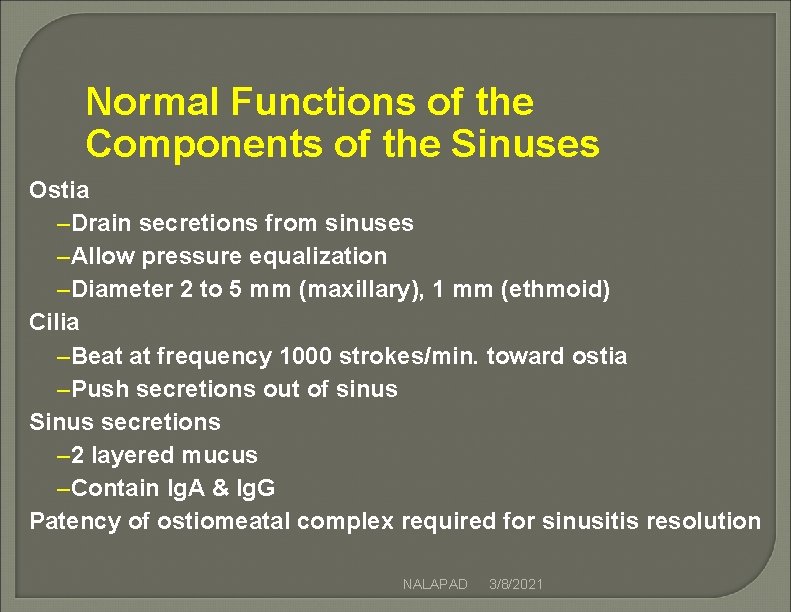
Normal Functions of the Components of the Sinuses Ostia –Drain secretions from sinuses –Allow pressure equalization –Diameter 2 to 5 mm (maxillary), 1 mm (ethmoid) Cilia –Beat at frequency 1000 strokes/min. toward ostia –Push secretions out of sinus Sinus secretions – 2 layered mucus –Contain Ig. A & Ig. G Patency of ostiomeatal complex required for sinusitis resolution NALAPAD 3/8/2021
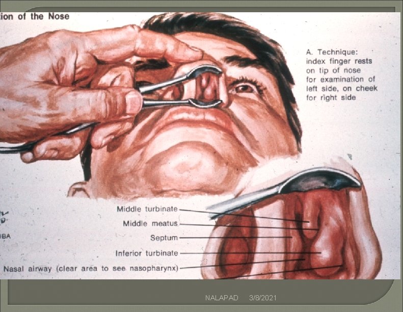
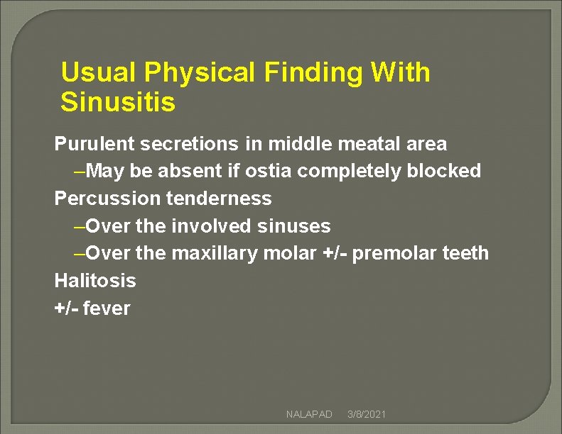
Usual Physical Finding With Sinusitis Purulent secretions in middle meatal area –May be absent if ostia completely blocked Percussion tenderness –Over the involved sinuses –Over the maxillary molar +/- premolar teeth Halitosis +/- fever NALAPAD 3/8/2021
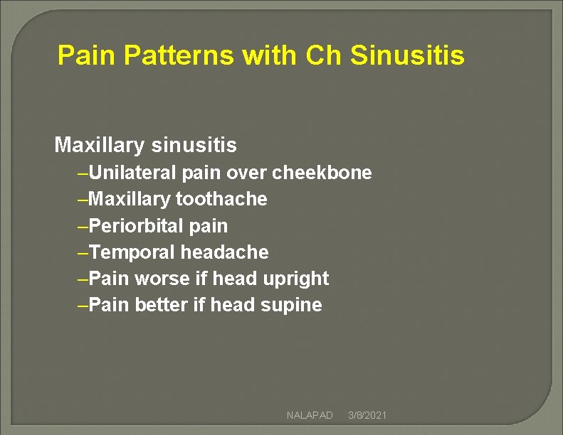
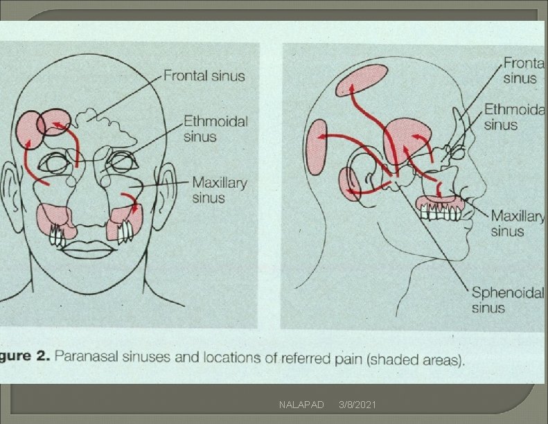
Pain Patterns with Ch Sinusitis Maxillary sinusitis –Unilateral pain over cheekbone –Maxillary toothache –Periorbital pain –Temporal headache –Pain worse if head upright –Pain better if head supine NALAPAD 3/8/2021
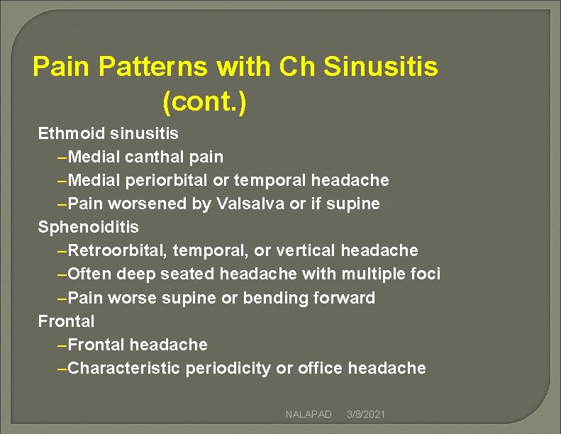
Pain Patterns with Ch Sinusitis (cont. ) Ethmoid sinusitis –Medial canthal pain –Medial periorbital or temporal headache –Pain worsened by Valsalva or if supine Sphenoiditis –Retroorbital, temporal, or vertical headache –Often deep seated headache with multiple foci –Pain worse supine or bending forward Frontal –Frontal headache –Characteristic periodicity or office headache NALAPAD 3/8/2021
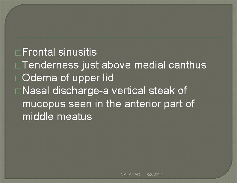
�Frontal sinusitis �Tenderness just above medial canthus �Odema of upper lid �Nasal discharge-a vertical steak of mucopus seen in the anterior part of middle meatus NALAPAD 3/8/2021
NALAPAD 3/8/2021
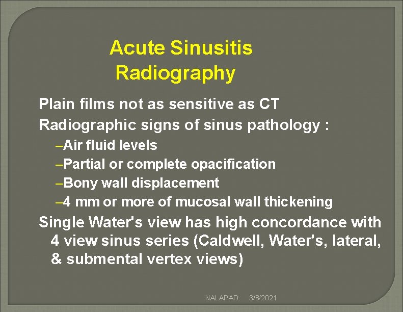
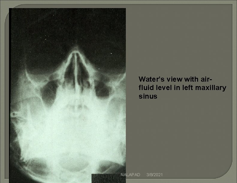
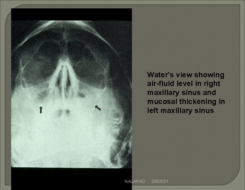
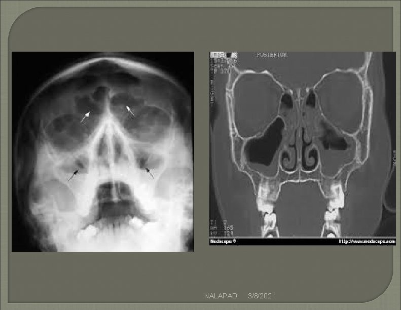
Acute Sinusitis Radiography Plain films not as sensitive as CT Radiographic signs of sinus pathology : –Air fluid levels –Partial or complete opacification –Bony wall displacement – 4 mm or more of mucosal wall thickening Single Water's view has high concordance with 4 view sinus series (Caldwell, Water's, lateral, & submental vertex views) NALAPAD 3/8/2021
Water’s view with airfluid level in left maxillary sinus NALAPAD 3/8/2021
Water’s view showing air-fluid level in right maxillary sinus and mucosal thickening in left maxillary sinus NALAPAD 3/8/2021
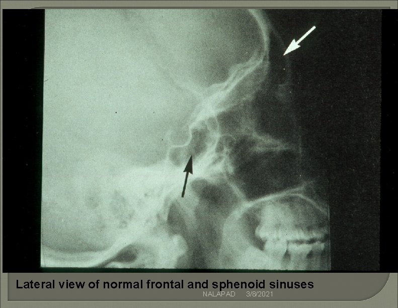
Lateral view of normal frontal and sphenoid sinuses NALAPAD 3/8/2021
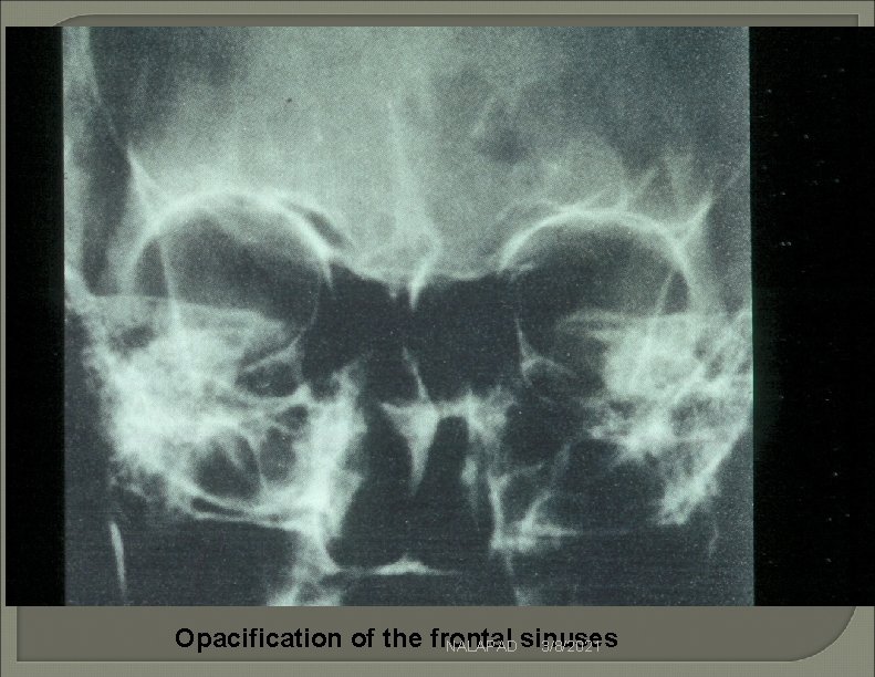
Opacification of the frontal NALAPAD sinuses 3/8/2021
NALAPAD 3/8/2021
Limitations of Plain Film Radiography for Sinusitis Poor visualization of ethmoid air cells Difficulty distinguishing between infection, tumor, or polyp if sinus is completely opacified NALAPAD 3/8/2021
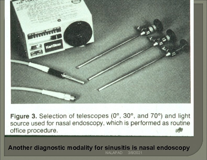
Another diagnostic modality for sinusitis is nasal endoscopy NALAPAD 3/8/2021
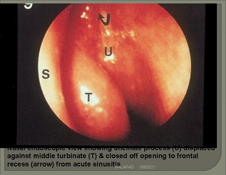
Nasal endoscopic view showing uncinate process (U) displaced against middle turbinate (T) & closed off opening to frontal recess (arrow) from acute sinusitis NALAPAD 3/8/2021
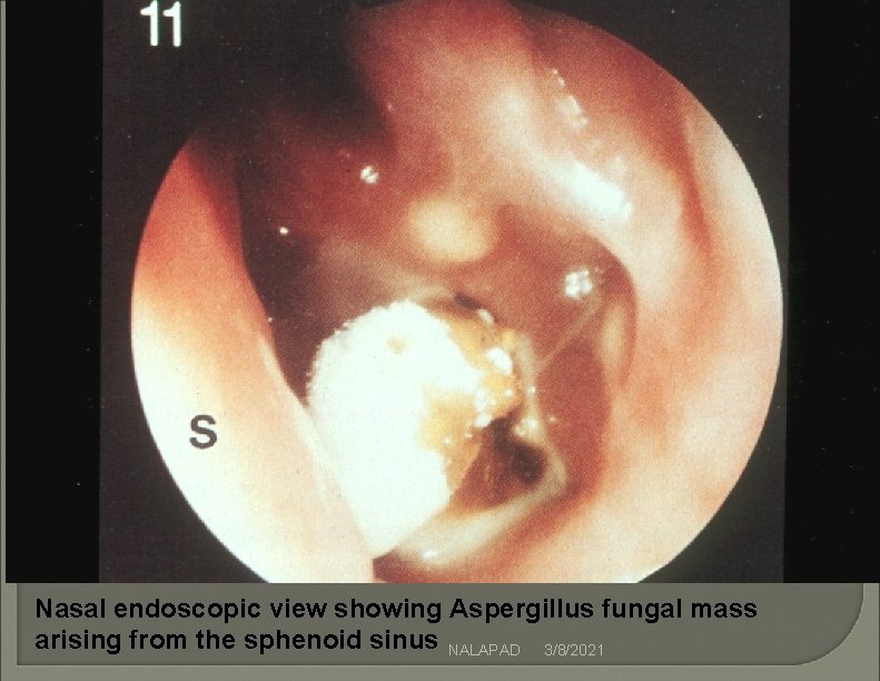
Nasal endoscopic view showing Aspergillus fungal mass arising from the sphenoid sinus NALAPAD 3/8/2021
Use of Computed Tomography (CT) for Diagnosis of Sinusitis Advantages of CT : –Visualizes ethmoid air cells –Evaluates cause of opacified sinus –Differentiates bony changes of chronic inflammation from osteomyelitis Indicated only if complications suspected or if diagnosis uncertain (not needed initially for most cases) NALAPAD 3/8/2021
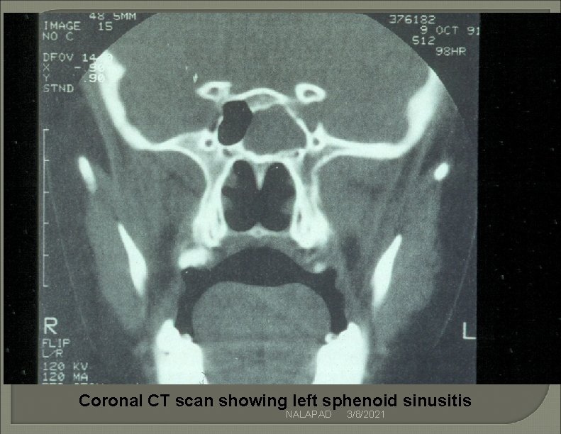
Coronal CT scan showing left sphenoid sinusitis NALAPAD 3/8/2021
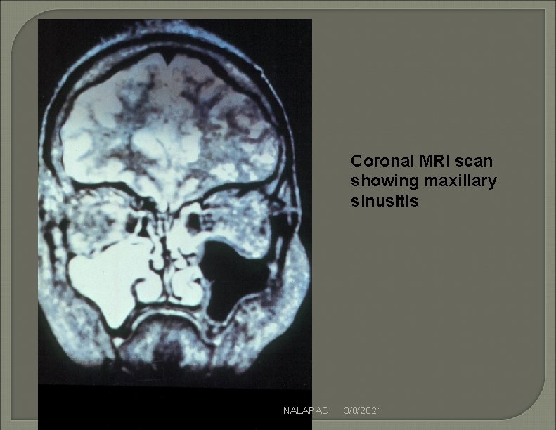
Coronal MRI scan showing maxillary sinusitis NALAPAD 3/8/2021
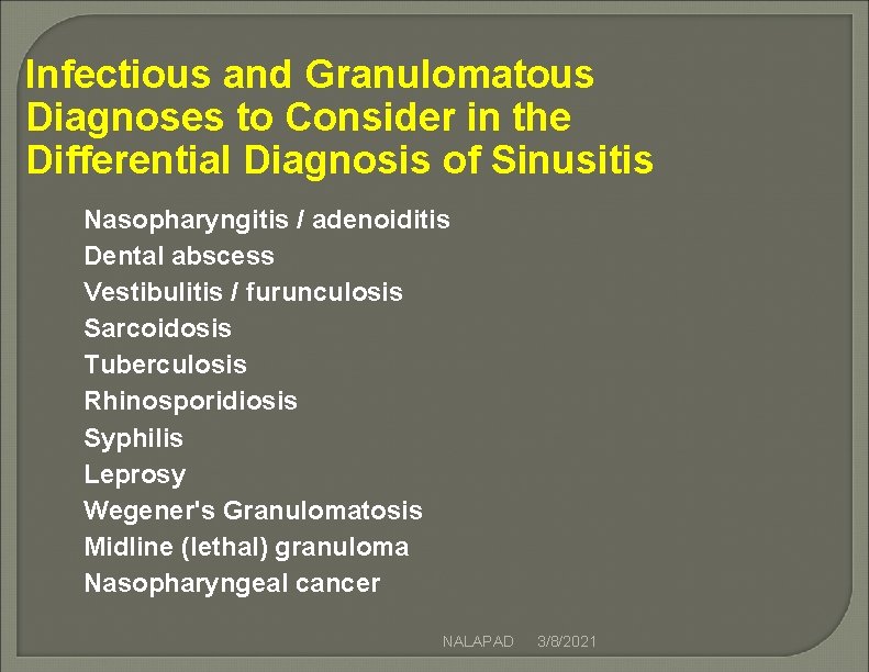
Infectious and Granulomatous Diagnoses to Consider in the Differential Diagnosis of Sinusitis Nasopharyngitis / adenoiditis Dental abscess Vestibulitis / furunculosis Sarcoidosis Tuberculosis Rhinosporidiosis Syphilis Leprosy Wegener's Granulomatosis Midline (lethal) granuloma Nasopharyngeal cancer NALAPAD 3/8/2021
Goals of Medical Therapy for Ch Sinusitis Control Infection Facilitate sinus ostial patency and drainage Provide relief of symptoms Evaluate and treat any predisposing conditions to prevent recurrences NALAPAD 3/8/2021
General Treatment for Ch Sinusitis Oral antibiotic Topical and systemic decongestants Pain medications Optional or secondary medications –warm nasal saline irrigations qid –Antihistamine orally : only in the small % of patients with true allergic component NALAPAD 3/8/2021
Fungal Sinusitis Increasing incidence in both immunocompetent and immunocompromised patients 3 types –Fulminant infection with soft tissue invasion –Progressive indolent invasive disease –Noninvasive localized disease ( mycetoma or allergic fungal sinusitis) NALAPAD 3/8/2021
Fungal Sinusitis Causative fungi: –Aspergillus (most common) –Rhizopus (mucormycosis) –Candida –Histoplasma NALAPAD 3/8/2021
Fungal Sinusitis Major risk factors: –Granulocytopenia –multiple prolonged courses of antibiotics or steroids –Diabetic –AIDS NALAPAD 3/8/2021
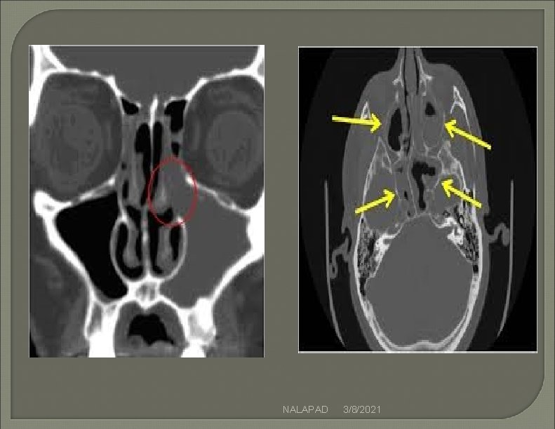
�Fungal ball –due to implantation of fungas in otherwise normal sinus , c/t shows a hyperdense area , maxillary sinus most commonly involved �Treatment- surgical removal of the fungal ball NALAPAD 3/8/2021
� Allergic fungal sinusitis – an allergic reaction to fungus causingsinonasal polyposis and mucin � More than one sinus involved � May be expansion of the sinus or bone erosion due to pressure but no fungal invasion � C/T shows mucosal thickening with hyperdense areas � Treatment- endoscopic surgical clearance, pre and post steriod spray NALAPAD 3/8/2021
�Chronic invasive sinusits- the fungus invade the sinus �There is bone erosion by the fungus �CT shows thickened mucosa , opacification and bone erosion �May intracranial/intraorbital invasion �Treatment- surgical removal, followed by amphotericin B , followed by itraconozole for one year NALAPAD 3/8/2021
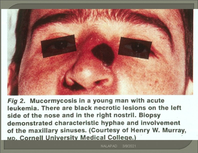
Presentation of Invasive or Acute Fulminant Fungal Sinusitis Facial soft tissue tenderness Cloudy rhinorrhea Fever Gray, friable, anesthetic nasal tissue May have necrotic black tissue May have bloody rhinorrhea NALAPAD 3/8/2021
NALAPAD 3/8/2021
NALAPAD 3/8/2021
Treatment of Invasive Fungal Sinusitis Always should be admitted Correct metabolic abnomalities High dose Amphotencin B +/fluconazole Surgical debidement NALAPAD 3/8/2021
NALAPAD 3/8/2021
NALAPAD 3/8/2021
NALAPAD 3/8/2021
NALAPAD 3/8/2021
NALAPAD 3/8/2021
NALAPAD 3/8/2021
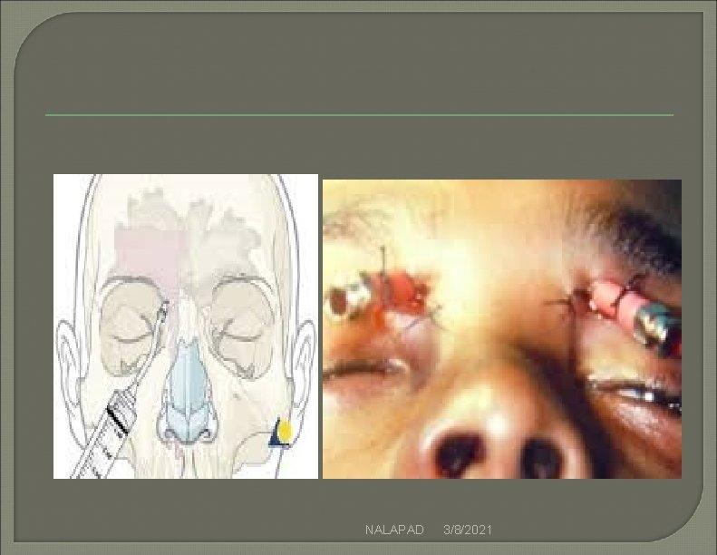
Chronic Frontal Sinusitis � 1) Intranasal drainage � 2)Trephination of frontal sinus � 3)External frontoethimodectomy � 4)Osteoplastic flap �Chronic sphenoidectomy- access to sphenoid obtained by removing the anterior wall , through external ethmoidectomy or transseptal NALAPAD 3/8/2021
NALAPAD 3/8/2021
Chronic ethmoid sinus �Intranasal ethmidectomy- ethmoid air cell are removed, with disease tissue �Frontal and shenoid sinus can also be drained through the procedure �External ethmoidectomy-etmoid is approached through a medial orbital incision, access can also be optained to shenoid and frontal sinus NALAPAD 3/8/2021
Functional endoscopic sinus surgery �Most of the pathology in the sinuses can be reversible if good drainage and ventilation are provided �Fess – surgery using rigid endosope, with illumination and good magnification with instruments which can reach precise site and limit surgery NALAPAD 3/8/2021
THANK YOU NALAPAD 3/8/2021