
SINUSITIS Dr Abdussalam M jahan ENT depart Misurata

SINUSITIS Dr. Abdussalam M jahan ENT depart, Misurata university, faculty of medicine
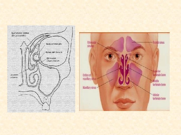
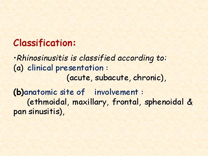
Classification: • Rhinosinusitis is classified according to: (a) clinical presentation : (acute, subacute, chronic), (b)anatomic site of involvement : (ethmoidal, maxillary, frontal, sphenoidal & pan sinusitis),
Acute Rhinosinusitis: • Having a duration of less than or equal to 4 weeks. Subacute Rhinosinusitis: • Having a duration of 4 to 12 weeks. Chronic Rhinosinusitis: • Lasting longer than 12 weeks.
Etiology: Acute Rhinosinusitis: • presence of bacteria in a sinus cavity with an obstructed ostium. • failure of the mucociliary clearance to removes the bacteria. • Quantity or consistency of the sinonasal secretions can affect mucociliary clearance and promote bacterial growth. • Once the ostium becomes occluded, a local hypoxia develops in the sinus cavity, and sinus secretions accumulate. This combination of low-oxygen tension and a rich culture medium of secretions allows bacterial growth to occur within the sinus.
Causative micro-organisms in Acute sinusitis: • In adults, The most common pathogens are Streptococcus pneumoniae and Haemophilus influenzae. • In children, similar organisms are seen, with the addition of Moraxella catarrhalis.
DIAGNOSIS: it is primarily clinical: Signs & symptoms: 7 Major factors 1. Facial pain & tenderness. 2. Headache severe and increase with leaning forward. 3. Facial fullness. 4. Nasal obstruction/blockage 5. Nasal & postnasal discharge (purulent, discolored). 6. Hyposmia/anosmia 7. Fever.
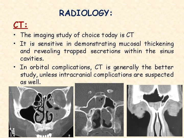
RADIOLOGY: CT: • The imaging study of choice today is CT • It is sensitive in demonstrating mucosal thickening and revealing trapped secretions within the sinus cavities. • In orbital complications, CT is generally the better study, unless intracranial complications are suspected as well.
• Viral respiratory tract infections and allergy will both cause mucosal thickening in the absence of infectious or chronic sinusitis. • About 40% of normal people without sinonasal complaints will have abnormalities of the sinus mucosa on CT scan that may be transient and not indicative of true disease.
• MRI: Indications: • If cranial or intracranial complications is suspected. It clearly demonstrate dural inflammation that would not be appreciable by CT.
COMPLICATIONS OF SINUSITIS: • In the antibiotic era, such complications have become less common, but they still have the potential for serious morbidity or even mortality. • Improved diagnostic modalities and advances in medical and surgical techniques have significantly reduced the risk of blindness or life-threatening intracranial infections.
I. Orbital complications: • Most orbital complications occur in young children, but those in older children and adults are typically more severe and necessitate surgery. • Ethmoiditis most commonly leads to orbital involvement.
• 5 Stages: 1 ST stage, pre-septal peri-orbital cellulites: • Consists of eyelid swelling anterior to the orbital septum (septum is a fibrous membrane dividing the eyelid into anterior and posterior chambers) without involvement of the orbital contents. 2 ND stage, orbital cellulites: • Orbital soft tissue becomes involved, a diffuse process of inflammation without abscess formation. • Patients with this complication are generally proptotic, with some degree of ophthalmoplegia and chemosis.
3 RD stage, sub-periosteal abscess: • Pus accumulates between bone and orbital periosteum. • This will displace the orbit inferolaterally and may cause some proptosis. • Unrecognized or untreated, the process can expand to cause extraocular muscle impairment, chemosis, and loss of visual acuity. 4 TH stage, orbital abscess: • Pus within the orbital tissue. • marked proptosis, limitation of extraocular movement, and visual loss are commonly observed.
5 TH stage, cavernous sinus thrombosis (CST): • Result from extension of ethmoid or sphenoid sinusitis directly or via thrombophlebitis of the ophthalmic vein. • Proptosis, chemosis, ophthalmoplegia, and decreasing visual acuity are the rule. • Process can extend to the opposite side, and bilateral findings are considered a diagnostic hallmark.
II. Intracranial complications: • Most intracranial infections arise from the frontal sinus. • The types of complications that may develop include osteomyelitis of the frontal bone, meningitis, epidural abscess, subdural empyema, and intracerebral abscess
TREATMENT OF SINUSITIS • Antibiotic better amoxil clavunic acid for two weeks. • Decongestant & mucolytic. • Analgesia Last one

Thanks for your attention www. zahrawi. ly ﺍﻟﺤﻤﺪ ﻟﻠﻪ
- Slides: 19