Assignment Topic Echo Cardio Graphy Presented To Dr


: n n A combination of an echocardiogram and an electrocardiogram")
: n. A specialized form of echocardiogram, producing images of your heart")


 Parasternal short axis: Once the long axis is imaged, the echo probe should")


- Slides: 37
Assignment Topic: Echo. Cardio. Graphy Presented To: Dr. Allah Nawaz Qazi Prepared by: M. Irfan M. Farrukh Gopal Kashif Iqbal Khair Muhammad
Goals n n n n Definition Anatomy of Heart Purpose Types Indication Contra Indication Procedure Orthogonal View After Care Risk Precaution Normal Result Abnormal Finding Links
Echocardigraphy n Gk, echo + kardia, heart, graphein, to record n Echocardiography is a non-invasive, imaging test that can be used to learn how the heart is pumping and what areas are not pumping normally. A scan of the heart is taken using ultrasound equipment
Heart n The human heart is a pear-shaped structure about the size of a fist weighs approximately 1 pound. Pumps about 5 quarts (4. 7 liters) of blood every minute. The heart is made of a special kind of muscle called myocardium, and is enclosed in a double-layered, membranous sac called a pericardium.
Heart 4 Chambers: Right atrium n Left atrium n Right ventricle n Left ventricle n Valves: n n Mitral valve Tricuspid Valve Pulmonary valve Aortic Valve
Purpose: n Echocardiography allows visualization of cardiac structures, cardiac walls and the velocity of blood flow at certain points in the heart. The technique is an extension of ultrasound examination using beams of sound at frequencies of 2. 55 MHz.
Classification of Echocardiography n n n Several types of echocardiography used clinically: M-mode, Two-dimensional (2 -D, B-mode or real time), and Doppler echocardiography. Echocardiography, Three-Dimensional Echocardiography, Four-Dimensional
M-mode n n The M-mode echocardiogram uses a high sampling rate and can yield cleaner images of cardiac borders, allowing the echo-cardiographer to obtain more accurate measurements of cardiac dimensions and more critically evaluate cardiac motion. Standard M-mode views are obtained from the right parasternal position. This technique can help us learn about the: size of one of the heart's chambers thickness of the chamber walls condition of the Mitral valve -- between the left upper chamber (atrium) and left lower chamber (ventricle)
Two-Dimensional Echocardiography Show results in cross sectional views of the heart, showing both length and width (two dimensions). The images are recorded in "real time. " It can help us learn about: n problems with the heart's valves n the relation of the heart's chambers to each other n how much blood is pumped from the heart n
Doppler Echocardiography n Doppler imaging allows evaluation of blood flow patterns, direction, and velocity; thus, it permits documentation and quantification of: – – – Valvular insufficiency or stenosis and Cardiac shunts. Estimations of blood flow and cardiac output can also be made – Two types of Doppler echocardiography are: 1. 2. Spectral Doppler Color Doppler
Doppler Spectral Doppler: n Records the flow of blood within the cardiovascular system, giving information about its speed Color Doppler: n Adds color to traditional Doppler studies, giving information about the direction and type of blood flow as well as problems such as valve leaks.
Echocardiography, Three. Dimensional n Echocardiography amplified by the addition of depth to the conventional two-dimensional ECHOCARDIOGRAPHY visualizing only the length and width of the heart.
Echocardiography, Four. Dimensional n Dynamic three-dimensional echocardiography using the added dimension of time to impart the cinematic perception of motion
Stress echocardiogram (stress echo): n n A combination of an echocardiogram and an electrocardiogram (or EKG), that gives a picture of your heart when it is working harder than normal (such as when you are physically active). It can help us learn about: Blockages Arrhythmias Blood pressure response
Transesophageal echocardiogram (TEE): n. A specialized form of echocardiogram, producing images of your heart from inside, rather than outside, your body. During a TEE, you swallow a small, flexible tube, through which the echo pictures are taken. This allows a view of the heart through the esophagus. ie. vegetation
Indication: Echocardiography can provide imaging of all aspects of cardiovascular physiology and pathology including. n n n n n Aortic, Mitral, Tricuspid and Pulmonary Valves including stenosis and regurgitation Left and Right Ventricular Function. Ischemic Ventricular Abnormalities Pericardial Disease Cardiac Tamponade Systolic and Diastolic Heart Failure Pulmonary Embolism Intracardiac and Pulmonary Shunts Myocardial Tumors Acending Aortic Pathology
Contra Indication n There are no frank contraindications to echocardiography.
Preparation n Take Consent of patient n The patient removes any clothing and jewelry above the chest. n Explain procedure
Procedure n Positioning the patient in the left lateral decubitus position will bring the heart forward to the chest wall and closer to the transducer. Obtaining images at end expiration will also bring the heart closer to the chest wall, and may be particularly useful in patients with significant obstructive lung disease. Air trapping in these patients may make imaging especially difficult. In patients that are unable to lie in the decubitus position or in very obese patients, sometimes the only images that can be obtained are in the subxiphoid windows.
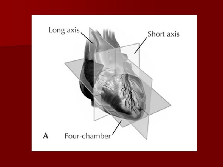
Three Orthogonal Planes: n Long Axis - transects the heart from aortic root to the left ventricular apex n Short Axis – runs from left mid clavicle to the right hip n Four Chamber Axis – runs from apex to the base; perpendicular to other 2 axis
Different Windows and View
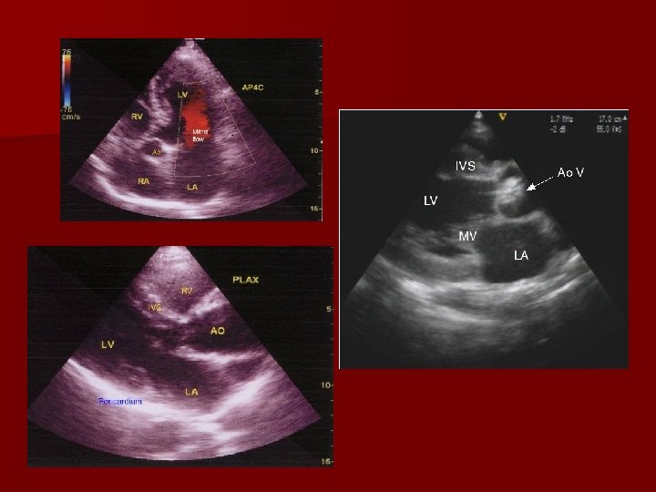
Parasternal Windows: Position the patient in the left lateral decubitus position. The first two windows are taken in the PARASTERNAL window located between the 3 rd-4 th intercostals space. 1)Parasternal Long axis: This image is obtained with the transducer notch facing the right shoulder. In this position the Mitral and aortic valve should be imaged. Pericardial effusions should be assessed.
2) Parasternal short axis: Once the long axis is imaged, the echo probe should be rotated 90 degrees, tilting the probe allows imaging of the left ventricle from the base to the apex. Important aspects and cardiac structures (Aortic and Mitral valves, the IAS and the tricuspid and pulmonic valves) are seen.
Apical Window n Once the parasternal images are obtained, the probe should be placed along the left chest wall at the cardiac apex in the APICAL WINDOW n Apical 2 -Chamber View: n Apical 3 -Chamber View
Sub-Xiphoid Window: n Once the Apical images are obtained, have the patient lie flat in the supine position and bend their knees to relax their abdominal musculature. The probe should then be placed under the xiphoid porcess and angles at the cardiac apex to show the SUBXIPHOID window.
Suprasternal Window: n The probe should then be placed in the suprasternal notch angled down toward the ascending aorta. 2 -D image of the aorta and great vessels if visible to assess origin or dilatation
Aftercare n No special measures need to be taken after echocardiography.
Risks n There are no known risks associated with the use of echocardiography.
Precautions n Echocardiography is an extremely safe procedure and no special precautions are required.
Normal results n. A normal echocardiogram shows a normal heart structure and the normal flow of blood through the heart chambers and heart valves. However, a normal echocardiogram does not rule out the possibility of heart disease.
Abnormal results An echocardiogram may show a number of abnormalities in the structure and function of the heart, such as: nthickening of the wall of the heart muscle (especially the left ventricle) nabnormal motion of the heart muscle nblood leaking backward through the heart valves (regurgitation) ndecreased blood flow through a heart valve (stenosis)
Conclusion n It is the safe and easy procedure to diagnose the pathology of heart. n So we go for this test for diagnostic and therapeutic purpose.
Links n http: //www. ncbi. nlm. nih. gov/sites/entrez? Db=mesh&Cm d=Show. Detail. View&Term. To. Search=68019561&ordinalpo s=1&itool=Entrez. System 2. PEntrez. Mesh_Results. Pa nel. Mesh_RVFull n http: //www. eechocardiography. com/chapters/mv/12. php n http: //ieeexplore. ieee. org/Xplore/login. jsp? url=/iel 5/651 3/17403/00804224. pdf? arnumber=804224 n http: //www. ncbi. nlm. nih. gov/pubmed/17914751