The Heart and Circulation The Electro Cardio Gram

 § The invasion of the cardiac muscle")


 give information about the")

- Slides: 14
The Heart and Circulation
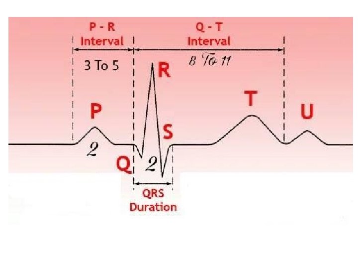
The Electro. Cardio. Gram (E. C. G) § The invasion of the cardiac muscle by the contraction wave is associated with electrical changes. § These potentials may be recorded at points remote from the heart and after electronic amplification may be displayed on a pen recorder or a cathode ray oscilloscope. § The wave form obtained is termed ECG. § The various waves present in the waveform are lettered starting with the letter P.
Each small box = 0. 04 sec Each large box = 0. 2 sec Each 5 large box = 1 sec
§ The P wave corresponds to the spread of excitation from the S. A. node over the atrial muscle and therefore represents atrial systole.
§ The propagation of the contraction wave down the bundle of His does not produce any detectable external electrical change. It is denoted on the E. C. G. by the iso-electric P-Q interval where there is no deflection. § The QRS complex at the commencement of the ventricular excitation, and the T wave at the end are all that remain of this algebraic summation. The relaxation phase T-P is iso-electric.
Leads I, Ill, a. VR, a. VL and a. VF § The detailed waveform depends on the site of the recording electrodes. § To record the standard limb leads, electrodes are applied to the left arm, right arm, and left leg. These electrodes usually consist of a metal plate which is strapped over the flat part of the limb. § Electrode fluid or jelly is used between the electrode and the skin to ensure a good electrical connexion.
§ The right leg is not used for recording the ECG, but a further electrode is often applied to this limb to 'earth' the subject, and thus minimize interference from the mains and electrical apparatus. § In order to make a recording, two input connexions must be made to the amplifier and there is usually a switch on the electrocardiograph apparatus which makes the appropriate connexions internally when a particular lead is chosen.
§ When the switch is set at Lead I, the electrodes on the right and left arms are connected to the amplifier. The amplifier then records the potential difference between the right arm and the left arm. § When the switch is set to Lead II, the potential difference between the right arm and left leg is recorded. §When set to Lead III, the potential difference between the left arm and left leg is recorded.
§ An alternative way of recording the ECG is to connect one input of the amplifier to a limb electrode, and to connect the other input of the amplifier to the other two limbs through two resistances. § Such an arrangement records the difference between the potential in one limb and the mean potential in the other two. § If the single limb is the right arm, the lead is termed 'a. VR', if the left arm, it is termed 'a. VL', and if the left leg (or foot), it is termed 'a. VF'. § Leads I, II III, a. VR, a. VL and a. VF are the six standard limb leads which are recorded clinically.
Cardiac Vector § The figure shows the ECG recorded using six limb leads. § They have been arranged to correspond to the figure below.
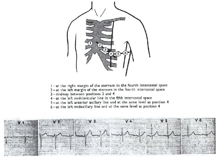
Chest Leads § The six limb leads (so far discussed) give information about the 'electrical axis of the heart' in the frontal plane. § To study the activity in a horizontal plane, chest leads are used. § These usually take the form of a single chest electrode which will adhere to the chest wall by suction, and which is connected to one input of the amplifier. § The other input of the amplifier is connected to an electrically neutral point V which is obtained by joining the three limb leads together through resistances.
§ It is found that the point V does not change its potential during the cardiac cycle and this potential may thus be used as a reference. § As the search electrode is moved across the chest, the ECG shows a dominant S -wave in chest positions 1 and 2, and a dominant R-wave in chest positions 5 and 6.