AP Unit 4 Lecture 6 and 7 External


")


")

 contractions No known")
- Slides: 53
A&P Unit 4 Lecture 6 and 7
External anatomy of the ear Helix Triangular Fossa Concha Pinna Tragus Antihelix Lobule Antitragus
Three Parts of the Ear
Overview of the anatomy of the external ear, middle ear and internal ear
Gross Anatomy of the Middle Ear
Gross Anatomy of the Inner Ear
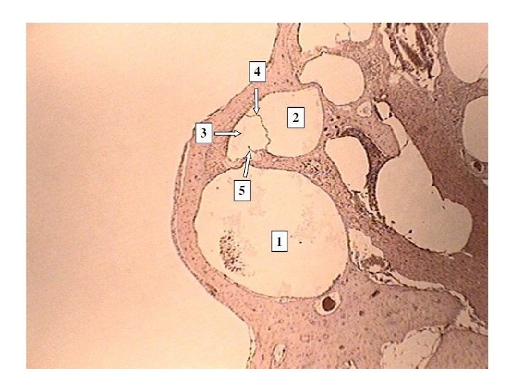
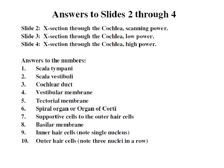
Anatomy of the Cochlea
Sectional View of the Cochlear as it will appear on a microscope slide
Internal Anatomy of the Cochlea with details of the Bony Labyrinth
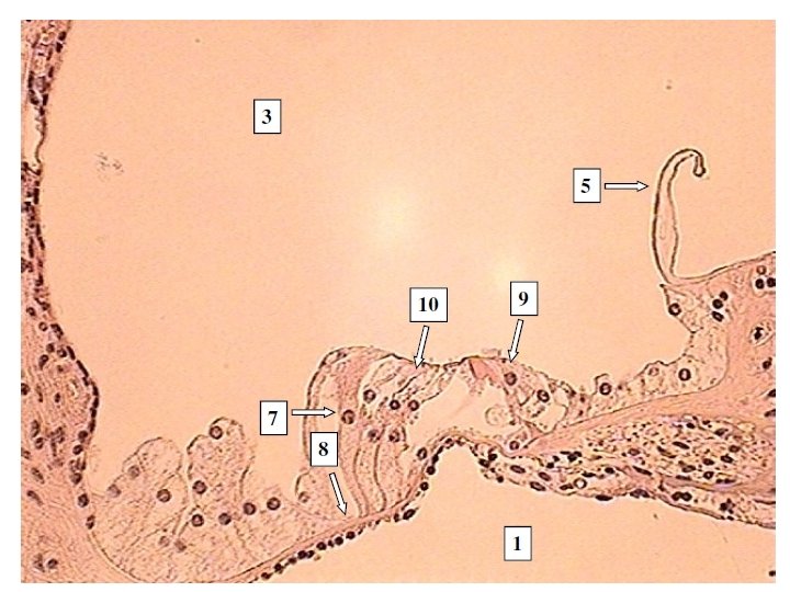
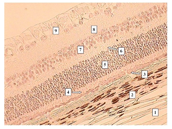
Internal Anatomy of the Bony Labyrinth with details of the Organ of Corti
Events involved in the creation of an Auditory action impulse 1. Pinna directs sound waves into the external auditory meatus. 2. Sound waves cause the tympanic membrane to vibrate. a. Slowly for lowfrequency sounds b. Rapidly for highfrequency sounds c. Distance the membrane travels during these vibrations relates to loudness or decibels.
Events involved in the creation of an Auditory action impulse 3. Vibrations are communicated from the tympanic membrane to the auditory ossicles. Malleus Incus Stapes 4. 5. Stapes vibrates back and forth in the oval window, thus vibrating the oval window membrane. Vibration of oval window membrane causes fluid pressure waves in the perilymph of the scala vestibuli.
Events involved in the creation of an Auditory action impulse 6. Perilymph pressure waves are transmitted to the scala tympani and eventually to the round window causing the secondary tympanic membrane to bulge outward. 7. Vibrations of the vestibular membrane cause vibrations of the endolymph within the cochlear duct. 8. Endolymph pressure waves cause the basilar membrane to vibrate.
Events involved in the creation of an Auditory action impulse 8 a. Vibrations of the basilar membrane cause the hair cells of the Organ of Corti to vibrate. 8 b. Hair cells vibrate upward, bending the stereocilia against the tectorial membrane. 8 c. Bending the stereocilia produces a receptor potential that ultimately leads to a action potential on Cochlear nerve.
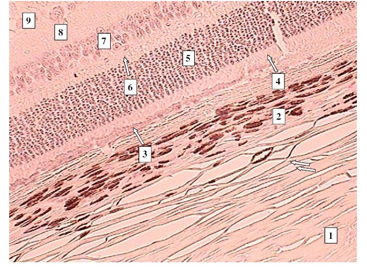
Histology of the retina of the eye
A&P Unit 4 Lecture 6 B
Two Kinds of Equilibrium 1. Static equilibrium: Refers to the maintenance of body (head) position relative to the force of gravity 2. Dynamic equilibrium: Refers to the maintenance of the body (head) in response to sudden movements such as rotation, acceleration, and deceleration
Static Equilibrium • The receptors for static equilibrium are the Utricle and the Saccule
Location and structure of the receptors in the macula for static equilibrium
How the macula functions in relation to gravity
Dynamic Equilibrium • The receptors for dynamic equilibrium are the semicircular canals
Anatomy of the Semicircular canal receptors called ampulla
How the Ampulla function to produce dynamic equilibrium sensations
Pathway for Vestibular System
Bio& 241 A&P Unit 4 Lecture 7
Organization of the Nervous System
Basic Patterns of the Autonomic Nervous System • Preganglionic fibers arise in the CNS and are myelinated “B” fibers • Postganglionic fibers arise in autonomic ganglia outside the CNS and are unmyelinated “C” fibers • These Postganglionic fibers produce either IPSP’s or EPSP’s at the organs they innervate • Consists of two antagonistic systems: Sympathetic and Parasympathetic nervous systems • The role of the ANS is to maintain homeostasis • Controlled by hypothalamic centers
Types of Nerve Fibers • “A” fibers: Largest diameter myelinated fibers, fastest saltatory conduction • “B” fibers: intermediate diameter myelinated fibers, slower saltatory conduction • “C” fibers: Smallest diameter unmyelinated fibers with slow continuous conduction
Comparison of the Somatic and Autonomic Nervous Systems
Role of the Sympathetic system in maintaining homeostasis • The Sympathetic system responds to “E” situations (exercise, emergency, excitement, and embarrassment) collectively called a “fight-or-flight response • During sympathetic stimulation, this system dominates or overrides the parasympathetic branch
Role of the Parasympathetic system in maintaining homeostasis • Parasympathetic system responses support body functions that conserve and restore body energy during times of rest and recovery. Its responses can be remembered by “SLUDD” (salivation, lacrimation, urination, digestion, and defecation) • Intense fear referred to as “paradoxical fear, ” the massive over-stimulation of parasympathetic system, which results in loss of control over urination or defecation
Basic patterns of the Sympathetic division of the ANS • Short Preganglionic fibers arise in the CNS and are myelinated “B” fibers • Preganglionic fibers arise as part of spinal nerves T 1 – T 12 and L 1 and L 2 (thoracolumber) • Long Postganglionic fibers arise in autonomic ganglia, either paravertebral or prevertebral ganglia, outside the CNS and are unmyelinated “C” fibers
Basic patterns of the Parasympathetic division of the ANS • Long Preganglionic fibers arise in the CNS and are myelinated “B” fibers • Preganglionic fibers arise as part of cranial nerves III, VII, IX, and X and S 2, S 3, And S 4 (Craniosacral) • Short Postganglionic fibers arise in autonomic ganglia, associated with visceral organ except for cranial nerves III, VII, and IX, outside the CNS and are unmyelinated “C” fibers
Comparison of the Sympathetic and Parasympathetic divisions of the ANS
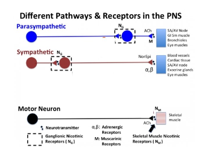
Neurotransmitters and Receptors of the Sympathetic division • Preganglionic fibers release Ach from their synaptic end bulbs • Postganglionic dendrites contain ligand-gated channels referred to as “nicotinic” receptors due to the effects of nicotine on these receptors • Postganglionic fibers release “NE” Norepinephrine from their synaptic end bulbs • Effector membranes contain ligand-gated receptors referred to as “adrenergic” receptors that occur in two forms (alpha 1 and alpha 2 receptors) which allows for IPSP and EPSP
Neurotransmitters and Receptors of the Parasympathetic division • Preganglionic fibers release Ach from their synaptic end bulbs • Postganglionic dendrites contain ligand-gated channels referred to as “nicotinic” receptors due to the effects of nicotine on these receptors • Postganglionic fibers also release Ach from their synaptic end bulbs • Effector membranes contain ligand-gated channels referred to as “muscarinic”receptors due to the effects of muscarine, the poison found in mushrooms, on these receptors
Summary of Neurotransmitters and receptors of the ANS
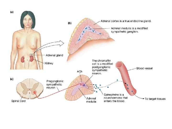
• • Enhancement of the Sympathetic branch by the Adrenal Medulla The Adrenal Medulla is one exception to the basic pattern of the Sympathetic branch Pregangloinic fibers innervate the adrenal medulla via the Celiac ganglion - No postganglionic fiber are present Chromaffin cells of the medulla release compounds that act as neurotransmitters but circulate in the blood like hormones These cells of the medulla release a mixture composed of 80% epinephrine, 20% norepinephrine and a trace of dopamine
Enhancement of the Sympathetic branch by the Adrenal Medulla • Effector membranes contain a second or different group of ligand-gated “adrenergic” receptors referred to as Beta receptors occur in three forms ( beta 1, beta 2, beta 3) which allow for both EPSP and IPSP • Epinephrine released by Chromaffin Cells targets beta receptors thus enhancing the overall sympathetic effect • Norepinephrine released by Chromaffin Cells targets alpha receptors also enhancing the overall sympathetic effect
Activities of the Sympathetic System Effector Radial iris muscle Circular iris muscle Ciliary Muscle Cardiac Muscle Sympathetic (+) contraction causes dilation No known effect (-) relaxation for far vision (+) Increased HR and force of contraction Parasympathetic No known effect (+) contraction causes constriction (+) contraction for near vision (-) Decreased HR and force of contraction
Activities of the Sympathetic System Effector GI Smooth muscle GI sphincters Gallbladder Pancreas Sympathetic (-) decreased contraction (+) increased contraction (-) relaxation Parasympathetic (+) increased contraction (-) decreased contraction (+)contraction and release of bile (-) Inhibits release (+) promotes release of enzymes, promotes secretion of glucagon of insulin
Activities of the Sympathetic System Effector Adipose tissue Liver Lungs Urinary bladder Sympathetic (+) Promotes lipolysis and release of triglycerides to blood (+) glycogenolysis gluconeogenesis (-) relaxation and airway dilation (-) relaxation of wall, (+) increases sphincter Parasympathetic No known effect (+) glycogenesis (+) contraction and airway constriction (+) contraction of wall, (-) relaxation of sphincter
Activities of the Sympathetic System Effector Sweat glands Arrector pili muscle Penis/ Clitoris Prostate Seminal vesicles Sympathetic Parasympathetic (+) increases No known effect sweating (+) contraction No known effect erection of hairs (-) vasocontriction (+) vasodilation and erection (+) contractions and ejaculation No known effect
Activities of the Sympathetic System Effector Vagina Uterus Sympathetic Parasympathetic (+) contractions No known effect and female orgasm (+) contractions in Minimal or pregnant uterus unknown effect