TRAUMA AND EMERGENCY IN OPHTHALMOLOGY Prof Dr Raciha


- Slides: 47

TRAUMA AND EMERGENCY IN OPHTHALMOLOGY Prof. Dr. Raciha Beril Küçümen 2016 -2017 Educational Year

1. Eyelid TRAUMA • Haematoma • Margin laceration • Canalicular laceration 2. Orbital blow-out fractures • Floor • Medial wall 3. Complications of blunt trauma • Anterior segment • Posterior segment 4. Complications of penetrating trauma 5. Management of intraocular foreign bodies 6. Chemical injuries

How to examine History : type of traumatic event, time of onset, nature of symptoms Time, place and type of injury Patients’ history of eye conditions, drug allergies and tetanus immunization Pearl: don’t delay prompt treatment for a detailed hx in an obvious injury (eg. Chemical burn) Visual Acuity Testing

How to examine External examination Palpation: orbital rims in blunt injury? Penlight inspection: signs of perforation, depth of AC, uveal prolapsus, hyphema Lid eversion: foreign body, chemical burn – Pearl: don’t manipulate eyelids in injury of the globe ? Fluorescein staining: foreign body sensation, hx of blunt or sharp injury. Topical anesthesia: relieve discomfort • Pearl: Don’t prescribe! Prolonged use can result in corneal ulceration and inadvertant injury Pupillary reactions: optic nerve injury Ocular motility testing: • Restricted in orbital hematoma. • Vertical restriction in blowout fracture. • Limitation of eye movements, bruit, proptosis: carotid-cavernous fistula

Ophthalmoscopy: Look for: edema, retinal hemorrhages, detachment, foreign body Absent red reflex: immediate REFERRAL Pearl: Don’t dilate pupils in patients with head trauma and shallow anterior chamber

Radiologic studies: Facial or orbital fracture Ocular or orbital foreign body Pearls: Don’t order MR if a metallic foreign body is suspected. Metal may heat, vibrate or move during the scan. Result in additional intraocular injury

Definitions and Classification in Ocular Trauma Eyewall: sclera and cornea Closed globe injury: no full thickness wound of eyewall Open globe injury: full thickness wound of eyewall Contusion: no wound, injury results from changes in the shape of the globe (eg. Angle recession) Lamellar laceration: partial-thickness wound Rupture: full-thickness wound of the eyewall caused by a blunt object Laceration: full-thickness wound of the eyewall caused by a sharp object Penetrating injury: entrance wound Perforating injury: entrance and exit wounds

Closed globe injuries: eyelid lacerations Non-marginal Marginal: referral Canalicular: referral Postop. Wound Care: - full thickness: pressure patching 1 week (caution in amblyogenic age) - antibiotic ointment 1 week Follow-up: 5 -7 days, remove sutures. (Margin sutures 2 weeks)

Lid margin laceration Carefully align to prevent notching Align with 6 -0 black silk suture Close tarsal plate with fine absorbable suture Place additional marginal silk sutures Close skin with multiple interrupted 6 -0 black silk sutures

Canalicular laceration • Repair within 24 hours • Locate and approximate ends of laceration • Bridge defect with silicone tubing • Leave in situ for about 3 months

Eyelid haematoma Usually innocuous but exclude associated trauma to globe or orbit Orbital roof fracture if associated with subconjunctival haemorrhage without visible posterior limit Basal skull fracture - bilateral ring haematomas (‘panda eyes’)

Closed globe injuries: orbital trauma Orbital blowout fractures Traumatic optic neuropathy Orbital hemorrhage and compartment syndrome Mild-moderate IOP elevations: glaucoma medications and observation Severe: lateral canthotomy and cantolysis surgical decompression

Pathogenesis of orbital floor Blow-out Fracture

Signs of orbital floor Blow-out Fracture • Periocular ecchymosis and oedema • Infraorbital nerve anaesthesia • Enophthalmos - if severe • Ophthalmoplegia typically in up- and downgaze (double diplopia)

Investigations of orbital floor blow-out fracture Coronal CT scan • Right blow-out fracture with ‘tear-drop’ sign Hess test • Restriction of right upgaze and downgaze • Secondary overaction of left eye

Surgical treatment of blow-out fracture a b c d (a) Subciliary incision • Coronal CT scan following repair of right blow-out fracture with synthetic (b) Periosteum elevated and entrapped material orbital contents freed (c) Defect repaired with synthetic material (d) Periosteum sutured

Medial wall Blow-out Fracture Signs Periorbital subcutaneous emphysema Ophthalmoplegia - adduction and abduction if medial rectus muscle is entrapped Treatment • Release of entrapped tissue • Repair of bony defect

Closed globe injuries: ocular surface Traumatic subconjunctival hemorrhage Corneal abrasions Corneal foreign bodies: removal Conjunctival lacerations: suture > 10 mm Lamellar corneal and scleral lacerations Topical antibiotics Shield Corneal glue? Chemical injuries

Subconjunctival hemorrhage Very common in ocular emergency Etiology: Spontaneous: valsalva maneuvers Acute bacterial or viral conjunctivitis Systemic hypertension and anticoagulation Traumatic: (peaked pupil, asymmetric AC depth, low IOP: think of occult globe violation) Conservative measures: lubricant eye drops, reassurance Change color over 10 -14 day course


Chemical Injuries True ocular emergency Needs immediate irrigation Alkali burns (ammonia, fresh lime in plaster and concrete, lye, bleach): More severe damage than acids Rapidly penetrate the eye, saponify cell membranes, denature collagen and thrombose vessels Acids: less damage Hydrogen ion precipitates protein Prevents further penetration to the cornea

Grading of severity of chemical injuries Grade I (excellent prognosis) • • Clear cornea Limbal ischaemia - nil Grade II (good prognosis) Grade III (guarded prognosis) • • Cornea hazy but visible iris details Limbal ischaemia < 1/3 • No iris details Limbal ischaemia - 1/3 • to 1/2 • Grade IV (very poor prognosis) • Opaque cornea • Limbal ischaemia > 1/2

Medical Treatment of Severe Injuries 1. Copious irrigation ( 15 -30 min ) - to restore normal p. H 2. Topical steroids ( first 7 -10 days ) - to reduce inflammation 3. Topical and systemic ascorbic acid - to enhance collagen production 4. Topical citric acid - to inhibit neutrophil activity 5. Topical and systemic tetracycline - to inhibit collagenase and neutrophil activity

Surgical treatment of severe chemical injurie Division of conjunctival bands Treatment of corneal opacity by Correction of eyelid deformitieskeratoplasty or keratoprosthesis

Closed globe injuries: anterior chamber Traumatic mydriasis Traumatic iritis Iris sphincter tears and iridodialysis Hyphema: blood accumulates in AC, may rebleed 3 -5 days after the initial injury Sx: pain, photophobia, decreased VA Tx: • • • Topical CS Cycloplegia : atropine Eyeshield Bed rest with minimal ambulation, head >45 degrees IOP elevation: appropriate antiglaucomatous agents Surgery: when necessary Angle recession: tear in the ciliary body cause iris insertion to appear posterior. IOP can increase.

Anterior segment complications of blunt trauma Hyphaema Sphincter tear Cataract Lens subluxation Iridodialysis Angle recession Vossius ring Rupture of globe


Closed globe injuries: lens Lens subluxation and dislocation Phacoanaphylactic uveitis Traumatic cataract Lens-induced glaucoma


Closed globe injuries: posterior segment Commotio retinae: confluent geographic areas of whitened retina. Macular involvement: Berlin’s edema. Damaged photoreceptor outer segments and rpe. No tx Traumatic vitreus hemorrhage Traumatic macular hole Choroidal rupture Sclopetaria: chorioretinal rupture Traumatic retinal detachment Traumatic optic neuropathy

Posterior segment complications of blunt trauma Commotio retinae Choroidal rupture and haemorrhage Avulsion of vitreous base and retinal dialysis Equatorial tears Macular hole Optic neuropathy


Open globe injuries: ruptures and lacerations Subconjunctival hemorrhage: 360 degrees Profound chemosis Relative asymmetry in the AC: Shallow often with peaking of the iris towards the wound Deep AC: in full-thickness injury of posterior segment Transillumination defects of the iris Traumatic cataract, lens dislocation Hyphema Vitreous hemorrhage

Complications of penetrating trauma Flat anterior chamber Uveal prolapse Vitreous haemorrhage Tractional retinal detachment Damage to lens and iris Endophthalmitis


Delayed complications of ocular injury Traumatic iritis Traumatic cataract Delayed trauma related glaucoma Retinal detachment Metallosis bulbi: intraocular foreign bodies should be removed! Traumatic endophthalmitis Sympathetic ophthalmia: least common, most feared Bilateral granulomatous uveitis after injury to the eye

Management of intraocular foreign bodies Localization with reference to radioopaque marker Removal with magnet or by pars plana vitrectomy

Management or Referral True emergency: initiate treatment within minutes! Chemical burns: • Immediate and profuse irrigation. • Referral to an ophthalmologist

Urgent situation: require therapy within a few hours Penetrating injuries of the globe: • • X-ray or CT scan Eye shield Don’t patch, don’t apply ointment REFERRAL Conjunctival or corneal foreign bodies: • • Topical anesthesia Cotton-tipped applicator Irrigation If remains embbedded: REFERRAL

Corneal abrasions: • • • Topical anesthesia Gross examination Fluorescein staining Instill antibiotic drops. Instill short-acting cycloplegic drops for the relief of pain. • Pressure patch for 24 hours (contraversial) • REFER severe cases Hyphema: Immediate REFERRAL.

Lid laceration: • Suture if not deep • Refer: – Lid margin – Canaliculi involvement Radiant energy burns, snow blindness: • • Topical anesthesia Examination Order topical antibiotic and cycloplegic agents Patching

Traumatic optic neuropathy: • Consider cranial or maxillofacial trauma • High resolution CT imaging of the orbital apex, optic canal, and cavernous sinus • May benefit from IV high-dose methylprednisolone if given in the first 8 hours. • Prompt referral

Semiurgent condition: refer to an ophthalmologist within 1 -2 days Orbital fracture Subconjunctival hemorrhage: unless globe rupture or intraocular hemorrhage is suspected

The End
 Dr glaukomflecken
 Glaukomflecken cataract
 Iridodialysis
 Emergency management of maxillofacial trauma
 Chapter 31 ophthalmology and otolaryngology
 Vanderbilt ophthalmology residency
 St john's hospital
 National tb control program
 Lacrim/o combining form
 Mr luke anderson ophthalmology
 Kreiger eye institute
 Ubc ophthalmology
 Ophthalmology code of ethics
 Ophthalmology
 Ubc ophthalmology
 Miosis
 Sump syndrome
 Veterinary ophthalmology
 Jama ophthalmology impact factor
 British journal of ophthalmology
 Online journal of ophthalmology
 Asia-pacific academy of ophthalmology
 Ophthalmology case study
 16710-17
 Alan bird ophthalmology
 Ophthalmology lecture
 Trauma awareness and treatment center utah
 Triune brain theory and trauma
 Limbic system and trauma
 Tarn database
 Trauma recovery and empowerment model
 Uccs veterans health and trauma clinic
 Vicarious felidae
 Trauma gestig behindert
 Trauma thorax
 Tscyc screening form
 Trauma sensitief lesgeven
 4 r's trauma informed care
 Trauma informed care lgbtq
 Dental mobility classification
 Miemss trauma decision tree
 Examples of little t trauma
 Trauma bonding definition
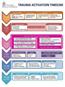
 Trauma timeline
 Dr anita ravi
 Golden hour of trauma
 Trauma-informed care cheat sheet
 Nethercutt emergency center


