
Ophthalmology JongMo Seo Eyespots Bipolar cell Epithelial invaginated
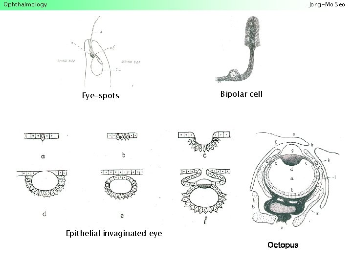
Ophthalmology Jong-Mo Seo Eye-spots Bipolar cell Epithelial invaginated eye Octopus
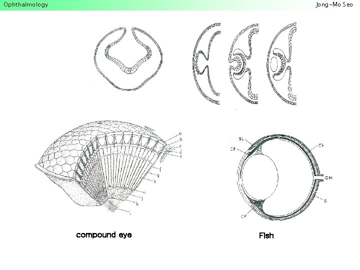
Ophthalmology Jong-Mo Seo compound eye Fish
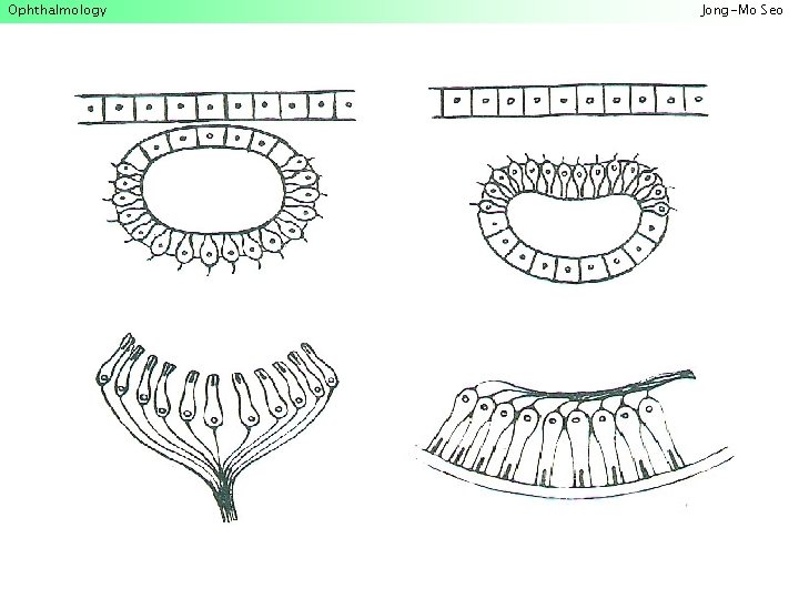
Ophthalmology Jong-Mo Seo
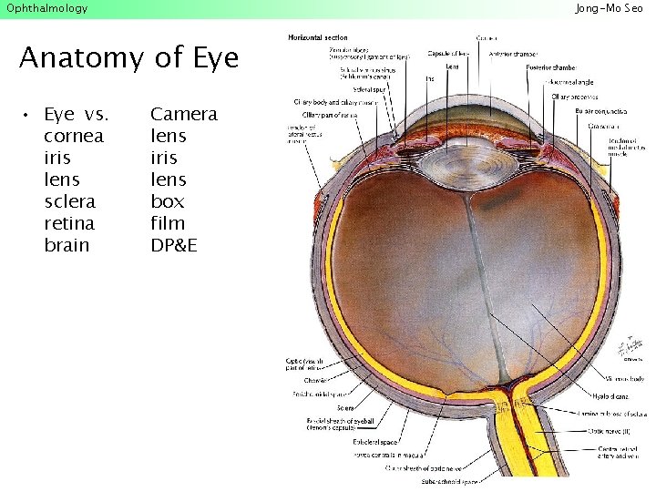
Ophthalmology Jong-Mo Seo Anatomy of Eye • Eye vs. cornea iris lens sclera retina brain Camera lens iris lens box film DP&E

Ophthalmology Diagnosis & Treatment • Diagnosis – Symptom • Manifested • Incipient – Sign • Structural; metamorphosis, dismorphysm, neoplasm • Functional change; mechanical, neuronal • Treatment – Medication; various ‘anti-’s, neurotransmitters, enzymes, . . . – Surgery; reconstruction, destruction, removal, ‘-plantation’ – Genetic approach Jong-Mo Seo
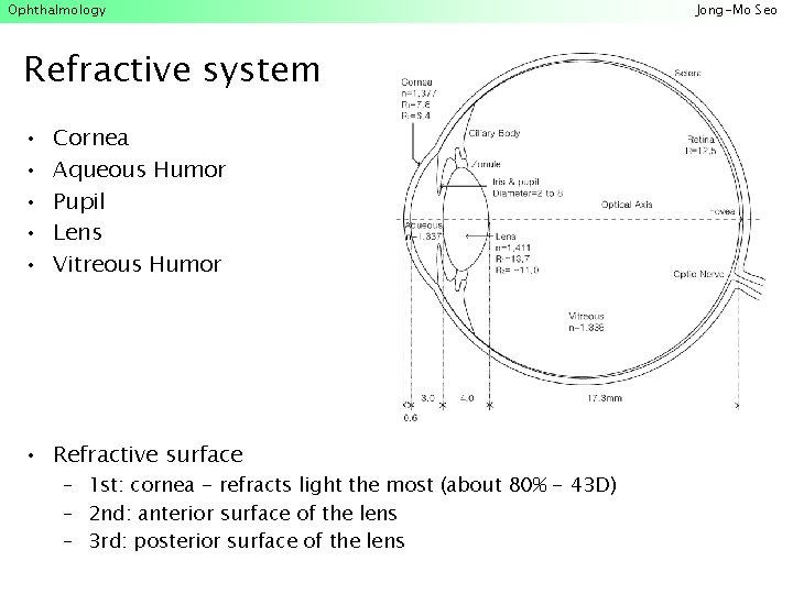
Ophthalmology Refractive system • • • Cornea Aqueous Humor Pupil Lens Vitreous Humor • Refractive surface – 1 st: cornea - refracts light the most (about 80% - 43 D) – 2 nd: anterior surface of the lens – 3 rd: posterior surface of the lens Jong-Mo Seo
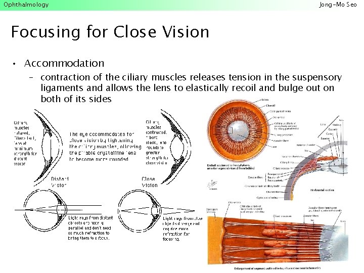
Ophthalmology Jong-Mo Seo Focusing for Close Vision • Accommodation – contraction of the ciliary muscles releases tension in the suspensory ligaments and allows the lens to elastically recoil and bulge out on both of its sides
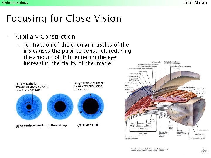
Ophthalmology Focusing for Close Vision • Pupillary Constriction – contraction of the circular muscles of the iris causes the pupil to constrict, reducing the amount of light entering the eye, increasing the clarity of the image Jong-Mo Seo
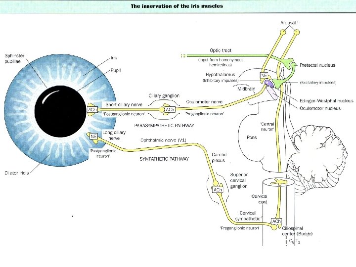
Ophthalmology Jong-Mo Seo
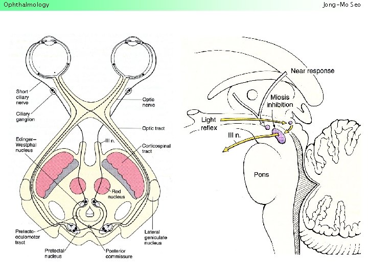
Ophthalmology Jong-Mo Seo
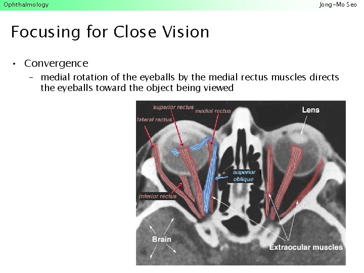
Ophthalmology Jong-Mo Seo Focusing for Close Vision • Convergence – medial rotation of the eyeballs by the medial rectus muscles directs the eyeballs toward the object being viewed
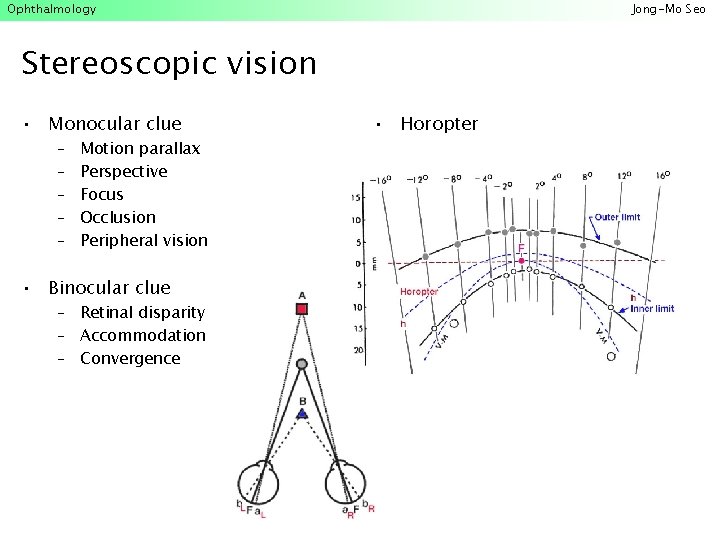
Ophthalmology Jong-Mo Seo Stereoscopic vision • Monocular clue – – – Motion parallax Perspective Focus Occlusion Peripheral vision • Binocular clue – Retinal disparity – Accommodation – Convergence • Horopter
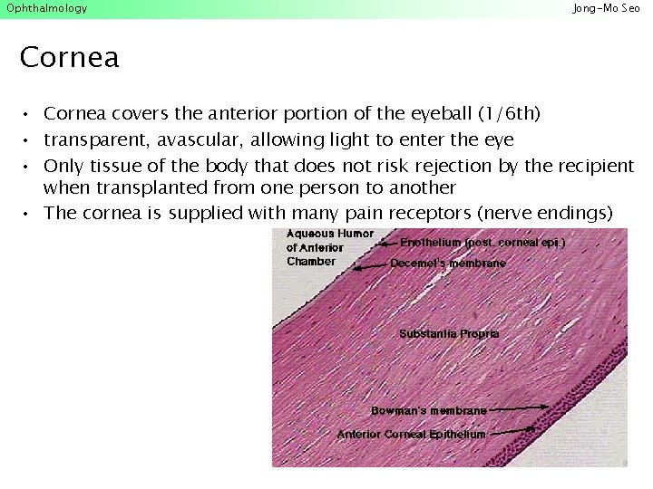
Ophthalmology Jong-Mo Seo Cornea • Cornea covers the anterior portion of the eyeball (1/6 th) • transparent, avascular, allowing light to enter the eye • Only tissue of the body that does not risk rejection by the recipient when transplanted from one person to another • The cornea is supplied with many pain receptors (nerve endings)
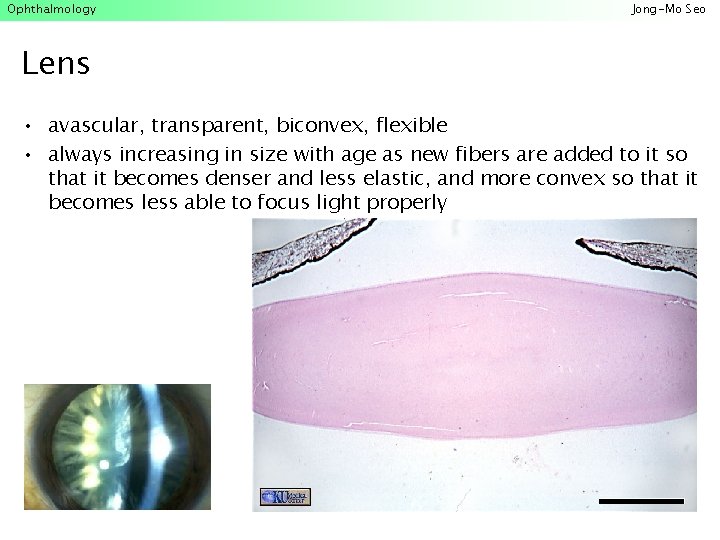
Ophthalmology Jong-Mo Seo Lens • avascular, transparent, biconvex, flexible • always increasing in size with age as new fibers are added to it so that it becomes denser and less elastic, and more convex so that it becomes less able to focus light properly
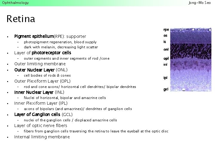
Ophthalmology Retina • Pigment epithelium(RPE): supporter • Layer of photoreceptor cells • • Outer limiting membrane Outer Nuclear Layer (ONL) • Outer Plexiform Layer (OPL) • Inner Nuclear Layer (INL) • Inner Plexiform Layer (IPL) • Layer of Ganglion cells (GCL) • Layer of optic nerve fibers • Internal limiting membrane – – photopigment regeneration, blood supply dark with melanin, decreasing light scatter – outer segments and inner segments of rod /cone – cell bodies of rods & cones – rod and cone axons/ horizontal cell dendrites/ bipolar dendrites – Nuclei of horizontal, bipolar and amacrine cells – axons of bipolars (and amacrines)/ dendrites of ganglion cells – nuclei of the ganglion cells / displaced amacrine cells – fibers from ganglion cells traversing the retina to leave the eyeball at the optic disc Jong-Mo Seo
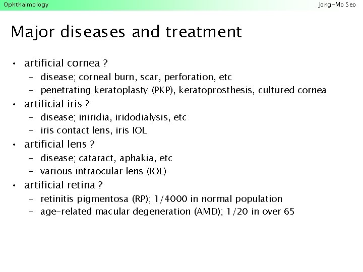
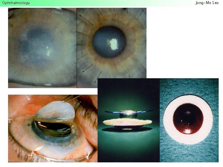
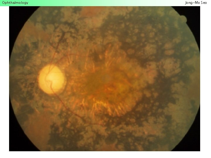
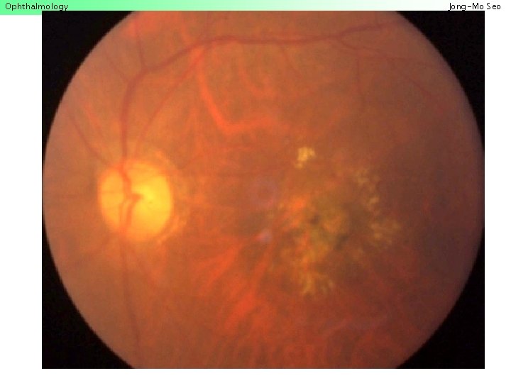
Ophthalmology Jong-Mo Seo Major diseases and treatment • artificial cornea ? – disease; corneal burn, scar, perforation, etc – penetrating keratoplasty (PKP), keratoprosthesis, cultured cornea • artificial iris ? – disease; iniridia, iridodialysis, etc – iris contact lens, iris IOL • artificial lens ? – disease; cataract, aphakia, etc – various intraocular lens (IOL) • artificial retina ? – retinitis pigmentosa (RP); 1/4000 in normal population – age-related macular degeneration (AMD); 1/20 in over 65
Ophthalmology Jong-Mo Seo
Ophthalmology Jong-Mo Seo
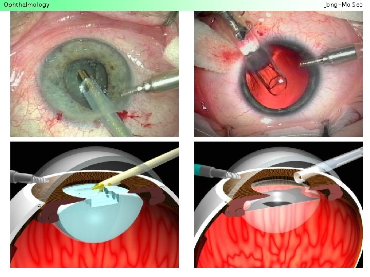
Ophthalmology Treatment modalities • • • Laser machine Phacoemusifier for cataract surgery Vitrectomy machine for retina surgery Operation microscope Microsurgical units Jong-Mo Seo
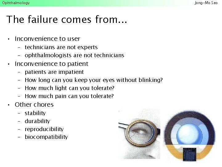
Ophthalmology The failure comes from. . . • Inconvenience to user – technicians are not experts – ophthalmologists are not technicians • Inconvenience to patient – – patients are impatient How long can you keep your eyes without blinking? How much light can you tolerate? How much pain can you tolerate? – – stability durability reproducibility biocompatibility • Other chores Jong-Mo Seo
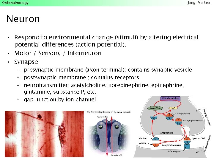
Ophthalmology Jong-Mo Seo Neuron • Respond to environmental change (stimuli) by altering electrical potential differences (action potential). • Motor / Sensory / Interneuron • Synapse – presynaptic membrane (axon terminal); contains synaptic vesicle – postsynaptic membrane ; contains receptors – neurotransmitter; acetylcholine, norepinephrine, glutamine, substance P, etc. – gap junction by ion channel
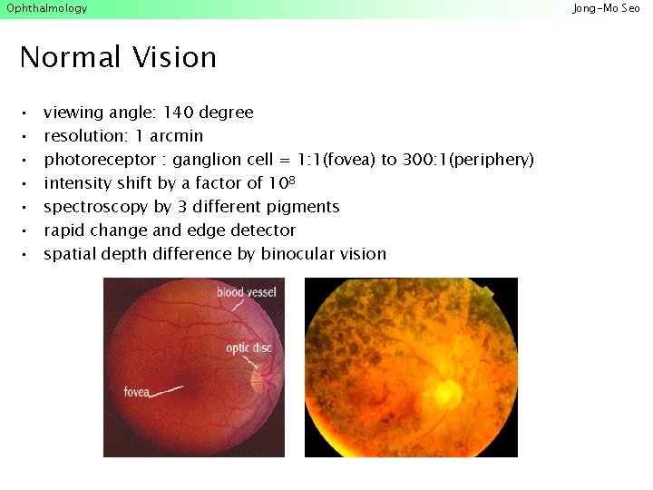
Ophthalmology Normal Vision • • viewing angle: 140 degree resolution: 1 arcmin photoreceptor : ganglion cell = 1: 1(fovea) to 300: 1(periphery) intensity shift by a factor of 108 spectroscopy by 3 different pigments rapid change and edge detector spatial depth difference by binocular vision Jong-Mo Seo
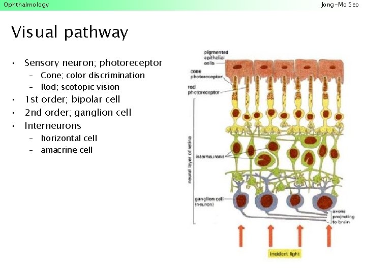
Ophthalmology Visual pathway • Sensory neuron; photoreceptor – Cone; color discrimination – Rod; scotopic vision • 1 st order; bipolar cell • 2 nd order; ganglion cell • Interneurons – horizontal cell – amacrine cell Jong-Mo Seo
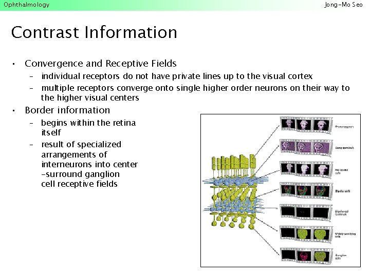
Ophthalmology Jong-Mo Seo Contrast Information • Convergence and Receptive Fields – individual receptors do not have private lines up to the visual cortex – multiple receptors converge onto single higher order neurons on their way to the higher visual centers • Border information – begins within the retina itself – result of specialized arrangements of interneurons into center -surround ganglion cell receptive fields
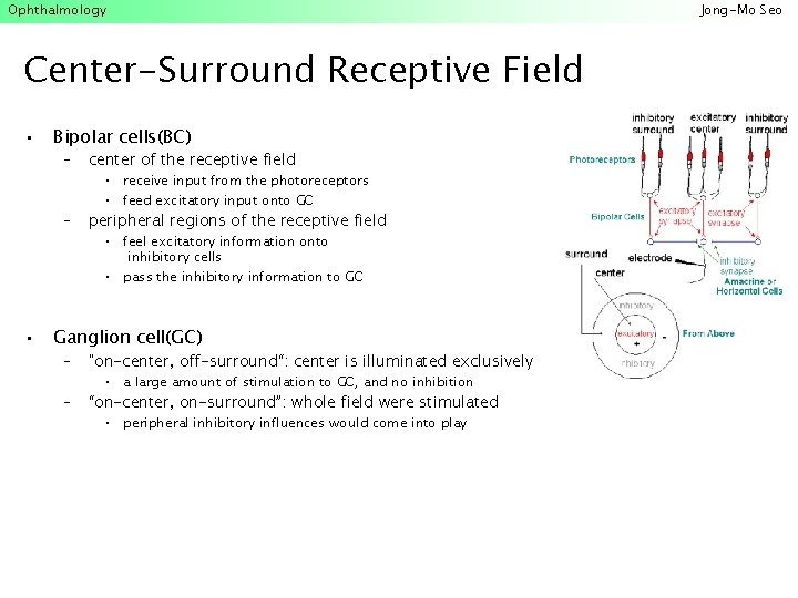
Ophthalmology Center-Surround Receptive Field • • Bipolar cells(BC) – center of the receptive field – peripheral regions of the receptive field • receive input from the photoreceptors • feed excitatory input onto GC • feel excitatory information onto inhibitory cells • pass the inhibitory information to GC Ganglion cell(GC) – "on-center, off-surround“: center is illuminated exclusively – “on-center, on-surround”: whole field were stimulated • a large amount of stimulation to GC, and no inhibition • peripheral inhibitory influences would come into play Jong-Mo Seo
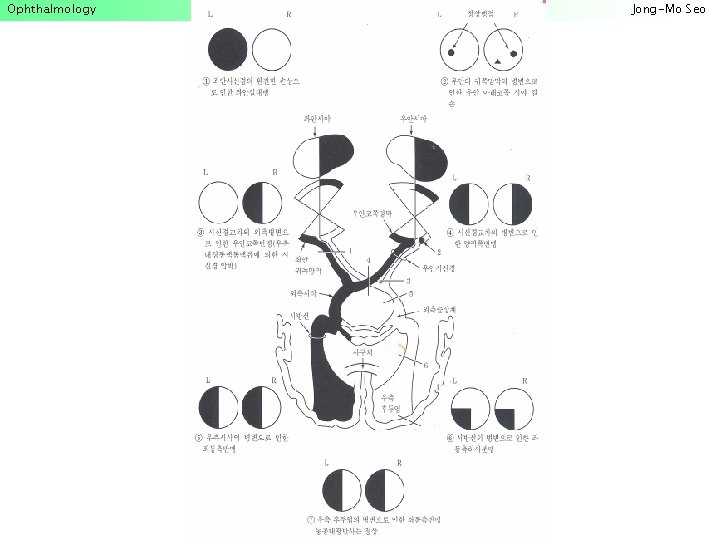
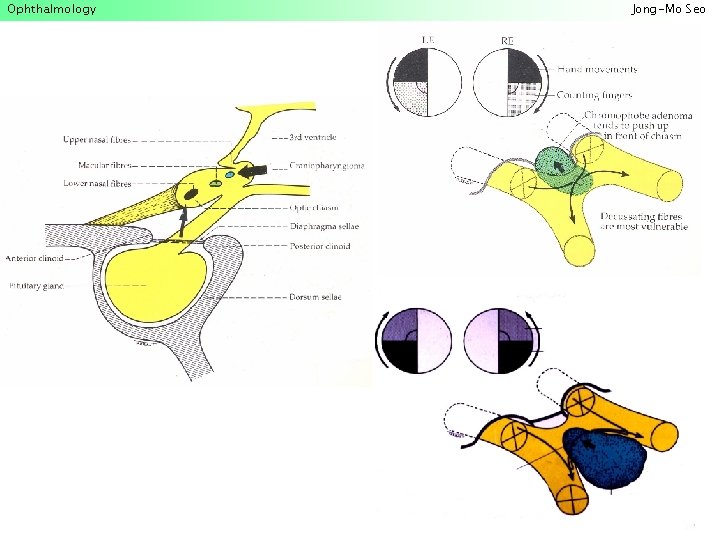
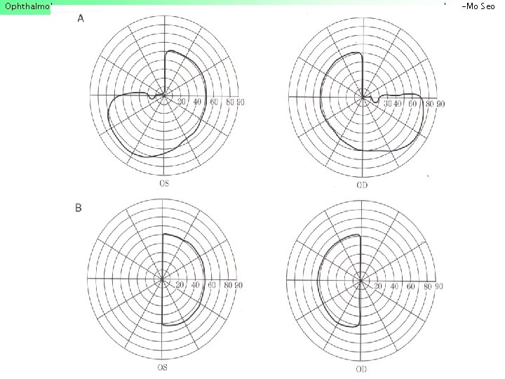
Ophthalmology Jong-Mo Seo Interaction with brain • Visual field; split in half and cross over • Optic nerve LGN primary visual(striate) cortex • Self-organization of the cortex: learning, organizing, testing • Artificial Neural Network: associative memory, task assignment problem, back propagation • Various "household chores" associated with vision – saccadic motion and compensation for it – the control of lens opening (pupillary reflex) – distance accommodation – binocular convergence
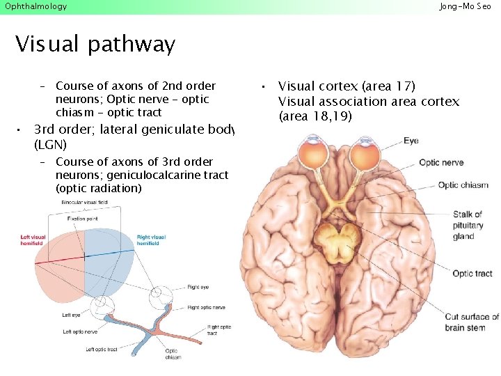
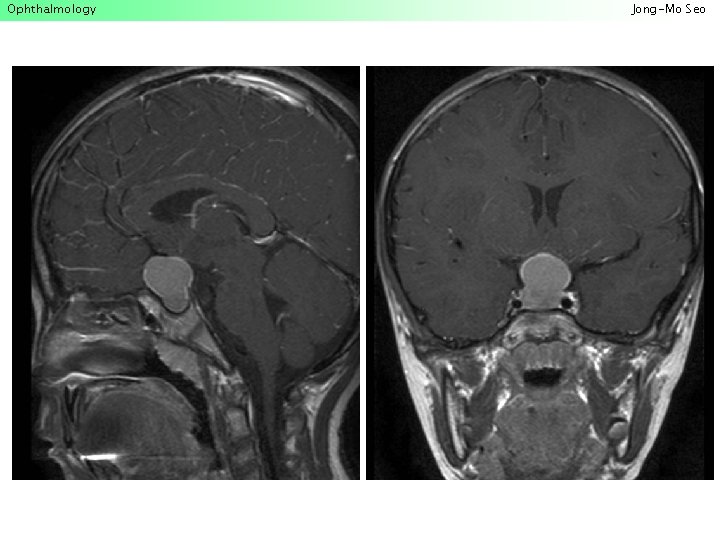
Ophthalmology Jong-Mo Seo Visual pathway – Course of axons of 2 nd order neurons; Optic nerve - optic chiasm - optic tract • 3 rd order; lateral geniculate body (LGN) – Course of axons of 3 rd order neurons; geniculocalcarine tract (optic radiation) • Visual cortex (area 17) Visual association area cortex (area 18, 19)
Ophthalmology Jong-Mo Seo
Ophthalmology Jong-Mo Seo
Ophthalmology Jong-Mo Seo
Ophthalmology Jong-Mo Seo
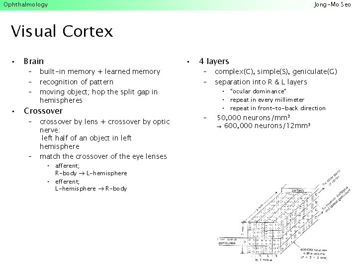
Ophthalmology Jong-Mo Seo Visual Cortex • • Brain – – – built-in memory + learned memory recognition of pattern moving object; hop the split gap in hemispheres Crossover – – crossover by lens + crossover by optic nerve: left half of an object in left hemisphere match the crossover of the eye lenses • afferent; R-body L-hemisphere • efferent; L-hemisphere R-body • 4 layers – – – complex(C), simple(S), geniculate(G) separation into R & L layers • "ocular dominance” • repeat in every millimeter • repeat in front-to-back direction 50, 000 neurons/mm 3 600, 000 neurons/12 mm 3
Ophthalmology Jong-Mo Seo Visual Cortex • Surface area of visual cortex – 105 neurons/mm 2 150 x 106 neurons/15 cm 2 – mainly in fovea; 150, 000 cones 1000 neurons/cone • Processing in primary visual cortex – mainly orientation response: direction of edge and line of visual images – fires when edge of particular orientation enters – all possible orientation; short line(9 neurons long) • Dissection and reconstruction of visual image – face recognition • dissection of smaller pieces of eyes, ears, nose • see eyes, ears, nose complete face grandmother – all in a fraction of second
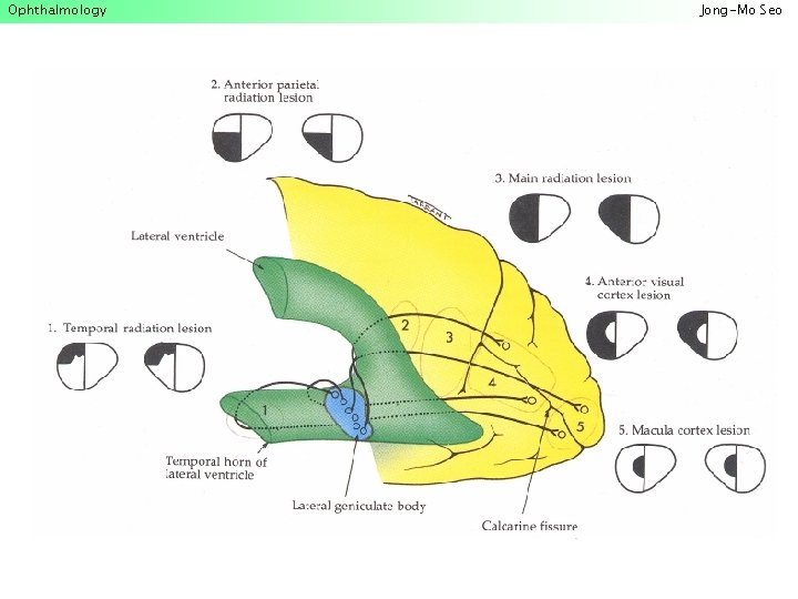
Ophthalmology Jong-Mo Seo
Ophthalmology Diagnostic tools • Subjective measurement – visual acuity, glare meter, contrast sensitivity, color test • Optical systems – Measuring cornea, anterior chamber, lens, vitreous to retina – slit lamp photography, keratometry, aberrometry, topography, pupillography, tonometry, optical coherent tomography • Radiological systems – Orbit CT, MRI, Ultrasonography • Electrophysiological systems – EOG, ERG, pattern ERG, multifocal VEP, VEP Jong-Mo Seo
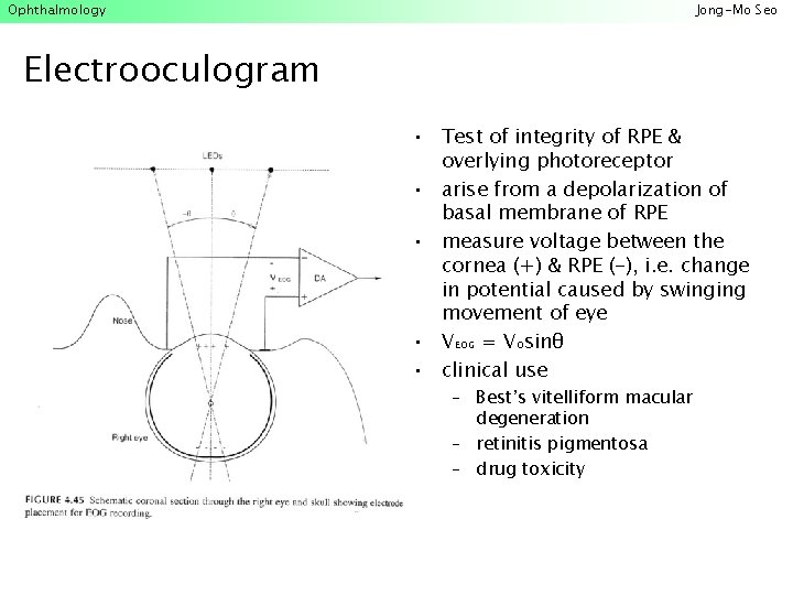
Ophthalmology Jong-Mo Seo Electrooculogram • Test of integrity of RPE & overlying photoreceptor • arise from a depolarization of basal membrane of RPE • measure voltage between the cornea (+) & RPE (-), i. e. change in potential caused by swinging movement of eye • VEOG = VOsinθ • clinical use – Best’s vitelliform macular degeneration – retinitis pigmentosa – drug toxicity
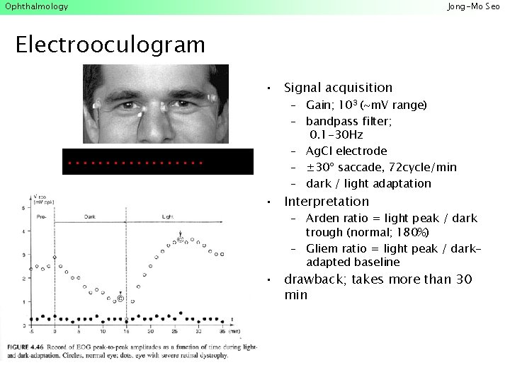
Ophthalmology Jong-Mo Seo Electrooculogram • Signal acquisition . . . . – Gain; 103 (~m. V range) – bandpass filter; 0. 1 -30 Hz – Ag. Cl electrode – ± 30º saccade, 72 cycle/min – dark / light adaptation • Interpretation – Arden ratio = light peak / dark trough (normal; 180%) – Gliem ratio = light peak / darkadapted baseline • drawback; takes more than 30 min
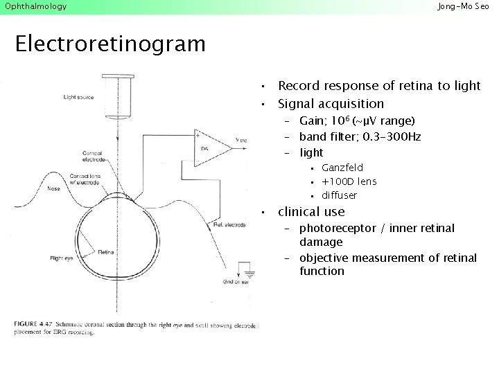
Ophthalmology Jong-Mo Seo Electroretinogram • Record response of retina to light • Signal acquisition – Gain; 106 (~μV range) – band filter; 0. 3 -300 Hz – light • Ganzfeld • +100 D lens • diffuser • clinical use – photoreceptor / inner retinal damage – objective measurement of retinal function
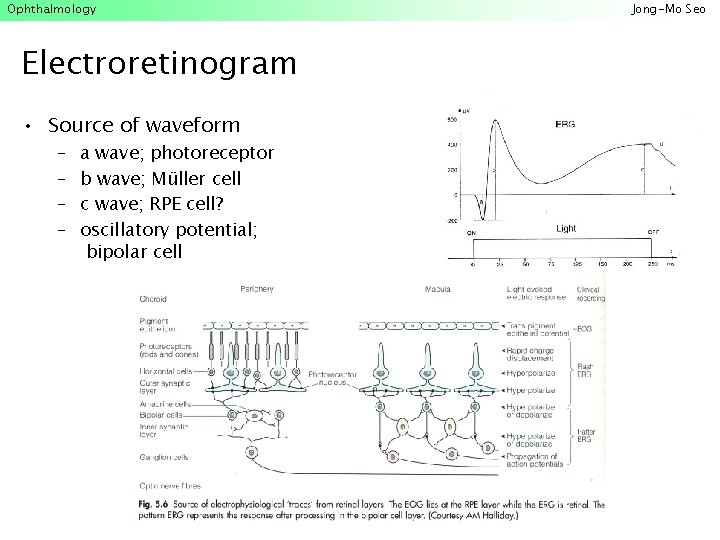
Ophthalmology Electroretinogram • Source of waveform – – a wave; photoreceptor b wave; Müller cell c wave; RPE cell? oscillatory potential; bipolar cell Jong-Mo Seo
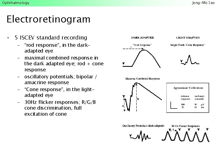
Ophthalmology Electroretinogram • 5 ISCEV standard recording – “rod response”, in the darkadapted eye – maximal combined response in the dark adapted eye; rod + cone response – oscillatory potentials; bipolar / amacrine response – “Cone response”, in the lightadapted eye – 30 Hz flicker responses; R/G/B cone discrimination, full excitation of cone Jong-Mo Seo
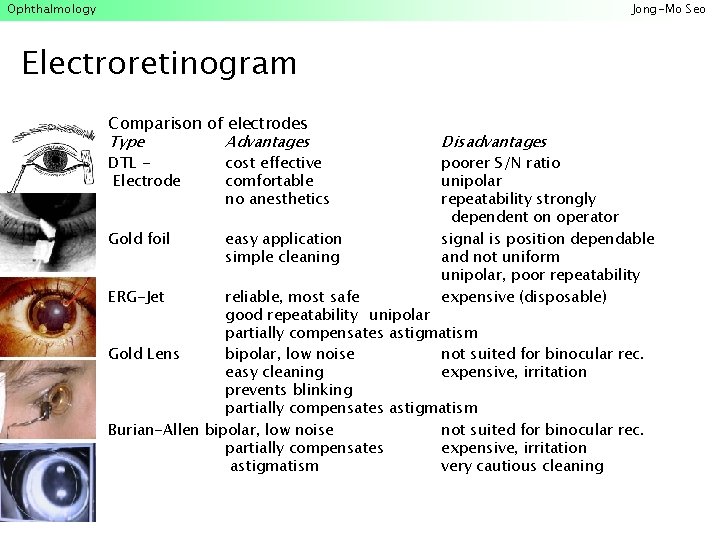
Ophthalmology Jong-Mo Seo Electroretinogram • • Comparison of electrodes Type Advantages DTL Electrode cost effective comfortable no anesthetics • Gold foil easy application simple cleaning • ERG-Jet • • Disadvantages poorer S/N ratio unipolar repeatability strongly dependent on operator signal is position dependable and not uniform unipolar, poor repeatability expensive (disposable) reliable, most safe good repeatability unipolar partially compensates astigmatism Gold Lens bipolar, low noise not suited for binocular rec. easy cleaning expensive, irritation prevents blinking partially compensates astigmatism Burian-Allen bipolar, low noise not suited for binocular rec. partially compensates expensive, irritation astigmatism very cautious cleaning
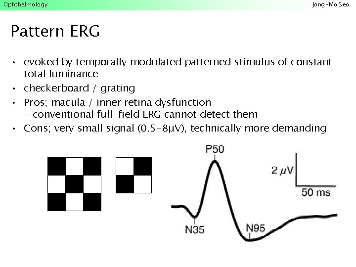
Ophthalmology Jong-Mo Seo Pattern ERG • evoked by temporally modulated patterned stimulus of constant total luminance • checkerboard / grating • Pros; macula / inner retina dysfunction - conventional full-field ERG cannot detect them • Cons; very small signal (0. 5 -8μV), technically more demanding
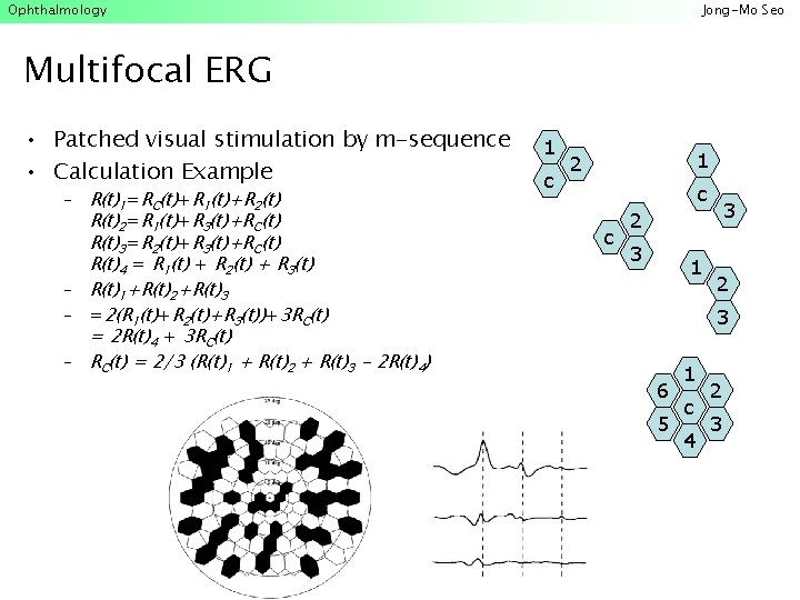
Ophthalmology Jong-Mo Seo Multifocal ERG • Patched visual stimulation by m-sequence • Calculation Example – R(t)1=RC(t)+R 1(t)+R 2(t) R(t)2=R 1(t)+R 3(t)+RC(t) R(t)3=R 2(t)+R 3(t)+RC(t) R(t)4 = R 1(t) + R 2(t) + R 3(t) – R(t)1+R(t)2+R(t)3 – =2(R 1(t)+R 2(t)+R 3(t))+3 RC(t) = 2 R(t)4 + 3 RC(t) – RC(t) = 2/3 (R(t)1 + R(t)2 + R(t)3 - 2 R(t)4) 1 c 1 2 c c 3 2 3 1 2 3 6 5 1 c 4 2 3
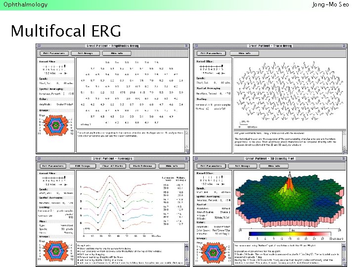
Ophthalmology Multifocal ERG Jong-Mo Seo
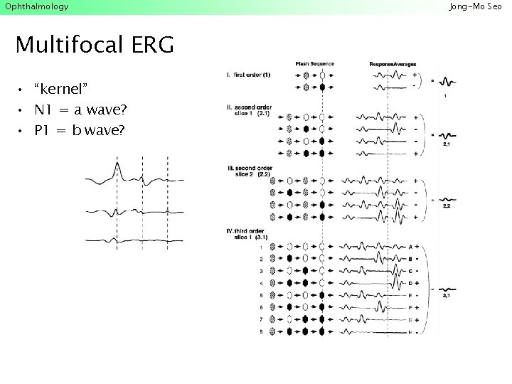
Ophthalmology Multifocal ERG • “kernel” • N 1 = a wave? • P 1 = b wave? Jong-Mo Seo
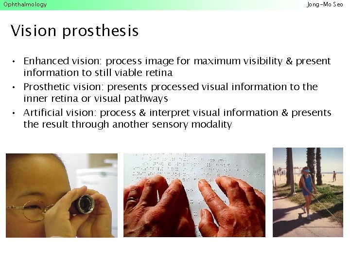
Ophthalmology Jong-Mo Seo Vision prosthesis • Enhanced vision: process image for maximum visibility & present information to still viable retina • Prosthetic vision: presents processed visual information to the inner retina or visual pathways • Artificial vision: process & interpret visual information & presents the result through another sensory modality
Ophthalmology Jong-Mo Seo Chronology • • • 1890 s 1906 1918 1928 1933 1943 1951 1953 1954 • • 1955 1959 • 1962 Animal-to-human organ grafts are tried, but failed Corneal transplantation succeeded Blood transfusions Iron lung developed for polio Human-to-human kidney transplant fails Kidney dialysis machine Artificial heart valve Heart-lung machine Kidney transplanted between identical twins; artificial hip implanted Heart-valve graft succeeded Kidney transplanted between nonidentical siblings Kidney transplanted from cadaver; silicone-filled breast implants
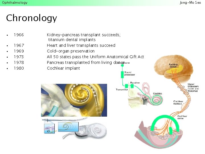
Ophthalmology Jong-Mo Seo Chronology • 1966 • • • 1967 1969 1973 1978 1980 Kidney-pancreas transplant succeeds; titanium dental implants Heart and liver transplants succeed Cold-organ preservation All 50 states pass the Uniform Anatomical Gift Act Pancreas transplanted from living donor Cochlear implant
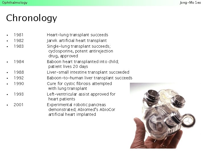
Ophthalmology Jong-Mo Seo Chronology • • • 1981 1982 1983 • 1984 • • • 1988 1992 1990 • 1993 • 2001 Heart-lung transplant succeeds Jarvik artificial heart transplant Single-lung transplant succeeds; cyclosporine, potent antirejection drug, approved Baboon heart transplanted into child; patient lives 20 days Liver-small intestine transplant succeeded Baboon-to-human liver transplant succeeds Cure for cystic fibrosis attempted with lung transplant Left-ventricular assist approved for heart patients Experimental robotic pancreas demonstrated; Abiomed’s Abio. Cor artificial heart implanted
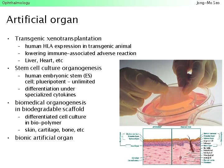
Ophthalmology Artificial organ • Transgenic xenotransplantation – human HLA expression in transgenic animal – lowering immune-associated adverse reaction – Liver, Heart, etc • Stem cell culture organogenesis – human embryonic stem (ES) cell; plueripotent - unlimited – differentiation under specialized cytokines • biomedical organogenesis in biodegradable scaffold – differentiated cell culture in bio-polymer – skin, cartilage, bone, etc • bionic artificial organ Jong-Mo Seo
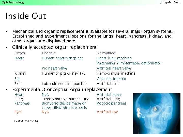
Ophthalmology Jong-Mo Seo Inside Out • Mechanical and organic replacement is available for several major organ systems. Established and experimental options for the lungs, heart, pancreas, kidney, and other organs are displayed here. • Clinically accepted organ replacement Organ Heart Kidney Ear Skin Organic Human heart transplant Pig heart valve Human or pig kidney TPL Lab-cultured skin patches Mechanical Heart-lung machine Pacemaker / Implantable defibrillator Artificial heart valve Hemodialysis machine Cochlear implant Artificial skin • Experimental/Conceptual organ replacement Heart Lung Pancreas Eyes SOURCE: Red Herring N/A Transplantable human lung Biohybrid device made of tubes filled with islet cells N/A Artificial heart Artificial lung Robotic pancreas Artificial Eye
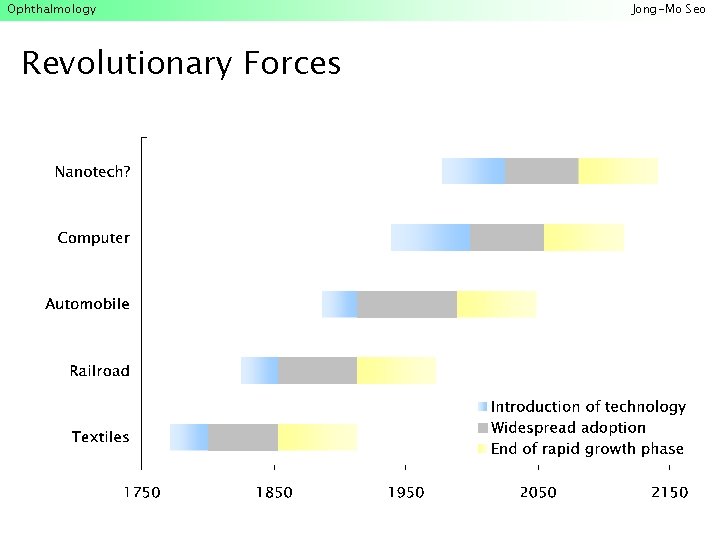
Ophthalmology Revolutionary Forces Jong-Mo Seo
Ophthalmology Jong-Mo Seo
Ophthalmology Jong-Mo Seo
Ophthalmology Jong-Mo Seo
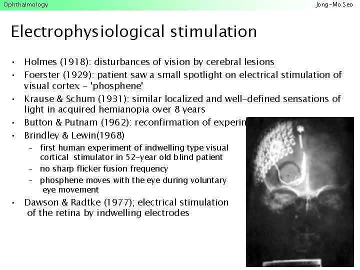
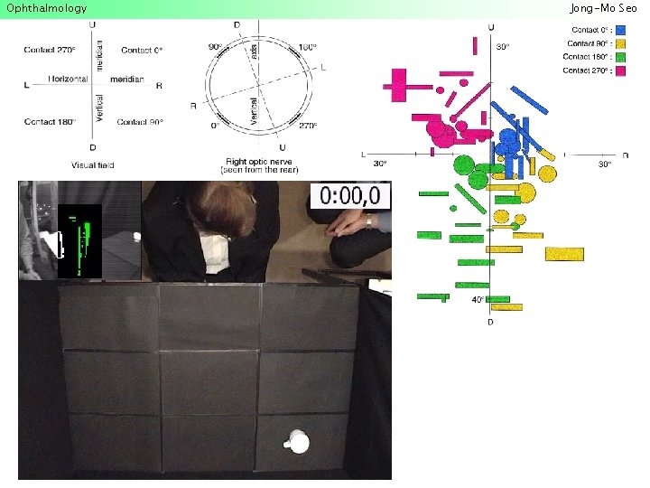
Ophthalmology Jong-Mo Seo Electrophysiological stimulation • Holmes (1918): disturbances of vision by cerebral lesions • Foerster (1929): patient saw a small spotlight on electrical stimulation of visual cortex - 'phosphene' • Krause & Schum (1931): similar localized and well-defined sensations of light in acquired hemianopia over 8 years • Button & Putnam (1962): reconfirmation of experiment • Brindley & Lewin(1968) – first human experiment of indwelling type visual cortical stimulator in 52 -year old blind patient – no sharp flicker fusion frequency – phosphene moves with the eye during voluntary eye movement • Dawson & Radtke (1977); electrical stimulation of the retina by indwelling electrodes
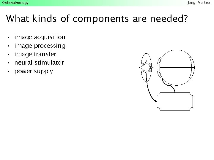
Ophthalmology What kinds of components are needed? • • • image acquisition image processing image transfer neural stimulator power supply Jong-Mo Seo
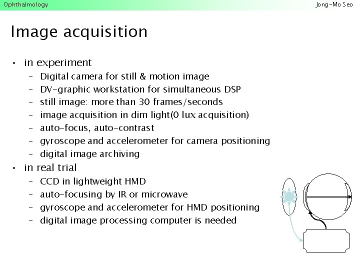
Ophthalmology Image acquisition • in experiment – – – – Digital camera for still & motion image DV-graphic workstation for simultaneous DSP still image: more than 30 frames/seconds image acquisition in dim light(0 lux acquisition) auto-focus, auto-contrast gyroscope and accelerometer for camera positioning digital image archiving – – CCD in lightweight HMD auto-focusing by IR or microwave gyroscope and accelerometer for HMD positioning digital image processing computer is needed • in real trial Jong-Mo Seo
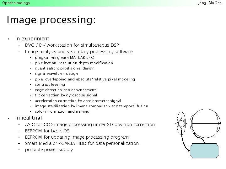
Ophthalmology Jong-Mo Seo Image processing: • • in experiment – – DVC / DV workstation for simultaneous DSP Image analysis and secondary processing software • • • programming with MATLAB or C pixelization: resolution depth modification quantization: pixel signal designal waveform design pixel overlapping and absolute/relative pixel modeling contrast leveling edge detection and enhancement tilt correction by gyroscope signal acceleration correction by accelerometer signal image stabilization by image comparison and temporal fusion color information and naming in real trial – – – ASIC for CCD image processing under 3 D position correction EEPROM for basic OS EEPROM for updating image processing program Smart Media or PCMCIA HDD for data personalization portable power supply
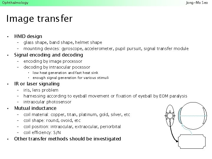
Ophthalmology Jong-Mo Seo Image transfer • HMD design • Signal encoding and decoding – – glass shape, band shape, helmet shape mounting devices: gyroscope, accelerometer, pupil pursuit, signal transfer module – – encoding by image processor decoding by intraocular pocessor • low heat generation and fast heat sink • enough signal generation for various stimuli • IR or laser signaling • Mutual inductance • Other transfer methods should be investigated – – – iris, lens problem harnessing according to eyeball movement or fixation of eyeball by EOM paralysis intraocular photosensor – – coil material: copper, titan, platinum, gold, silver, etc shape: round, ovoid, etc position: intraocular, extraocular, periorbital efficiency: S/N
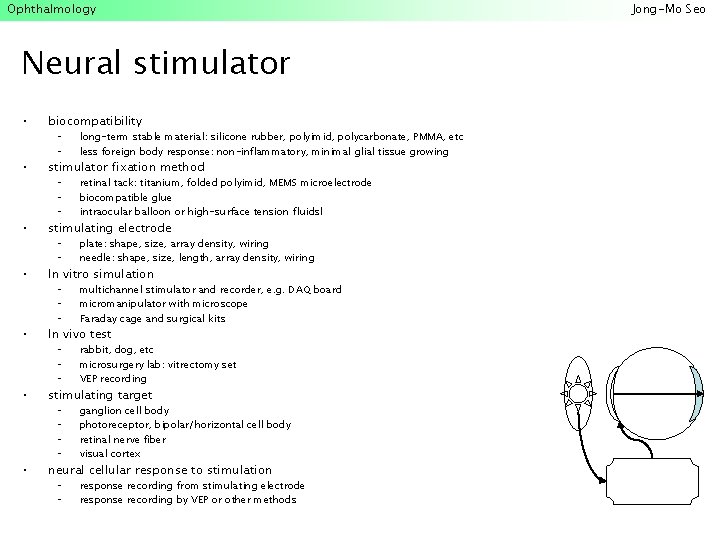
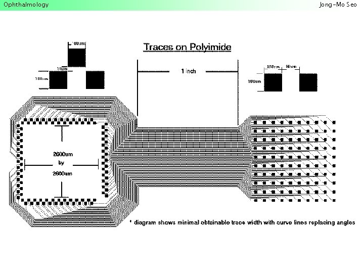
Ophthalmology Neural stimulator • biocompatibility • stimulator fixation method • stimulating electrode • In vitro simulation • In vivo test • stimulating target • neural cellular response to stimulation – – long-term stable material: silicone rubber, polyimid, polycarbonate, PMMA, etc less foreign body response: non-inflammatory, minimal glial tissue growing – – – retinal tack: titanium, folded polyimid, MEMS microelectrode biocompatible glue intraocular balloon or high-surface tension fluidsl – – plate: shape, size, array density, wiring needle: shape, size, length, array density, wiring – – – multichannel stimulator and recorder, e. g. DAQ board micromanipulator with microscope Faraday cage and surgical kits – – – rabbit, dog, etc microsurgery lab: vitrectomy set VEP recording – – ganglion cell body photoreceptor, bipolar/horizontal cell body retinal nerve fiber visual cortex – – response recording from stimulating electrode response recording by VEP or other methods Jong-Mo Seo

Ophthalmology Power supply • microcomputer power supply • intraocular or cortical stimulator power supply – IR or laser – Mutual inductance – others Jong-Mo Seo
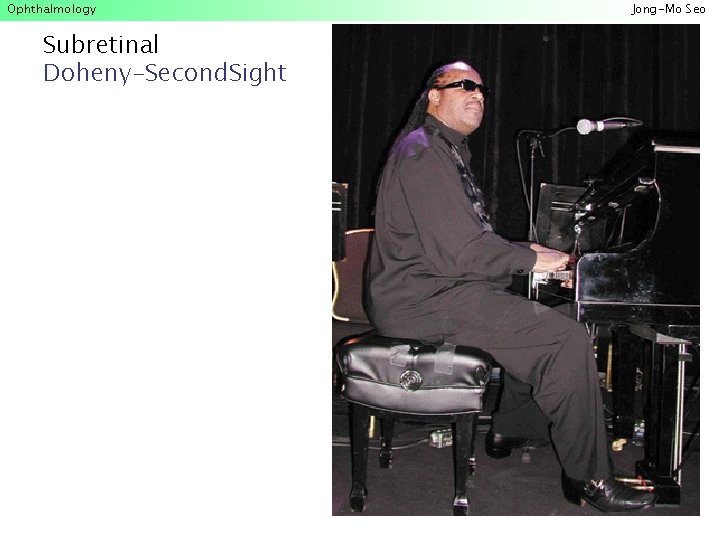
Ophthalmology Subretinal Doheny-Second. Sight Jong-Mo Seo
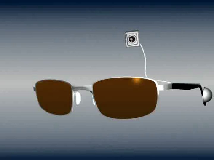
Ophthalmology Jong-Mo Seo
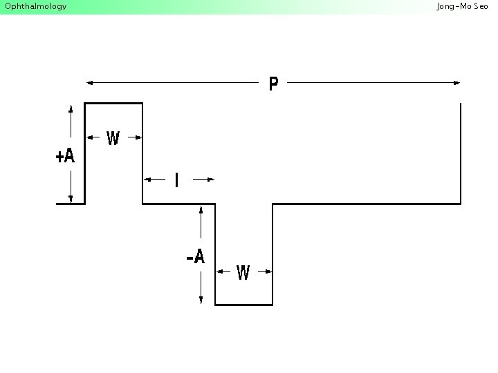
Ophthalmology Jong-Mo Seo
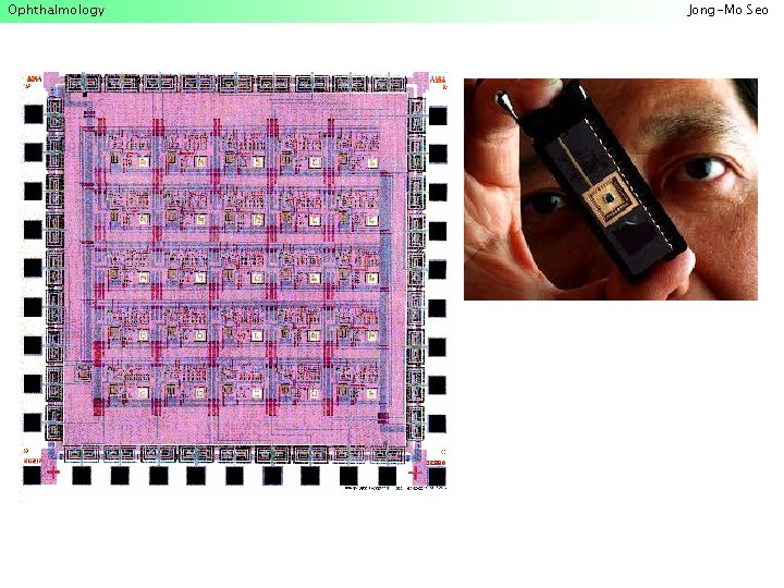
Ophthalmology Jong-Mo Seo
Ophthalmology Jong-Mo Seo
Ophthalmology Jong-Mo Seo
Ophthalmology Jong-Mo Seo
Ophthalmology Jong-Mo Seo
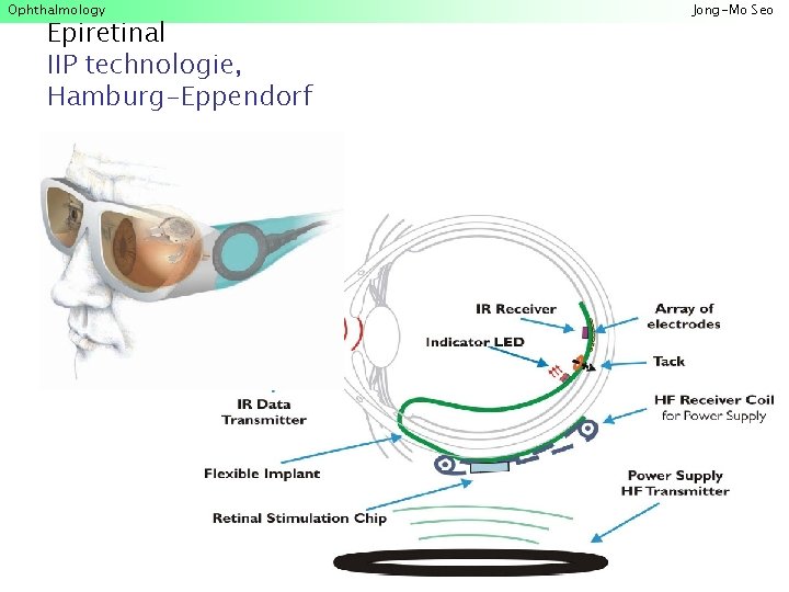
Ophthalmology Epiretinal IIP technologie, Hamburg-Eppendorf Jong-Mo Seo
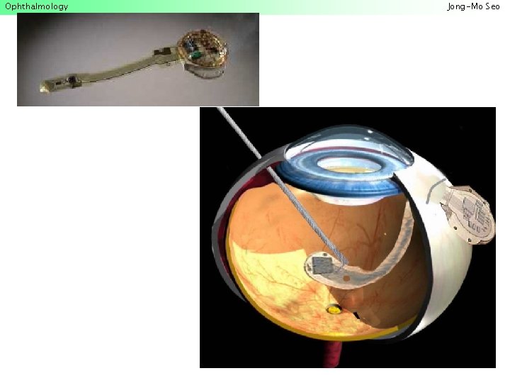
Ophthalmology Jong-Mo Seo
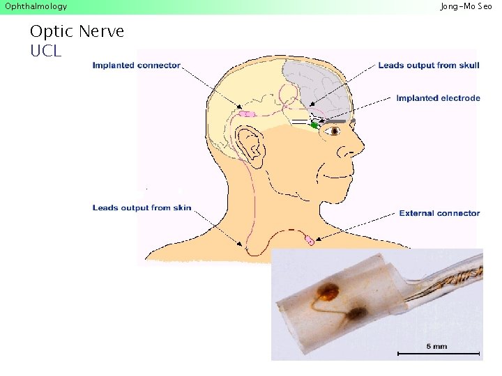
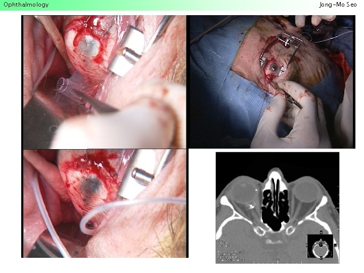
Ophthalmology Optic Nerve UCL Jong-Mo Seo
Ophthalmology Jong-Mo Seo
Ophthalmology Jong-Mo Seo
Ophthalmology Jong-Mo Seo
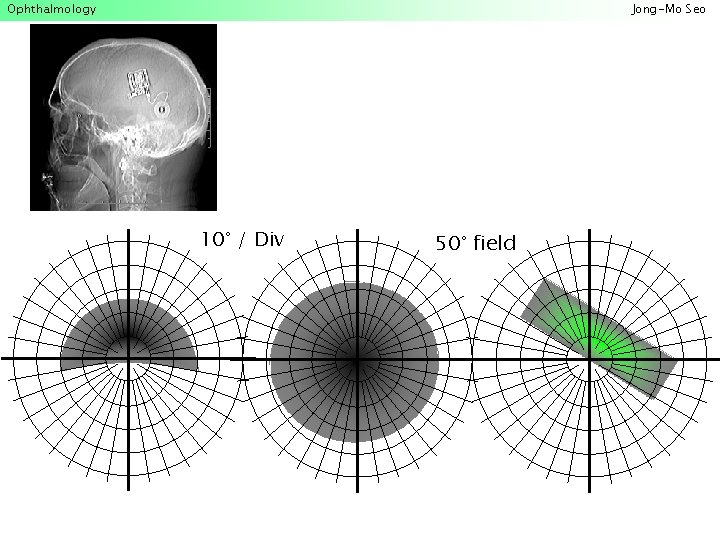
Ophthalmology Jong-Mo Seo 10° / Div 50° field
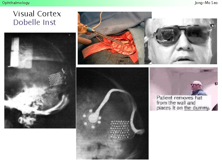
Ophthalmology Visual Cortex Dobelle Inst Jong-Mo Seo
- Slides: 78