
Chronic Visual Loss UBC Ophthalmology Club 2012 Approach
Chronic Visual Loss UBC Ophthalmology Club 2012
Approach � History, physical, tests � Patient population tends to be the elderly, but 2% of adults in the US over age 40 have vision <20/40 (Congdon et al. 2004. Arch Ophthalmol 122(4): 477 -85. ) � Prevalence increases with age � Early detection may lead to early intervention and preservation of vision � Primary care is the first screen, know when to refer
Case 1 � 55 M comes to GP for routine physical � Has HTN, currently on thiazide � Denies visual loss, eye pain, headaches � Sister was taking an eye drop but not sure what that’s for � Exam: � OD 20/30 OS 20/30 (both 20/25 2 years ago) � Pupils equal reactive � No RAPD � EOM full � Confrontational VF grossly intact
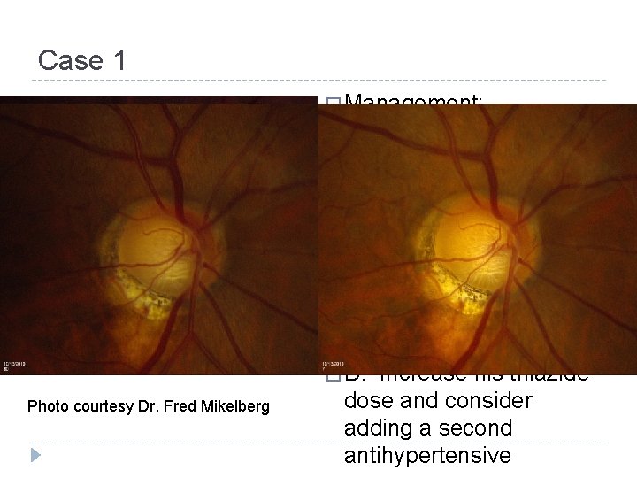
Case 1 � Management: � A. Photo courtesy Dr. Fred Mikelberg This pt needs urgent treatment to lower his IOP � B. Refer pt to ophthalmologist 1 -2 weeks � C. Reassess pt in 3 months � D. Increase his thiazide dose and consider adding a second antihypertensive

Transillumination defect. (Kuo & Noecker, AAO 2009) Pseudoexfoliation. (Shaw, AAO 2003)
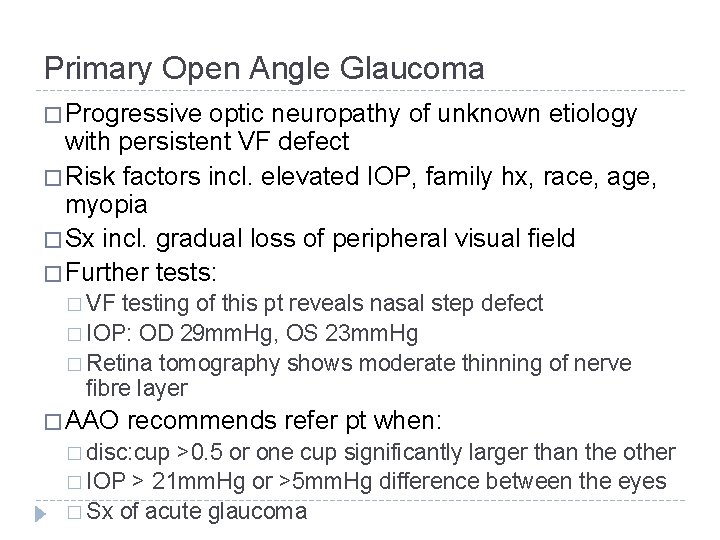
Primary Open Angle Glaucoma � Progressive optic neuropathy of unknown etiology with persistent VF defect � Risk factors incl. elevated IOP, family hx, race, age, myopia � Sx incl. gradual loss of peripheral visual field � Further tests: � VF testing of this pt reveals nasal step defect � IOP: OD 29 mm. Hg, OS 23 mm. Hg � Retina tomography shows moderate thinning of nerve fibre layer � AAO recommends refer pt when: � disc: cup >0. 5 or one cup significantly larger than the other � IOP > 21 mm. Hg or >5 mm. Hg difference between the eyes � Sx of acute glaucoma
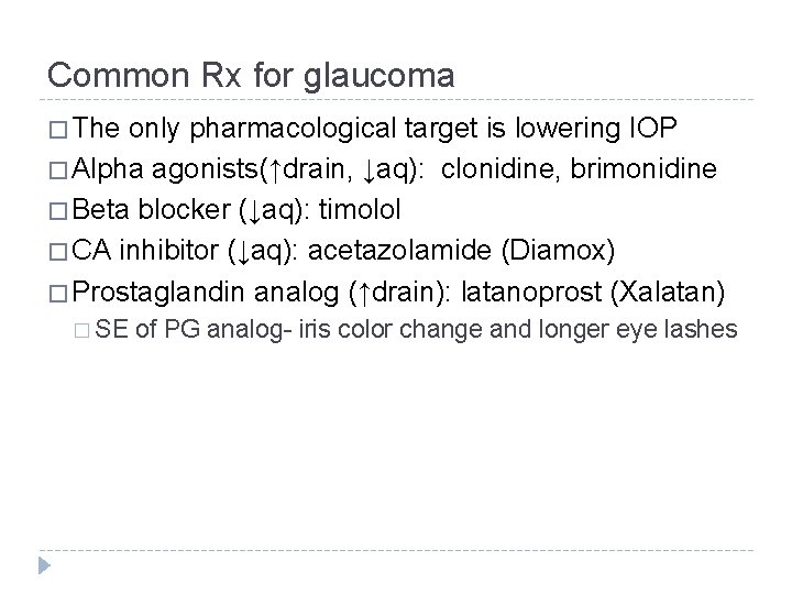
Common Rx for glaucoma � The only pharmacological target is lowering IOP � Alpha agonists(↑drain, ↓aq): clonidine, brimonidine � Beta blocker (↓aq): timolol � CA inhibitor (↓aq): acetazolamide (Diamox) � Prostaglandin analog (↑drain): latanoprost (Xalatan) � SE of PG analog- iris color change and longer eye lashes
Case 2 � 70 M c/o decreasing vision in both eyes over last 6 months to GP � This is particularly bothersome as he is having more trouble reading and watching TV � No eye conditions in the past � Hx significant for obesity and 50 pkyr smoking, quit 5 years ago � Family history unremarkable
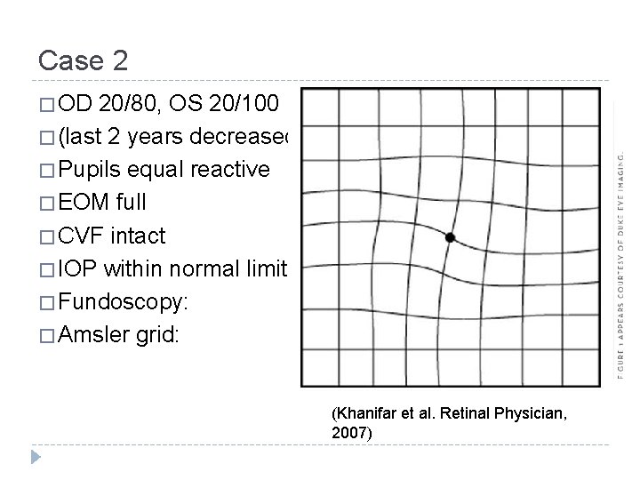
Case 2 � OD 20/80, OS 20/100 � (last 2 years decreased) � Pupils equal reactive � EOM full � CVF intact � IOP within normal limits � Fundoscopy: � Amsler grid: (Khanifar et al. Retinal Physician, 2007)
� What do you tell this patient? � A. he has missed the window for effective intervention � B. he needs immediate antioxidant and zinc supplement � C. his children are at increased risk of this disease � D. his condition probably won’t cause complete blindness
Age Related Macular Degeneration � 2 forms: atrophic (dry) and exudative (wet) � Leading cause of blindness in adults >75 yr, mostly from exudative form � Multifactorial disease, see characteristic drusen � Early diagnosis enables detection of exudative form, which can be effectively treated with anti-VEGF agents � Screening in primary care: � Visual distortions, especially in central vision � Presence of drusen in macula, retinal pigment breakdown � Refer to ophthalmologist for full evaluation
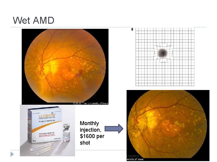
Wet AMD Monthly injection, $1600 per shot
Case 3 � 68 F comes to GP with c/o decreased vision in her L eye � She denies double vision or glares, in fact she said she can read better with her L eye than her R eye now; she wants to know if her reading glasses are still necessary � No eye disease or trauma � No family hx of eye diseases � Meds include prednisone 20 mg daily for last 2 months for RA flare
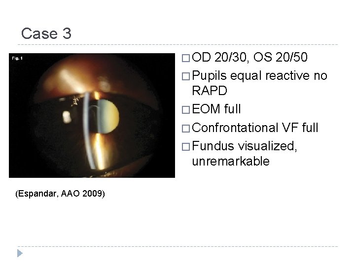
Case 3 � OD 20/30, OS 20/50 � Pupils equal reactive no RAPD � EOM full � Confrontational VF full � Fundus visualized, unremarkable (Espandar, AAO 2009)
Management � What’s your course of action? � A. Inform pt that her cataract is the result of her prednisone use � B. This pt needs to see an ophthalmologist STAT because of risk of irreversible visual loss � C. This pt’s presbyopia is improving so she should be followed up in 6 months at your office � D. Referral to ophthalmologist for evaluation and treatment options
Cataract � Etiology: opacified lens � Most commonly associated with increasing age, but also congenital, DM, steroid use, trauma, radiation � Pt complain of painless gradual unilateral vision decrease � “Second sight” refers to myopic shift as cataract increases power of lens; this is temporary � Referral to ophthalmologist when decrease in vision becomes symptomatic and/or interfere with function � Cataract removal+IOL implant is one of the most frequently-performed and successful procedures in all of surgery
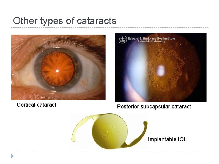
Other types of cataracts Cortical cataract Posterior subcapsular cataract Implantable IOL
Case 4 � 63 M with 17 yr hx of Type 2 DM comes to GP to c/o decrease in vision in both eyes � Denies pain, distortions, double vision � Hb A 1 c 7. 5% despite being on metformin and gliclazide � Also has dyslipidemia, on atorvastatin � No previous eye complaints
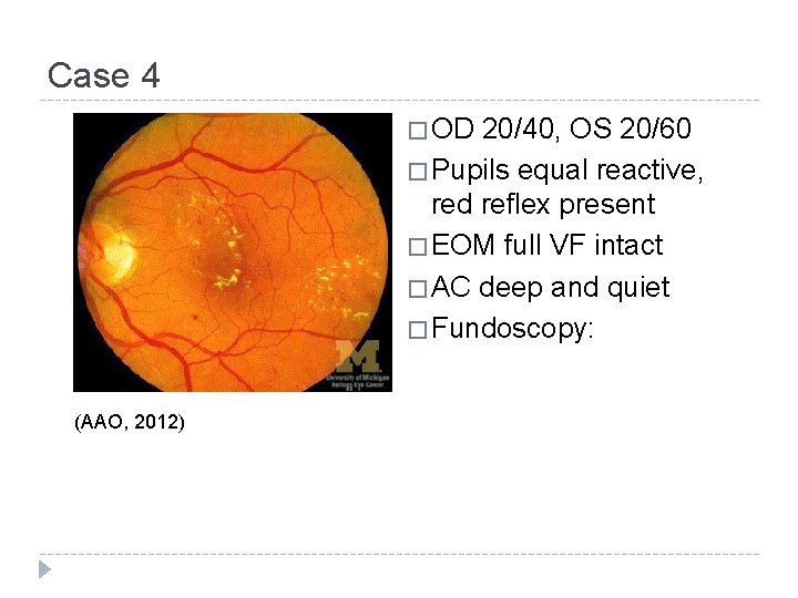
Case 4 � OD 20/40, OS 20/60 � Pupils equal reactive, red reflex present � EOM full VF intact � AC deep and quiet � Fundoscopy: (AAO, 2012)
Diagnosis � What is the cause of this pt’s decreased vision? � A. Non-proliferative diabetic retinopathy � B. Age related macular degeneration � C. Proliferative diabetic retinopathy � D. Branch retinal vein occlusion
Diabetic retinopathy � Microvascular complication of DM � Most common cause of vision loss in adults 25 -74 yr � In NPDR, vision loss arise from macular edema � In PDR, vision loss can be rapid, secondary to scarring and vitreous hemorrhage � Ophthalmologist referral when: � Newly diagnosed DM patient � Eye exam every 1 -2 years after � Patient who develop rapid vision change � Glycemic control is the cornerstone of systemic management. DR is managed with laser and anti. VEGF
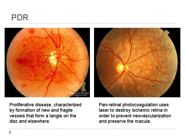
PDR Proliferative disease, characterized by formation of new and fragile vessels that form a tangle on the disc and elsewhere. Pan-retinal photocoagulation uses laser to destroy ischemic retina in order to prevent neovascularization and preserve the macula.
Summary � 4 most common causes of chronic visual loss and their features: � Open angle glaucoma- insidious, treat IOP � Age related macular degeneration- distortions, most common � Cataract- often unilateral, good result with surgery � Diabetic retinopathy- check in all DM pt, bilateral visual loss � All are either reversible or can be managed well (slow/stop vision loss) if detected early � Therefore, primary care’s role is vital in screening of chronic eye diseases
Questions? � Edited by: Steven Schendel, PGY-4 � Reviewed by: Drs. Fred Mikelberg, David Maberley, Francis Law � Contact: �R Tom Liu � rztom. liu@gmail. com
- Slides: 24