THORAX DANIK AGUSTIN P DEPARTEMENT OF ANATOMYHISTOLOGY MEDICAL


 Intercostalis Internus Expiration")


 1. Medial")
 1. Lower")
 1.")


 · Two phases · Inspiration – flow of air")


- Slides: 32
THORAX DANIK AGUSTIN P. DEPARTEMENT OF ANATOMY-HISTOLOGY MEDICAL FACULTY BRAWIJAYA UNIVERSITY
THORAX • THORAX : The part of the body between the neck and abdomen • FUNCTION : n Breathing n Protection of vital organs n Conduit
A. THE BORDER OF THORAX n CRANIAL: The line that connect incisura jugularis sterni articulatio coraco clavicularis processus spinosus of VIIth vertebra cervicalis n CAUDAL: The line that connect procecessus xyphoideus arcus costarum end part of x-xii th thoracalis vertebra
n ANTERIORLY : Sternum Cartilago costa Bagian ventral costa. n POSTERIORLY : Vertebra thoracalis I – XII. Bagian posterior costae n LATERALLY : Corpus costae.
B. THE SHAPE OF THORAX The Shape Of Thorax Are Determined By : The n bone of thorax muscle of thorax age sex
DEVELOPMENT OF THORAX SHAPE At birth : the ratio of anteroposterior distance and transversal distance is 1 n At infant age : ap < transversal. n At adult age : ap << transversal. n n Index thoracis: antero posterior distance transversal distance
THE ANOMALIS AND DEFORMITIES OF THORAX SHAPE THORAX PARALITICUS: a long narrow chest with emacipation so that the ribs standout sharply uner the skin. BARREL CHEST : a rounded, bulging chest, showing little movement on respiration seen in emphysema. PHITINOID/ ALAR/ FLAT CHEST : a chest in which it is flattened from before back: so called as indicating a tubercular diathese. FUNNEL CHEST : a chest in which there is funnel shaped depression in the middle of the anterior thoracic wall, the deepest part beeing in sternum, called also pectus excavatus. PIGEON BREAST: a chest in which there is prominence of sternum
C. CAVUM THORACIS n Enclosed by the thoracic wall & the diaphragm n Subdivided into 3 compartments : - 2 pleural cavity - mediastinum n RELATIONS Cranially : Communicates with neck ( colli ) by Apertura thoracis superior Caudally : Communicate with abdomen by Apertura thoracis inferior
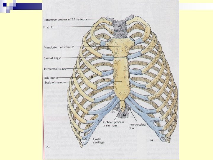
n APERTURA THORACIS SUPERIOR : • posterior : the 1 st thoracal vertebrae • lateral : the 1 st pair of ribs • anterior : Incisura jugularis sterni n APERTURA THORACIS INFERIOR : • posterior : the 12 th thoracal vertebrae • posterolat. : the 11 th & 12 th pair of ribs • anterolateral: joined costal cartilages of ribs 7 -10 • anterior : articulatio xiphysternalis.
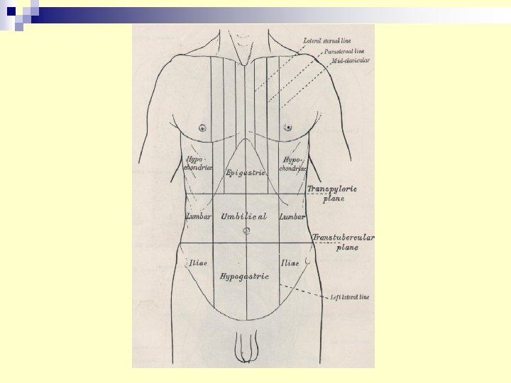
D. THORACIC WALL LINES AT THORACIS WALL : n n n n n Mid Sternal Line Parasternal Line Mid Clavicular Line. Anterior Axillary Line Midaxillary Line Posterior Axillary Line. Scapular Line Vertebral Line.
THORACIC WALL FASCIA : n Fascia pectoralis superficialis n Fascia thoracica externa n Fascia thoracica interna n Fascia endothoracica SKELETON : n 12 pairs of ribs n 12 thoracic vertebrae n Sternum MUSCLES :
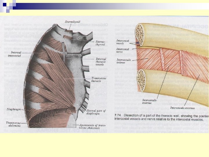
THE MUSCLES OF THORACIC WALL Function : n Alter the position of the ribs and sternum n Change thoracic volume during breathing
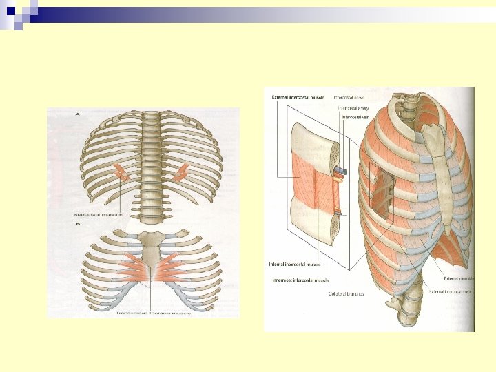
THORAX MUSCLES MUSCLE INNERVATION ACTION Intercostalis Externus Inspiration (Move Ribs Superiorly) Intercostalis Internus Expiration (Move Ribs Inferiorly) Act With Intercostalis internus Intercostalis Intima Nn. Intercostales Subcostalis Depress Ribs Transversus Thoracis Depress Costal Cartilages
VASCULARISATION & INNERVATION OF THORACIC WALL VASCULARISATION : n A. Intercostalis n A. Subcostalis n A. Thoracica Interna INNERVATION : n N. Intercostalis n N. Subcostalis
LYMPHATIC DRAINAGE OF THORACIC WALL n 1. 2. 3. Lymphatic node of thorax wall are Parasternal Phrenic. Intercostal.
PARASTERNAL NODE Location : along the upper part Receive lymph from. (Afferent) 1. Medial part of the breast. 2. Diaphragm. 3. Intercostalspace. 4. Costal pleura. Drains lymph to. (Efferent) Truncus bronchomediastinalis
PHRENIC NODE Location. Thoracic surface of diaphragm Receive lymph from ( afferent) 1. Lower intercostal space. 2. Pericardium. 3. Diaphragm. 4. Liver. Drain the lymph to (efferent) Parasternal node.
INTERCOSTAL NODE Location : vertebral end of intercostal space Receive lymph from (Afferent) 1. The structure along the adjacent blood vessel. 2. The pleura. Drain lymph into the thoracic duct.
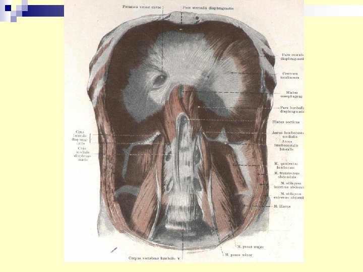
DIAPHRAGM Diaphragm divided into n Pars sternalis : have origin at posterior surface of sternum. n Pars costalis : have origin at inner surface of cartilage of VII - X th rib and XI – XIIth rib. n Pars lumbalis : consist of : - arcus lumbo-costalis lateralis and medialis - crura diaphragmatica dexter et sinister
MAJOR OPENINGS AT DIAPHRAGM n Hiatus aortae: Transmit aorta, n. splanchnicus major and ductus thoracicus n Hiatus oesophagii Transmit n. vagus and oesophagus. n Foramen venae cavae Transmit v. cava inferior and n. phrenicus dextra.
VASCULARISATION & INNERVATION OF DIAPHRAGM Blood Supply : n a. musculophrenica n a. pericardiacophrenica Innervation : N. Phrenicus
Mechanics of Breathing (Pulmonary Ventilation) · Two phases · Inspiration – flow of air into lung · Expiration – air leaving lung
Inspiration · Diaphragm and intercostal muscles contract · The size of the thoracic cavity increases · External air is pulled into the lungs due to an increase in intrapulmonary volume
Inspiration
Expiration · A passive process · depends on natural lung elasticity · As muscles relax, air is pushed out of the lungs · Forced expiration can occur by contracting internal intercostal muscles
Expiration Figure 13. 7 b
Nonrespiratory Air Movements · Can be caused by reflexes or voluntary actions · Examples · Cough and sneeze – clear lungs of debris · Laughing · Crying · Yawn · Hiccup